dental anatomy exam pulp & root sample question pool
 Call Kai
 Learn
 Practice Test
 Spaced Repetition
 Match
 Flashcards
 Knowt Play
1/53

There's no tags or description
Looks like no tags are added yet.
54 Terms
1. Maxillary canine
Of the following teeth, which is least likely to have two roots?
1. Maxillary canine
2. Mandibular canine
3. Maxillary first premolar
4. Maxillary second premolar
5. Mandibular first premolar
4. mesiobuccal root of the maxillary permanent first molar.
Two pulp canals are most frequently found in the
1. root of the mandibular second premolar.
2. lingual root of the maxillary permanent first molar.
3. distal root of the mandibular permanent second molar.
4. mesiobuccal root of the maxillary permanent first molar.
2. mandibular canine.
The anterior tooth most likely to have a bifurcated root is the
1. maxillary canine.
2. mandibular canine.
3. maxillary central incisor.
4. mandibular lateral incisor.
5. mandibular central incisor.
2. Maxillary first premolars.
There is a sharp demarcation between pulp chambers and pulp canals in which of the following teeth?
1. Mandibular second premolars.
2. Maxillary first premolars.
3 Maxillary lateral incisors.
4. Mandibular canines.
5. precollagenous and collagenous fibers.
The pulp consists of concentrated connective tissue cells located in a stroma of
1. Korff's fibers.
2. odontoblastic processes.
3. yellow elastic fibers.
4. oblique principal fibers.
5. precollagenous and collagenous fibers.
4. apical to the cervical level of the crown.
In a molar, root canals usually join the pulp chamber
1. at the level of the furcation.
2. at varying levels, dependent upon age.
3. within the cervical third of the crown.
4. apical to the cervical level of the crown.
4. odontoblasts.
The cells of the dental pulp which are responsible for the deposition of reparative dentin are
1. odontoclasts.
2. cementoblasts.
3. fibroblasts.
4. odontoblasts.
4. mesial root of mandibular permanent first molars.
Two pulp canals are commonly found in the
1. roots of maxillary permanent molars.
2. distal root of mandibular permanent first molars.
3. buccal root of maxillary first premolars.
4. mesial root of mandibular permanent first molars.
3. Mandibular canine
In which of the following roots would a divided pulp canal be most likely to occur?
1. Maxillary central incisor
2. Maxillary canine
3. Mandibular canine
4. Lingual root of maxillary first molar
5. Distofacial root of maxillary second molar
1. form dentin.
The primary function of the dental pulp is to
1. form dentin.
2. provide nutrition.
3. provide sensation.
4. assure root end closure.
5. protect the periodontium.
5. five.
At eruption, the number of pulp horns in a permanent mandibular first molar is usually
1. one.
2. two.
3. three.
4. four.
5. five.
5. cervical third of the crown.
A faciolingual, longitudinal section of a maxillary canine has its widest dimension of the pulp cavity faciolingually in the
1. middle third of the root.
2. apical third of the root.
3. incisal third of the crown.
4. middle third of the crown.
5. cervical third of the crown.
3. Mesiofacial
Which root of a permanent maxillary first molar is flattened
mesiodistally and has root depressions on its mesial and distal surfaces?
1. Lingual
2. Distofacial
3. Mesiofacial
4. Distolingual
5. Mesiolingual
3. flattened mesiodistally.
A cross section at mid root of a permanent mandibular central incisor is likely to show that the pulp cavity is
1. almost round.
2. divided into two canals.
3. flattened mesiodistally.
4. flattened faciolingually.
5. shaped much the same as that of a maxillary central incisor.
2. irregularly oval.
In cross section, the root of a mandibular canine is described as
1. roughly conical.
2. irregularly oval.
3. flattened in a mesiodistal direction.
4. broader mesiodistally on the lingual than on the labial.
1. Distal
Which pulp horn of a permanent mandibular first molar is the smallest?
1. Distal
2. Mesiofacial
3. Distofacial
4. Mesiolingual
5. Distolingual
4. forming secondary dentin.
Direct functions of the mature dental pulp include
1. forming enamel.
2. forming cementum.
3. forming primary dentin.
4. forming secondary dentin.
5. any aspect of the root.
Accessory root canals, when present, are located at
1. the bifurcation only.
2. the trifurcation only.
3. the apical region only.
4. the cervical region only.
5. any aspect of the root.
1. Maxillary first
In a cervical cross section, which premolar(s) sometimes exhibit a root outline and pulp chamber floor outline that are both kidney-shaped?
1. Maxillary first
2. Mandibular first
3. Maxillary second
4. Mandibular second
5. All premolars
2.
Which of the following cross section drawings, at mid root, would be most typical of a maxillary central incisor?
1.
2.
3.
4.

1. mesial aspect of a maxillary canine.
The sketch below represents a pulp cavity if a fully formed permanent tooth. It is most typical of the
1. mesial aspect of a maxillary canine.
2. mesial aspect of a mandibular second premolar.
3. facial aspect of a maxillary lateral incisor.
4. distal aspect of the distal root of a mandibular first molar.
5. None of the above

4. mesial aspect of a mandibular first molar.
The pulp cavity in the illustration below would probably be the
1. mesial aspect of a maxillary first molar.
2. buccal aspect of a mandibular first molar.
3. mesial aspect of a maxillary first premolar.
4. mesial aspect of a mandibular first molar.
5. lingual aspect of a maxillary second molar.

1. Right first
The mid root cross-sectional diagram below illustrated the root of which maxillary molar?
1. Right first
2. Left first
3. Right third
4. Left third

3. maxillary molar.
The sketches below of a cross section at the cemento-enamel junction are typical of a
1. mandibular second premolar.
2. maxillary first premolar.
3. maxillary molar.
4. mandibular molar.
5. number of different teeth and cannot be identified.

3. Maxillary left central incisor
The drawing below is a cross section at the cemento-enamel junction of what permanent tooth?
1. Maxillary right canine
2. Mandibular left canine
3. Maxillary left central incisor
4. Maxillary left canine
5. Maxillary right central incisor

5. maxillary first premolar.
The cross-sectional cut at mid root pictured below is most probably that of a
1. lingual root of a maxillary molar.
2. distal root of a mandibular first molar.
3. mandibular canine.
4. mandibular first premolar.
5. maxillary first premolar.

4. maxillary central incisor.
From a facial view, the sketch below is typical of a permanent
1. maxillary canine.
2. mandibular canine.
3. mandibular first premolar.
4. maxillary central incisor.

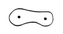
5. Mesial of a mandibular molar
The drawing below represents a cross section at mid root of which of the following teeth?
1. Mandibular canine
2. Maxillary canine
3. Distofacial of a maxillary molar
4. Lingual of a maxillary molar
5. Mesial of a mandibular molar
3. Maxillary right central incisor
The drawing below is a cross section at the cemento-enamel
junction of what permanent tooth?
1. Maxillary right canine
2. Mandibular right canine
3. Maxillary right central incisor
4. Mandibular right central incisor

2. Mandibular first molar
The drawing below of a cross section at the cemento-enamel junction is typical of which permanent tooth?
1. Maxillary first molar
2. Mandibular first molar
3. Maxillary second premolar
4. Mandibular second premolar
5. None of the above

3. Mandibular central incisor
In the drawings below, the outline of a cross section made at the cervix belongs to which of the following permanent teeth?
1. Maxillary canine
2. Maxillary lateral incisor
3. Mandibular central incisor
4. Mandibular second premolar

2. Left first
The mid root cross-sectional diagram below illustrates the root structure of which maxillary molar?
1. Right first
2. Left first
3. Right third
4. Left third

4. Maxillary right first premolar
The sketch below represents a cross section at mid root of which of the following permanent teeth?
1. Mandibular left canine
2. Mandibular right canine
3. Maxillary left first premolar
4. Maxillary right first premolar
5. Maxillary left second premolar

c. maxillary right central incisor
This is a mid-root cross section, incisal view. What tooth is it?
a. maxillary right lateral incisor
b. mandibular left cuspid
c. maxillary right central incisor
d. maxillary left central incisor
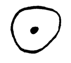
d. maxillary left central incisor
This is a mid-root cross section, apical view. What tooth is it?
a. maxillary right lateral incisor
b. mandibular left cuspid
c. maxillary right central incisor
d. maxillary left central incisor

d. maxillary central incisor
This is a mid-root cross section. What tooth is it?
a. maxillary lateral incisor
b. mandibular cuspid
c. maxillary first premolar
d. maxillary central incisor
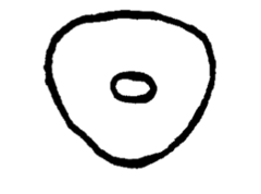
d. central incisor
This is a diagram of a mesiodistal pulp outline, it was most likely taken from a permanent maxillary
a. lateral incisor
b. cuspid
c. first premolar
d. central incisor

d. mandibular central incisor
This is a mid-root cross section of which tooth?
a. maxillary lateral incisor
b. maxillary cuspid
c. mandibular first premolar
d. mandibular central incisor

c. maxillary right first premolar
This is a mid-root cross section, apical view, of which tooth?
a. maxillary right central incisor
b. maxillary left cuspid
c. maxillary right first premolar
d. mandibular left second premolar

c. maxillary left first premolar
This is a mid-root cross section, occlusal view, of which tooth?
a. maxillary right central incisor
b. maxillary left cuspid
c. maxillary left first premolar
d. maxillary right second premolar

c. maxillary first premolar
This is a mid-root cross section of which tooth
a. maxillary central incisor
b. maxillary cuspid
c. maxillary first premolar
d. maxillary second premolar

a. mandibular first molar
c. 30
This is a cross section at the CEJ of which tooth
a. mandibular first molar
b. mandibular second molar
c. maxillary first molar
d. maxillary second molar
What is the tooth number?
a. 18
b. 15
c. 30
d. 31
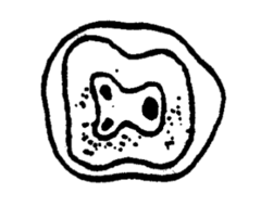
c. maxillary first molar
This is a cross section at the CEJ of which tooth?
a. mandibular first molar
b. mandibular second molar
c. maxillary first molar
d. maxillary second premolar
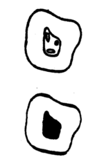
d. maxillary right first molar
This is a mid-root cross section occlusal view of which tooth
a. mandibular left first molar
b. mandibular left second molar
c. maxillary left first molar
d. maxillary right first molar

c. maxillary left first molar
This is a mid-root cross section apical view of which tooth
a. mandibular left first molar
b. mandibular left second molar
c. maxillary left first molar
d. maxillary right first molar

a. mesial root of mandibular molar
This is a mid-root cross section view of which tooth
a. mesial root of mandibular molar
b. mandibular second premolar
c. maxillary first molar
d. maxillary first premolar

b. mesiobuccal root canal
This diagram is a mid-root cross section. The root canal pointed to by arrow "A" is correctly identified as the
a. lingual root canal
b. mesiobuccal root canal
c. mesiolingual root canal
d. distobuccal root canal
e. distolingual root canal
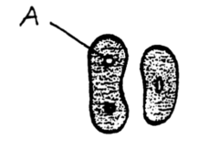
a. lingual surface
This diagram is a mid-root cross section. The outline pointed to by arrow "A" is most likely the
a. lingual surface
b. mesial surface
c. buccal surface
d. distal surface

17. maxillary canine - mid-root section - D
18. maxillary canine - mesiodistal section - A
19. maxillary second premolar - buccolingual section - B
17. maxillary canine - mid-root section
18. maxillary canine - mesiodistal section
19. maxillary second premolar - buccolingual section

20. maxillary canine - faciolingual section - C
21. maxillary central - cervical section - E
20. maxillary canine - faciolingual section
21. maxillary central - cervical section

b. Calcium phosphate
Which of the following is contained in greatest proportion in tooth enamel?
a. Calcium fluoride
b. Calcium phosphate
c. Calcium carbonate
d. Potassium carbonate
e. Potassium phosphate
b. Cementum, dentin, enamel
Which of the following is the correct sequence of dental tissues from softest to hardest?
a. Dentin, cementum, enamel
b. Cementum, dentin, enamel
c. Dentin, enamel, cementum
d. Cementum, enamel, dentin
e. deposition of secondary dentin.
Following the initial period of functional activity, an appreciable alteration in size of the pulp chamber is a direct result of
a. pulp fibrosis.
b. morphodifferentiation.
c. dentinal dysplasia.
d. deposition of primary dentin.
e. deposition of secondary dentin.
c. 20-30%.
The percentage of dentin that is organic is
a. 3-5%.
b. 10-15%.
c. 20-30%.
d. 40-50%.
e. 50-60%.