Muscle and Tendon Histology SDL (NMSK)
 Call Kai
 Learn
 Practice Test
 Spaced Repetition
 Match
 Flashcards
 Knowt Play
1/39

Earn XP
Description and Tags
Distinguish between smooth, skeletal and cardiac muscle on histological preparations Identify the main features of muscle, tendon and tendon sheaths on histological preparations
40 Terms
•LO: Distinguish between smooth, skeletal and cardiac muscle on histological preparations
•LO: Identify the main features of muscle, tendon and tendon sheaths on histological preparations
smooth muscle level of differentiation
Simple + least differentiated form
smooth muscle shape
•Narrow spindle-shaped fibres of up to 200 µm in length and 8 µm in diameter.
•A single centrally located nucleus per fibre
•No striations within the sarcoplasm (cytoplasm)
smooth muscle how is it controlled?
Autonomic nervous system
smooth muscle rate + strength of contraction
slow contraction + very powerful
smooth muscle - does it tire easily?
no, can undergo many hours of tension
are actin and myosin filaments ordered?
no, termed ‘smooth‘ not striated
what organs/ structures contain smooth muscle?
airways
blood vessels
digestive system
Apart from neural controls, how else can smooth muscle be made to contract?
hormones, neurotransmitters, and other receptors to control smooth muscle spontaneously.

what is this?
longitudinal section of smooth muscle

what is this?
transverse section of smooth muscle

what is this?
longitudinal section of smooth muscle in colon muscular layers in a monkey
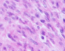
what is this?
transverse section of smooth muscle in colon muscular layers in a monkey
skeletal muscle fibre shape?
•Narrow spindle-shaped fibres that can be up to 40 cm in length and 100 µm in diameter
skeletal muscle - how many nuclei? why? where?
multinucleate - due to embryonic fusion of myocytes to create myofibre
located at periphery of cell (outer edges of myofibre)
structural arrangement in skeletal muscle?
transverse striations in cytoplasm (visible on longitudinal sections)
due to special arrangement of actin and myosin myofilaments
nominated as: bands, zones, lines
What are the different physiological features of the two different types of skeletal muscle? (Just find 2-3 examples).
slow twitch, fast twitch muscle !!!!!!!!!!!!!!!!!!!!!!!!!!!!!!!!!!!!!

what is this and label it?
skeletal muscle
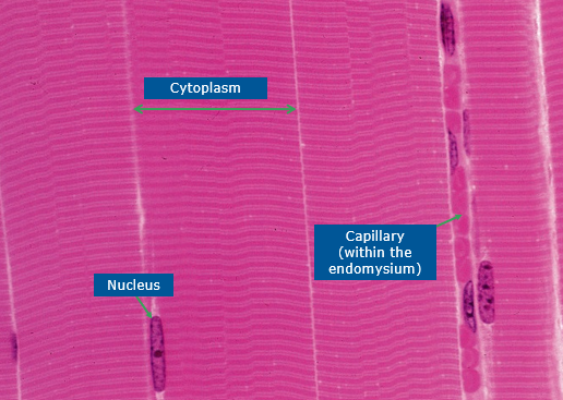

label the Z line and the A band on this image


label this image of skeletal muscle
(why is it a different shape than im used to?)
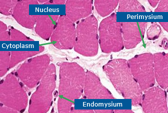

what is this?
skeletal muscle - some should be satellite cells but not distinguishable with HE

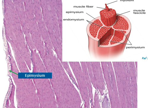
label the epimysium
cardiac muscle alternative name
myocardium
cardiac muscle structure
striations
cardiac muscle vs skeletal muscle
•Shorter (up to 90 µm x 15 µm in diameter)
•Presence of one or two nuclei per cell (less than in skeletal muscle), being located in the centre
•Cardiomyocytes are often branched to allow multiple cell-cell connections
•These connections are seen as intercalated disks: junctional complexes viewed as darkly stained transverse lines
What physiological ability is present in cardiomyocytes?
automatic: generate an electrical impulse spontaneously without external nervous stimulation. While specialized pacemaker cells (like those in the SA node) dominate this role, all cardiomyocytes possess varying degrees of latent automaticity.
in sync: mechanically and electrically coupled so closely that they contract together as a single unit.
refractory period ability

what is this? and label?
cardiac muscle


what is this and label?

tendons function
connect muscle to bone
what are tendons made of?
dense regular connective tissue
tendons composition
•Closely packed abundant extracellular fibres (mainly type I collagen), parallel and regularly oriented
how do tendons structure aid their function?
dense regular connective tissue, regularly oriented = strong/ resistant to stress
what cell types are most numerous in tendons
fibrocyte and fibroblast (sometimes called tenocytes)
tenocyte features and how this aids function
long and multiple fine cytoplasmic processes = allow them to generate fibre crimp
How do the physical properties of tendons aid locomotion?
They are strong and slightly elastic which allows a certain level of spring during movement.
strong = resistant to tearing and damage.
Describe a tendon sheath. Why are tendon sheaths near to joints?
they protect the tendons and prevent them from rubbing the bone in the high impact area. ???????????

what is this and label?
tendon

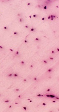
what is this?
transverse section of tendon

what is this and label?
tendon sheath
