Cardio - Blueprint
 Call Kai
 Learn
Learn Practice Test
 Spaced Repetition
 Match
Match Flashcards
 Knowt Play
1/347

There's no tags or description
Looks like no tags are added yet.
Name | Mastery | Learn | Test | Matching | Spaced | Call with Kai | Chat |
|---|
No analytics yet
Send a link to your students to track their progress
348 Terms
gACC/AHA 2017 Hypertension categories
Normal: < 120/80 mm Hg
Elevated BP: systolic 120–129 mm Hg, diastolic < 80 mm Hg
Stage 1 HTN: systolic 130–139 mm Hg, diastolic 80–89 mm Hg
Stage 2 HTN: systolic ≥ 140 mm Hg OR diastolic ≥ 90 mm Hg
first-line tx for high BP (general)
ALWAYS lifestyle modifications
meds start at >130/80 AND clinical CVD or 10 year CVD risk >10%
thiazides, CCB, ACEI, ARB
first line BP tx black pt
CCB or thiazide
(d/t efficacy in lowering BP in this pop)
first line BP for CKD/albuminuria
ACEI/ARB
first-line BP tx for pts with MI
BB
ACEI
ACEI MOA
Renoprotective effect although can see a slight decrease in kidney function before becomes protective
used in HTN, kidney dz, HF
blocks conversion of angiotensin I → II
“pril”
ACEI adverse effects
cough
angioedema
hyperkalemia
adverse effects if with ARB
orthostatic (postural hypotension) etiology
nadequate autonomic reflexes,
volume depletion (dehydration, hemorrhage, vomiting),
certain medications (antihypertensives, CCBs,
antidepressants, opiates)
orthostatic hypotension
Decrease in systolic blood pressure ≥ 20 mm Hg OR
Decrease in diastolic blood pressure ≥ 10 mm Hg
orthostatic hypotension tx
1st: stop causative meds → INC salt, stockings, exercise
#2: low dose fludricortisone or micodrine
hypertensive urgency def + sx
>180/110-120 and NO signs of acute organ damage
sx: HA, atypical CP, epistaxis, dizziness
hypertensive urgency tx
PO meds (captopril, labetalol, clonidine [relaxes BP], prazosin)
Follow up → 1-7 days
clonidine = centrally acting alpha-2 adrenergic agonist
hypertensive emergency acute organ damage
hemorrhagic or ischemic stroke, acute coronary syndrome, aortic dissection, diffuse microvascular injury (anemia, thrombocytopenia, acute kidney injury, new onset retinopathy), hypertensive encephalopathy
hypertensive emergency tx
Admit to ICU → BP stabilization with IV antihypertensives
Lower BP (reduce MAP) by no more that 20-25% over 1 hour, then 160-100 within 6 hours, then to target BP in 48 hours
Nicardipine and labetalol → shown to be most effective at lowering BP
Once stable → start a chronic PO med
renal artery stenosis (RAS)
MC cx of secondary HTN (along with 1º aldosteronism) in adults
Pursue screening in patients with: drug resistant HTN, unusual presentation (YA), abdominal bruit, low serum potassium (indicating 1º aldosterone)
RAS causes
atherosclerosis, fibromuscular dysplasia (FMD), kidney trasplant
if FMD = young, F <60
atherosclerosis = old, CV RF
kidney transplant: surgical, native dz, immune
neonates = congenital
possible drug resistant hypertension
RAS PE
abdominal bruit
RAS dx - “next appropriate”, highest sensitivity, gold standard
Doppler U/S,
MRA (highest sensitivity),
CTA,
renal arteriography (gold standard for dx)
Fibromuscular dysplasia →
Multifocal type: “string of beads” d/t alternating stenosis & dilation
Focal type: circumferential or tubular stenosis
tx options for RAS
Antihypertensive therapy (e.g., ACEI or ARB)
Revascularization in select patients (e.g., those with a short duration of hypertension, intolerance of medical Tx, recurrent flash pulmonary edema/refractory CHF (Percutaneous angioplasty w/ or w/o stent placement)
secondary CVD prevention (e.g., aspirin, statin, lifestyle modifications)
CAD → atherosclerosis process
Begins w endothelial dysfunction → cause fatty streak formation (initial xanthomas accumulate lipid laden macrophages (foam cells)) → grows in size → a stable fatty plaque with a fibrous cap atheroma → gains O2 supply through intimal neovascularization (originating from vasa visorum) → risk for a plaque rupture thrombosis
CAD RF
family history of premature coronary heart disease, smoking, hypertension, high-density lipoprotein levels below 40 mg/dL, and age ≥ 45 in men or ≥ 55 in women
protective factor CAD
having HDL levels ≥ 60
CAD sx
pattern of chest pain (unstable angina, NSTEMI, STEMI, heart failure)
CAD dx
2 lead EKG and exercise stress EKG, stress echocardiography, cardiac catheterization (dx and tx), cardiac enzymes (troponin & creatinine kinase-MB)
NSTEMI - same manifestations as unstable angina w/ election in troponin
STEMI - elevated troponins, ST segments elevation, and unstable angina symptoms
cardiac enzymes for NSTEMI
same as unstable w/ troponin
cardiac enzymes for STEMI
elevated troponin
ST segment elevation
unstable angina
creatinine kinase-MB and whats it best for determining
rise within 3 hours, peak within 24, normalized within 48-72 → more helpful when determining reinfarction
troponin is best for
initial eval of a possible MI, trop I is the most specific for MI
CAD/atherosclerosis FIRSTLINE and other tx
#1: BB
(alts: CCB, nitrates) + ASA
if recent stent placement → dual antiplt therapy (ASA and P2y12 inhibitor like ticagrelor or clopidogrel)
statins, lifestyle changes, comorbidities
Myocarditis cause
infectious - viral
((e.g., parvovirus B19, coxsackievirus, influenza, adenovirus, coronavirus), autoimmune, toxins, MIS-C))
→ ventricular enlargement, dilated cardiomyopathy
myocarditis sx
viral prodrome (fatigue, fever, chest discomfort, dyspnea, palpitations)
Can present with symptoms of fulminant heart failure
S3/S4, abnormal ECG, elevated troponin, unexplained sinus tachycardia, exercise fatigue
myocarditis PE
tachycardia disproportionate to fever, tachypnea, hepatomegaly, pulmonary edema, jugular venous distention
Triad: fever, new onset/worsening murmur, + culture
myocarditis dx + gold standard
ECG, inflammatory markers, echocardiogram
Echocardiogram will show decreased ventricular ejection fraction with hypokinesis and wall motion abnormalities
Cardiac MRI, endomyocardial biopsy (gold standard)
myocarditis tx
diuretics, ACEI, B-Blocker if EF <40%, afterload reduction, inotropes
VADs, ECMO may be required for severe disease
HEART score (for pts with ACS)
History (slightly, moderately, highly suspicious)
ECG (normal, nonspecific disturbance, ST depressions)
Age (< 45 years, 45–65 years, > 65 years)
Risk factors (none, 1–2, ≥ 3)
Troponin (≤ normal limit, 1–2x normal limit, > 2x normal limit)
Results:
Low risk 0-3: low risk of major cardiovascular event (MACE) in next 6 wks
Moderate risk 4-6: moderate risk of MACE in next 6 wks
High risk 7-10: high risk of MACE in next 6 weeks
MI RF
advanced age, HTN, hyperlipidemia, DM, obesity, smoking, prior CV disease
MI sx and what pop can have atypical presentatoin
chest pain (MC) tightness/pressure and worse with exertion, dyspnea, N/V, weakness, palpitation, syncopes
Women, pts with diabetes, older pts are more likely to have an atypical presentation w/o chest pain
MI dx (short)
ECG (new ST elevation at the J point in two contiguous leads), troponin w/in 10 mins of first contact (cardiac injury biomarker)
MI tx
1. supplemental O2 if <90% sat or in respiratory ditress
2. aspirin (if no contra.),
anticoagulation therapy,
nitroglycerin to alleviate chest pain (contra. if taken a phosphodiesterase inhibitor (sildenafil/viagra) w/in 24 hours or if hypotensive),
B-Blocker and statins (if not contra.
reperfusion,
thrombolysis (If PCI not available), medical therapy
MI most important part of tx
reperfusion -
Fibrinolytics
PCI (percutaneous coronary intervention): more direct effect (preferred), increased survival, decreased ICH and recurrent MI
If have PCI center (cath lab): < 90 minutes door-to-device time
Non-PCI center: transfer for PCI if door-to-device time can be < 120 minutes
if >120 mins → thrombolysis has to be given in 30 mins and 12 hours within sx onset but CI in ICH, intracranial malignancy, stroke within 3 months, aortic dissection
medical therapy for MI (pt 1)
Oxygen: indicated for saturations < 90%, dyspnea, heart failure
Nitroglycerin: relieves ongoing chest pain, lowers BP
Morphine: relieves pain, reduces work of breathing in setting of pulmonary edema
Only used if nitroglycerin fails to relieve the pain
Can be associated with negative outcome
Beta-blockers: prevent recurrent ischemia and dysrhythmia
Metoprolol or esmolol
medical therapy for MI (pt 2 - anticoags/antiplts)
Antiplatelet agents: reduce recurrent coronary artery thrombosis, stent thrombosis, and death
Aspirin: given before PCI and continued indefinitely
P2Y12 inhibitor (clopidogrel, prasugrel, ticagrelor): continue for 1 year if stent placed
GPIIb/IIIa antagonists for patients undergoing PCI
Anticoagulation: if LV thrombus or Afib present and for all patients receiving thrombolytic therapy
ACE inhibitors: reduce cardiovascular events, prevent LV remodeling
High-dose statin: lipid lowering, and lowers risk of death, recurrent MI, and stroke
Pt with hx of MI and current dx of HTN → B-Blockers and ACEI’s are 1st line
troponin (long)
Highest sensitivity and specificity
Time detectable from onset: 3–12 hours
Peak: 24–48 hours
returns to baseline in 5-14 days
CK-MB
Time detectable from onset: 3–12 hours
Peak: 24 hours
Return to baseline: 48–72 hrs
Useful for Dx of reinfarction
myoglobin
First to appear, first to peak, first to decline. But lacks specificity
Cardiac Electrical Conduction System
SA node → AV node → bundle of His → bundle branches → Purkinje fibers
ST Elevation Myocardial Infarction (STEMI) presentation
Patient presents with substernal chest pain that radiates to the neck and arm so severe that nitro or even opioids give no relief
STEMI dx
elevated troponin I or troponin T and CK, ST segment elevations > 1 mm in two or more contiguous leads
ECG: should be performed within 10 minutes of medical contact
Hyperacute T waves, ST elevation, Q wave development, T wave inversion, NEW LBBB
STEMI tx
<12 hours since sx = PCI (if 90 mins) or fibrinolysis
thrombolytic therapy in 3 hrs (CI if hemorrhage, stroke, brain cancer, internal bleeding, AD)
ASA OR p2y12 receptor blocker (ticagrelor)
BB
anticoag (UFH, LMWH, fondaparinux)
MI walls + arteries
Anterior wall ST elevation in leads V1 through V4 → LAD artery
Inferior wall ST elevation in leads II, III, and aVF → R coronary artery or L circumflex
Lateral wall ST elevation in leads I, aVL, V5, and V6 → L circumflex
Posterior wall ST depressions in leads V1 through V3 and elevations in leads V8 and V9 (posterior lead placement)
Right Ventricular Infarction ECG and sx
assoc with anterior STEMI
hypotension, JVD, clear lungs
ECG
ST elevation in lead V1
ST elevation in lead III > lead II
ST elevation in V4R–V6R
right ventricular infarction tx and what to avoid
patient is preload-dependent and has impaired filing of left ventricle
Reperfusion, IVF
Avoid nitrates (decreases preload which a R Vent. infarction is dependent on)
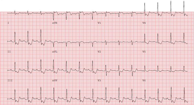
Non-ST Elevation Myocardial Infarction (NSTEMI) sx
Sx: substernal chest pain that often radiates to left arm or jaw, dyspnea, nausea
Commonly presents with T wave inversions or ST segment depressions on ECG and elevated troponin
NSTEMI medical therapy
Oxygen: indicated for SpO2 < 90%, dyspnea, heart failure
Nitroglycerin: relieves ongoing chest pain, lowers BP
Morphine: relieves pain and anxiety, reduces work of breathing in setting of pulmonary edema
Beta-blockers: prevent recurrent ischemia and dysrhythmia
Antiplatelet agents: aspirin and P2Y12 inhibitor (clopidogrel, prasugrel, or ticagrelor)
GPIIb/IIIa antagonists may be considered in certain patients (e.g., high-risk patients undergoing PCI)
Anticoagulation: while hospitalized or until PCI is performed
High-intensity statin: lowers lipids, risk of death, recurrent MI, and stroke
ACE inhibitor: indicated for patients with LVEF < 40%, DM, heart failure, CKD
Treat underlying condition if NSTEMI is type 2
NSTEMI revascularization strategy
Within 2 hours: heart failure, hemodynamic instability, angina at rest, mechanical complications, sustained VT or VF
Risk scores (e.g., TIMI, GRACE) stratify risk of further cardiovascular events and may aid in determining need for or timing of revascularization
PCI or CABG
Dressler Syndrome (aka postmyocardial infarction syndrome) hx
History of MI → pericarditis occurring 1-2 weeks after an acute MI or cardiac procedure
Most likely d/t autoimmune inflammatory response following necrosis
dressler syndrome sx (short)
pleuritic chest pain that is improved when leaning forward
dressler syndrome PE
pericardial friction rub, tachycardia, fever, leukocytosis, CRP, ESR

dressler syndrome ECG
PR depression, PR elevation (aVR), diffuse ST segment elevation (concave)
dressler syndrome tx
NSAIDs, colchicine
First line → Aspirin and Colchicine
Glucocorticoids may be considered in refractory cases
pericarditis causes
idiopathic, viral (mc after an acute viral illness in US i.e. URI), neoplastic, uremic, rheumatologic
pericarditis sx
pleuritic chest pain radiating to the back that is worse when lying back and improved when leaning forward
3Ps of Chest Pain in Pericarditis: Pleuritic, Persistent, and Postural
pericarditis PE
tachycardia and pericardial friction rub (sensitive and specific), distant heart sounds
pericarditis dx
ECG: PR depression, PR elevation (aVR), diffuse ST segment elevation (concave)
1 week after onset → ECG is isoelectronic, diffuse T wave inversions
Resolution of abnormal ECG findings in 2-6 weeks
pericarditis tx
NSAIDs (ASA) + colchicine
corticosteroids in refractory cases, NSAID contraindication, or certain rheumatologic disease
constrictive pericarditis cause and RF
Caused by thickening and scarring of the pericardium → resulting in loss of elasticity of pericardial sac
Risk factors: history of viral or idiopathic pericarditis, prior radiation therapy or cardiac surgery, prior infection (e.g., tuberculosis), autoimmune conditions, malignancy
constrictive pericarditis sx
sx of RHF (edema), INC JVD
pericardial knock
kussmaul sign
pulsus paradoxicus (BP drops when you breathe in)
pericardial knock definition
extra heart sounds heard just prior to S3 and is sometimes palpable - caused by sudden cessation of ventricular filling d/t thickened and noncompliant pericardium

constrictive pericarditis echo
thickened pericardium, abnormal septal motion, dilated IVC and hepatic veins
pericarditis tx
depending on whether early or chronic disease) include anti-inflammatory agents (NSAID and colchicine), Tx of the underlying cause, pericardiectomy (is hemodynamically unstable or persistent symptoms)
Pericardial Tamponade cause
penetrating trauma, pericarditis, iatrogenesis (central line placement, pericardiocentesis, pacemaker placement), post MI with free wall rupture, recent viral illness
pericardial tamponade sx
dyspnea and chest pain
pericardial tamponade PE
muffled heart sounds, JVD, hypotension (Beck’s triad), pulsus paradoxus (an abnormal drop in systolic BP ≥10 mmHg on inspiration), tachypnea, tachycardia,

pericardial tamponade ECG
low-voltage QRS
electrical alternans (beat to beat change in amplitude of QRS or t wave)
pericardial tamponade CXR
enlarged cardiac silhouette (water bottle sign) if effusion > 200mL
Echocardiography (sensitive and specific)
Diastolic collapse of RV (less sensitive but very specific)
Early systolic collapse of RA (sensitive and specific)
Plethoric IVC (sensitive)
pericardial tamponade tx
pericardiocentesis or pericardial window
angina definition
Chest pain or pressure with exertion or stress that is relieved by rest
Duration of pain → typically < 30 minutes (if > 30 mins suggestive of acute coronary syndrome (MI or unstable angina))
stable: caused by activity, stops when activity stops
unstable: CP >10 mins, less responsive to nitro, ST segment depression, normal troponin
angina diagnosis is made by
exercise ECG
Angiogram needed to confirm and assess degree of CAD
ECG: horizontal or downsloping ST segment depression or T wave flattening/inversion that resolve after sx of angina disappear → a/w stable angina
angina tx
lifestyle modifications, statins, antiplt (ASA or clopidogrel), BB, nitrates, CCB, ranolazine, surgery
Acute 1st: sublingual nitroglycerine** (TOC) for stable angina for immediate sx relief & 325 mg aspirin (unstable)
Chronic 1st: B-Blockers → decrease recurrence of MI
If not tolerated then try CCB, long acting nitrate, or ranolazine
AVOID B-Blockers in a cocaine induced MI
ASA and B-Blockers are only known meds to lower mortality in patients with stable angina
vasospastic angina (variant or prinzmetal) cause
coronary (MC right coronary) artery spasm with minimal fixed coronary lesions (often late PM - midnight or early AM)
Vasospasm at rest with preservation of exercise capacity
Associated with other vasospastic disorders, i.e. raynaud’s
vasospastic angina RF
smoking/cocaine, female, < 50 years, HTN, DM, obesity, emotional stress, medications (B-blockers and -triptans)
Triggers: sympathomimetic use, migraine therapies, ergot derivatives, hyperventilation, extreme cold
vasospastic angina sx
squeezing, pressure-like chest discomfort at rest but have preservation of exercise capacity
Episodes commonly occur from midnight to early AM (overnight)
vasospastic angina ECG and dx
transient ST segment elevations
dx - cannot differentiate from STEMI but cardiac enzymes normal, angiographic evidence of spasm, no lesions
vasospastic angina - tx ; absence vs presence of stenosis
NO coronary stenosis = CCB and nitrates, and ACEI NO BB!!!
YES: PCI
rheumatic fever hx*
hx of GAS infx (ie strep throat, 5-15 yo)
rheumatic fever - sx and MC affected valve
fever, red skin lesions on the trunk and proximal extremities, and small nontender lumps located over the joints, migratory arthritis of large joints
MC affected valve is the mitral valve
rheumatic fever PE
Jones criteria: joints, oh no—carditis!, nodules, erythema marginatum, Sydenham chorea
Initial Dx: 2 major OR 1 major and 2 minor
Recurrent dx: 2 major OR 1 major and 2 minor OR 3 minor
rheumatic fever labs
antistreptolysin O, anti-DNase B, positive throat culture, or positive rapid antigen test
rheumatic fever tx
antibiotics, NSAIDs
Secondary prophylaxis against GAS
Penicillin G benzathine → administer IM q21-28 days
If have rheumatic fever with carditis and echocardiographic evidence of persistent valvular disease → continue prophylaxis for 10 years or until age 40 (whichever is longer)
jones major criteria
Joints (Polyarthritis)
♥- Carditis (Pancarditis - MC mitral regurgitation)
Nodules (subcutaneous)
Erythema Marginatum (nonpruritic rash, central clearing, face is spared)
Sydenham’s Chorea
jones minor criteria
fever, poly or monoarthralgia, elevated ESR or CRP, prolonged PR interval on ECG
Most common cyanotic congenital heart disease in childhood
tetralogy of fallot
tetralogy of fallot hx
episodes of cyanosis (tet spells) and squatting for relief (decreases R ➔ L shunt, increases oxygenation)
Test spells are preceded with irritability/crying or feeding - sudden onset, deepening of dyspnea, irritation, alteration in consciousness, decrease/disappearance of systolic murmur (d/t increased obstruction to the point of no flow through valve and increased VSD R → L shunt)
Spells frequently occur between 4-6 months and may resolve spontaneously
“Pink tet” → mild to moderate RV outflow obstruction
tetralogy of fallot sx
easy fatigue, dyspnea on exertion, cyanotic episodes
tetralogy of fallot PE
palpable Right ventricular lift, nail clubbing
tetralogy of fallot dx
TOC - Echocardiogram: pulmonic stenosis, right ventricular hypertrophy, overriding aorta, and VSD
CXR: boot-shaped heart, RVH
ECG: RVH and right axis deviation
tetralogy of fallot - tx
Tet spell → supplemental O2, place in knee to hest position to reduce shunting across VSD
Corrective surgery → for infants w/ severe obstruction
If less severe can delay operation until up to 2 years old
Palliative surgery (stent placement to maintain PDA or to creat a shunt with similar effect) → Neonates that cannot yet undergo corrective surgery
tetralogy of fallot - PROVe
Pulmonic stenosis (RV outflow tract obstruction)
Rough, crescendo-decrescendo systolic ejection murmur heard best at the LSB in the 3rd ICS with radiation to the back
Right ventricular hypertrophy
Overriding aorta
VSD (ventricular septal defect)
pathologic murmurs
Grade ≥ 3 intensity
Holosystolic timing, diastolic, friction rub (pericarditis), gallop rhythm (S3 or S4), systolic click
Harsh or blowing quality, maximum intensity along Left upper sternal border
Increased intensity with upright positions (sitting or standing from supine)
innocent murmurs
Grade ≤ 2 intensity (Still or flow murmur)
Short systolic duration (not holosystolic and not diastolic)
Minimal radiation
Musical or vibratory quality
Soften in intensity when patient is sitting/standing compared to supine
Exception is venous hum murmur (louder when sitting)