Parasitology Collection
 Call Kai
 Learn
Learn Practice Test
 Spaced Repetition
 Match
Match Flashcards
 Knowt Play
1/49

There's no tags or description
Looks like no tags are added yet.
Name | Mastery | Learn | Test | Matching | Spaced | Call with Kai | Chat |
|---|
No analytics yet
Send a link to your students to track their progress
50 Terms
What is the ideal number of samples, and when are they collected?
Three
Monday/Wednesday/Friday
Why are the three stool samples spaced out?
Because they are collected on an intermittent basis
How soon should the stool sample be examined after collection?
Within 30 mins
What form of parasite is likely to be missed if the specimen is not examined in 30 mins?
Trophozoite
What is a purged specimen?
A stool sample obtained after administration of a laxative, used to enhance detection of intestinal parasites.
What laxatives are useful for parasite purging?
Sodium sulfate
Buffered Phosphosoda cathartic
Why should purging be avoided?
Parasites do not return to the stool for a week after
Should you use enemas for collection?
No, since parasites are rarely located in the colon
What kind of parasite causes dry stool? What kind causes wet stool?
Dry: Cysts
Wet: Trophozoites
What does a chalky-white stool indicate, and what should you do with the sample?
Indicates patient had a barium x-ray
Cancel specimen because this is not acceptable.
What kind of stool does Giardia cause? Why is this?
Clay-colored or yellow
Malabsorption syndrome leading to increased fats
Where does the bleeding occur if stool is red? Where does it occur if black?
Red: Lower GI tract or rectum
Black: Higher GI tract (invasive diarrhea)
What should you do with mucus?
Test this area since organisms might be trapped.
What other macroscopic feature should you look for?
Helminths (adult worms or proglottids may be present)
What preservative is used?
Formalin
What stain is used?
Iodine
What color does the iodine stain the cytoplasm, glycogen, and chromatin?
Cytoplasm: yellow
Glycogen: Reddish-brown
Chromatin: Dark brown
What is the procedure for the wet prep?
Scan entirety of coverslip on 10X
Scan 1/3 of coverslip on 40X
Use immersion oil as needed
What are permanently stained smears prepared from?
PVA-fixed specimens
How much of the slide (how long) should be examined in a PVA-fixed specimen?
All (15-20 mins)
How many fields should be examined under oil immersion?
10-15
What is the most common permanent stain?
Trichome
What color does Trichome stain the background?
Green
What color does Trichome stain the cytoplasm?
Blue-green tinged with purple (cysts are more purple)
What color does Trichome stain the nuclear and chromatoid structures?
Purple
What color does Trichome stain the ingested RBC’s and bacteria found in parasites?
Purplish-red
What color does trichome stain the ingested yeast in parasites?
Green
What color does trichome stain eggs and larvae?
Red
What are the three characteristics you should use to distinguish between parasites and artifacts?
Size
Cell wall
Internal structure
What are examples of artifacts?
Meat and plant fibers
Plant cells
PMN’s
Starch granules
Pollen grains
Other organisms
What is an ocular micrometer?
A glass disc with an engraved scale placed within the ocular.
What is an ocular unit based on?
The magnification
How is the value of ocular units determined?
Through calibration
How is calibration performed?
Ocular micrometer is compared with a scale of known dimensions which is placed under the various objective lenses.
What is a micrometer?
A scale with a slide of an engraved ruler used in the calibration of the ocular micrometer
What is the value of an ocular unit for 10X?
10 micrometers
What is the value of an ocular unit for 40X?
2.5 micrometers
What is the value of an ocular unit for 100X?
1 micrometer
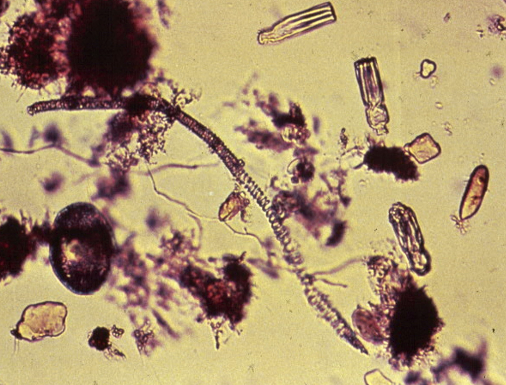
Amorphous particles with vegetable spiral (string beans)
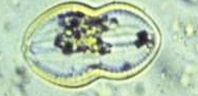
Diatom (harmless organism in fresh water)
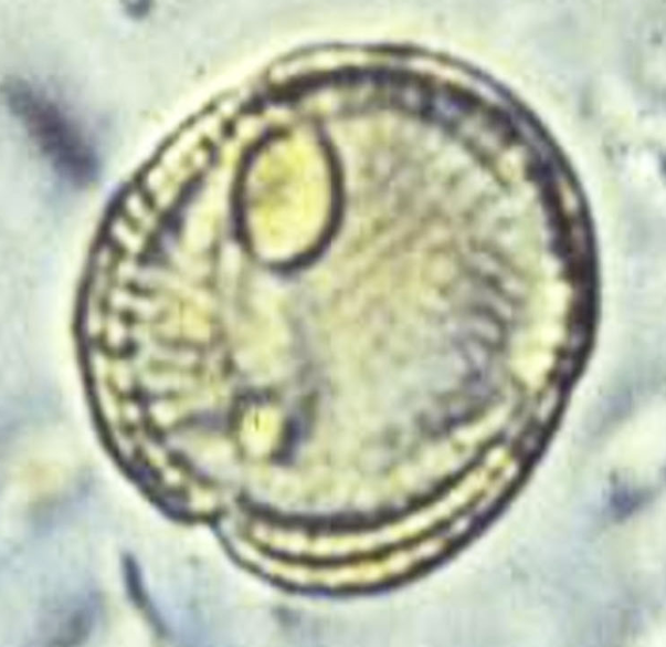
Pollen grain
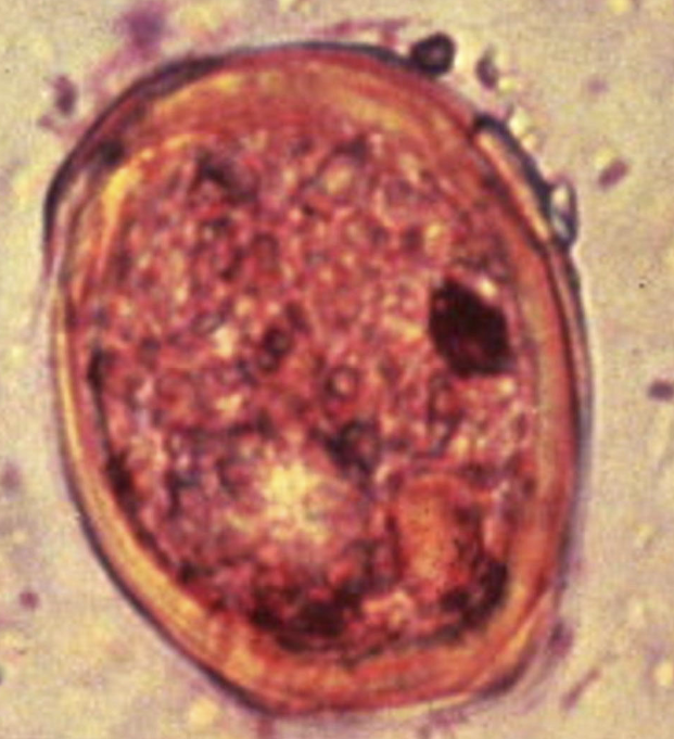
Plant cell
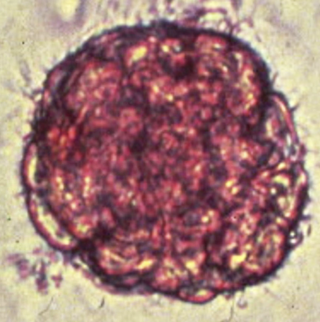
Plant cell
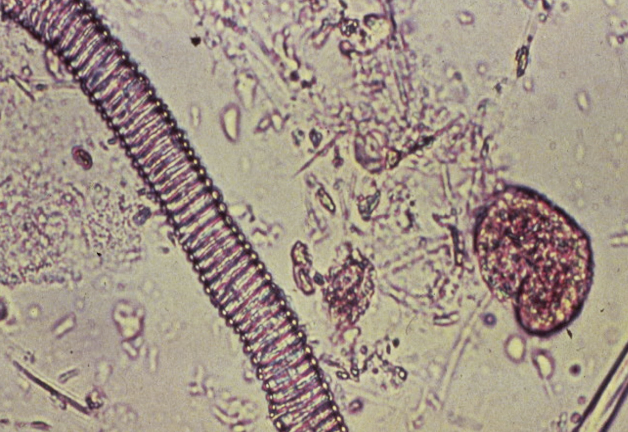
Meat fiber on left with plant cell on right
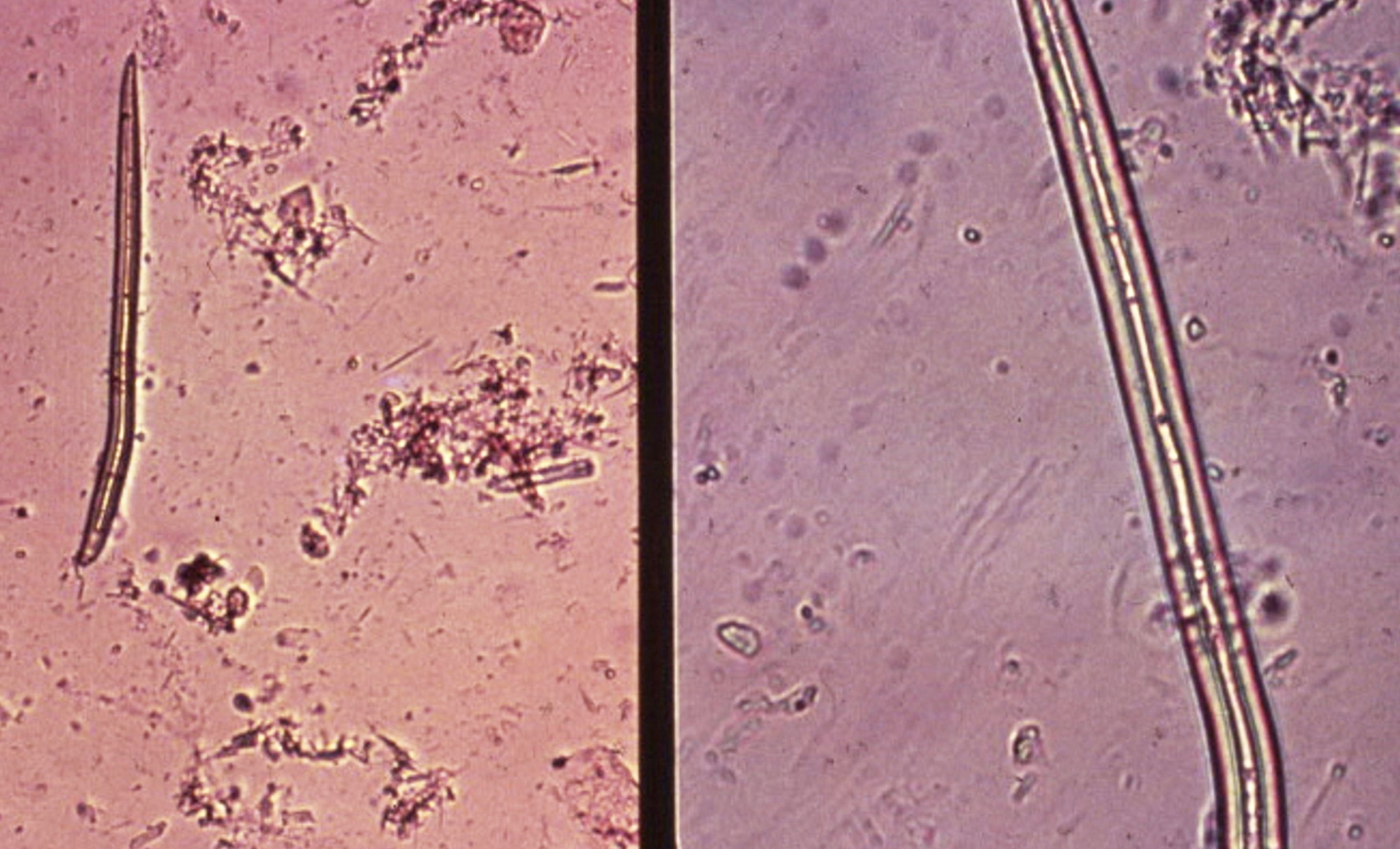
Plant fiber (root hair - note strightness)
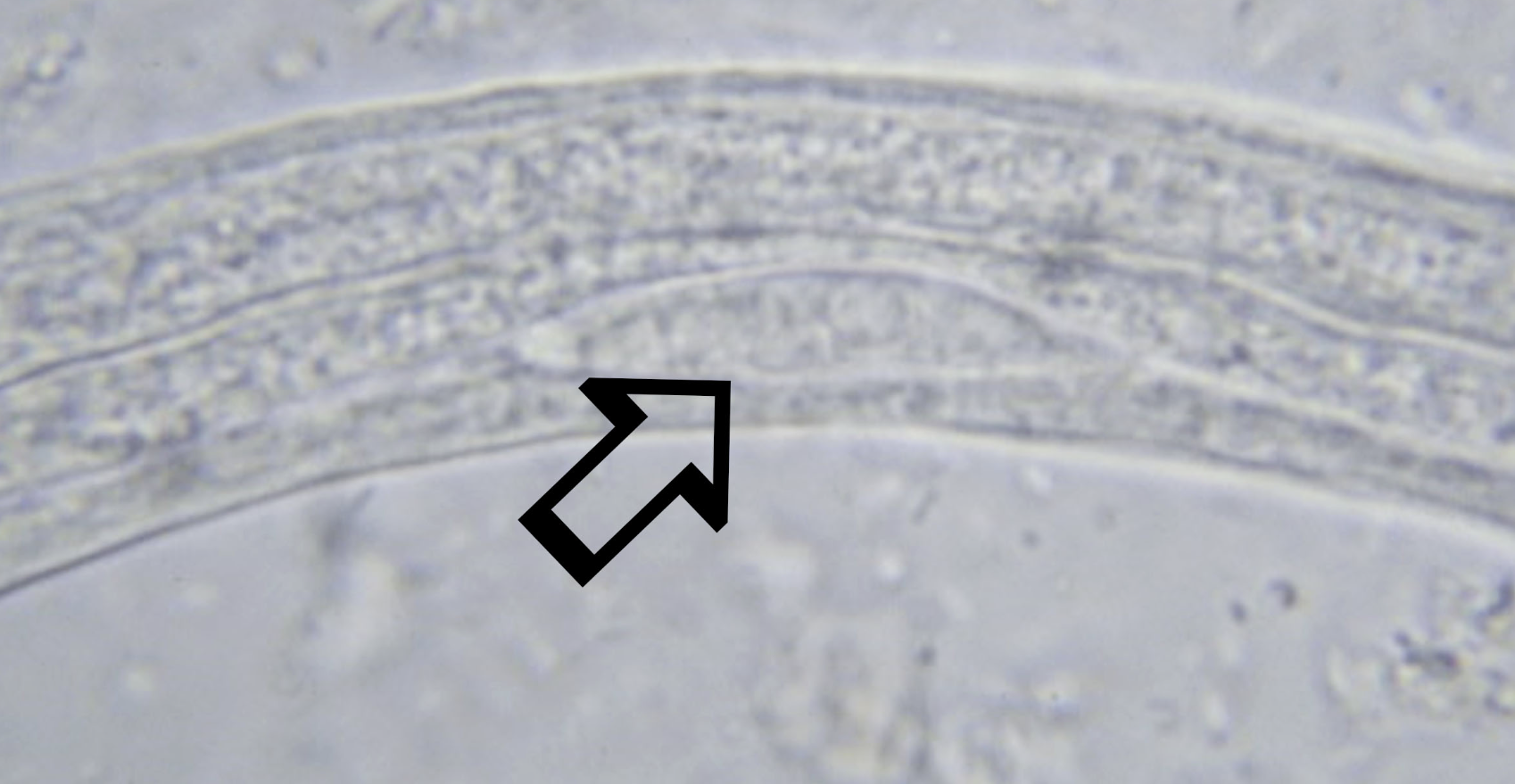
What is indicated by the arrow?
Genital primordium (distinguishing characteristic of Strongyloides stercoralis)
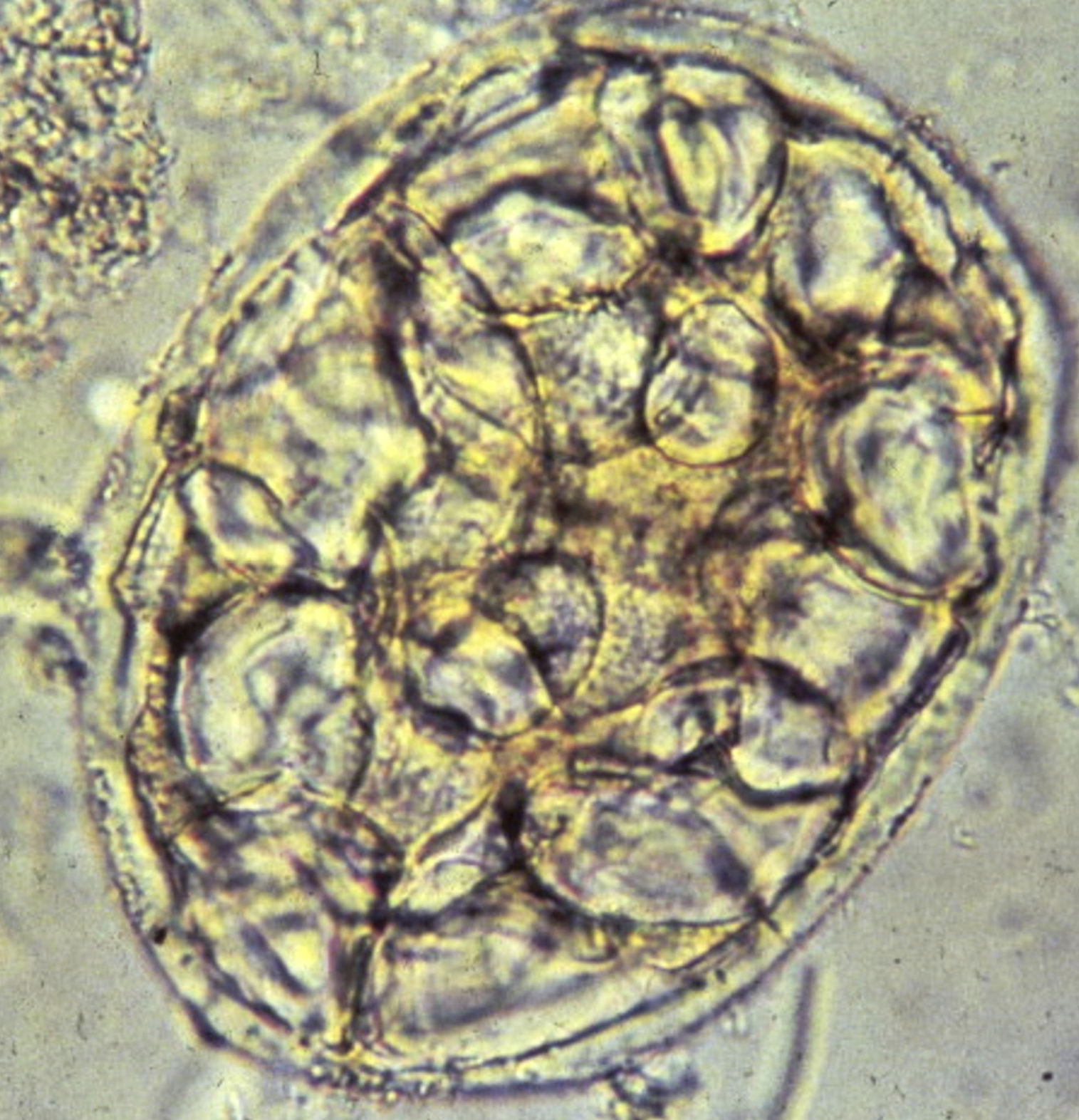
Cell with starch granules
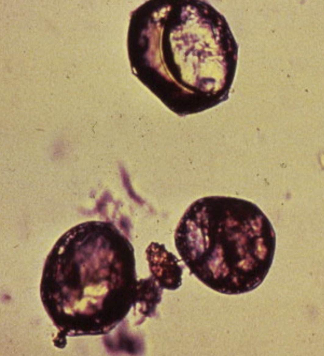
Free-floating starch granules made purple by iodine
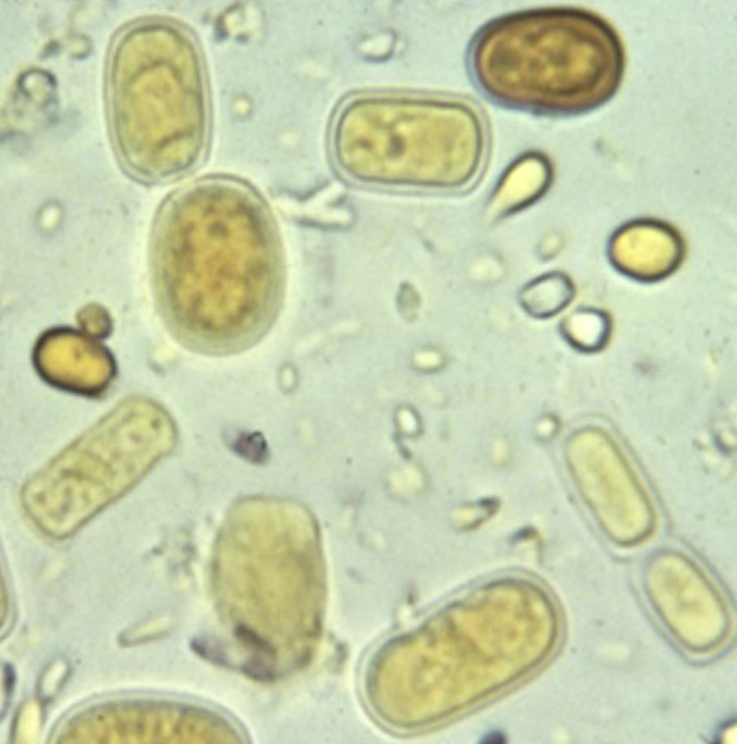
Geotrichum (a fungus not significant in human stool)
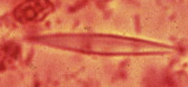
What are these? How are they formed? What do they indicate?
Charcot-leyden crystals
Breakdown product of eosinophils
Parasitic infection