Hemodynamics 11/23
 Call Kai
 Learn
 Practice Test
 Spaced Repetition
 Match
 Flashcards
 Knowt Play
1/94

There's no tags or description
Looks like no tags are added yet.
95 Terms
What are the mean gradient values for aortic valve pressure?
Mild <20mmHg
Moderate 20-40mmHg
Severe >40mmHg
What is the normal area of the aortic valve?
2.5-4.5cm²
What is a mild area of the aortic valve?
>1.5cm²
What is a moderate area of the aortic valve?
1.0-1.5cm²
What is a severe area of the aortic valve?
<1.0 cm²
What is a severe AVA index?
<0.6
What is a mild jet velocity of the aortic valve?
2.6 - 2.9 m/s
What is a moderate jet velocity of the aortic valve?
3.0 - 4.0 m/s
What is a severe jet velocity of the aortic valve?
>4.0 m/s
In aortic insufficiency what is a mild and severe jet diameter/LVOT diameter %
Mild <25
Severe >65
In aortic insufficiency what is a mild and severe jet area/LVOT area %
Mild <30
Severe >50
In aortic insufficiency what is a mild and severe vena contracta (cm)?
Mild <0.3
Severe >0.6
In aortic insufficiency what is a mild and severe regurgitant volume (ml/beat)?
Mild <30
Severe >60
In aortic insufficiency what is a mild and severe regurgitant fraction (%)
Mild <30
Severe >50
In aortic insufficiency what is a mild and severe EROA cm²
Mild <0.1
Severe >0.30
In aortic insufficiency what is a mild and severe pressure half time (msec)?
Mild >500
Moderate 200 - 500 msec
Severe <200
What are the mean pressure gradient values for the mitral valve?
Mild <5 mmHg
Moderate 5-10 mmHg
Severe >10 mmHg
What is a normal area for the mitral valve?
4 - 5 cm²
What is a mild area for the mitral valve?
>1.5 cm²
What is a moderate area for the mitral valve?
1.0 - 1.5 cm²
What is a severe area for the mitral valve?
<1.0 cm²
What is a normal pressure half time for the mitral valve?
30-60 msec
What is an abnormal pressure half time for the mitral valve?
90-400 msec
What is a severe pressure half time for the mitral valve?
>220 msec
In mitral regurgitation what is a mild and severe jet area (cm²)
Mild <4
Severe >8
In mitral regurgitation what is a mild and severe jet area/LA area (%)
Mild <20
Severe >40
In mitral regurgitation what is a mild and severe vena contracta (cm)
Mild <0.3
Severe >0.7
In mitral regurgitation what is a mild and severe PISA?
Mild <0.4
Severe >0.9
In mitral regurgitation what is a mild and severe regurgitant volume (mL)
Mild <30
Severe >60
In mitral regurgitation what is a mild and severe regurgitant fraction (%)
Mild <30
Severe >50
In mitral regurgitation what is a mild and severe EROA (cm²)
Mild <0.2
Severe >0.40
When you think of aortic stenosis what do you think of?
Limited ability of valve to open: degeneration, calcification

What is this showing?
Late stage aortic valve stenosis (limited EF)
Can’t differentiate cusps, thick, echo bright = calcification
Color doppler on this = turbulent, aliasing
Secondary finding = hypertrophy of myocardium
What is aortic stenosis?
Narrowing of the aortic valve
Is aortic stenosis a retrograde or antegrade flow problem?
Antegrade: problem getting blood out of the heart
What are the three causes of aortic stenosis?
Degenerative aortic stenosis: generally in 5th to 8th decade of life patients
Congenital: bi/unicuspid aortic valve
Rheumatic heart disease: can cause stenosis, most commonly affects MV first
What kind of murmur is present in aortic stenosis?
Harsh, systolic ejection murmur
What does a Wigger’s diagram of aortic stenosis look like?
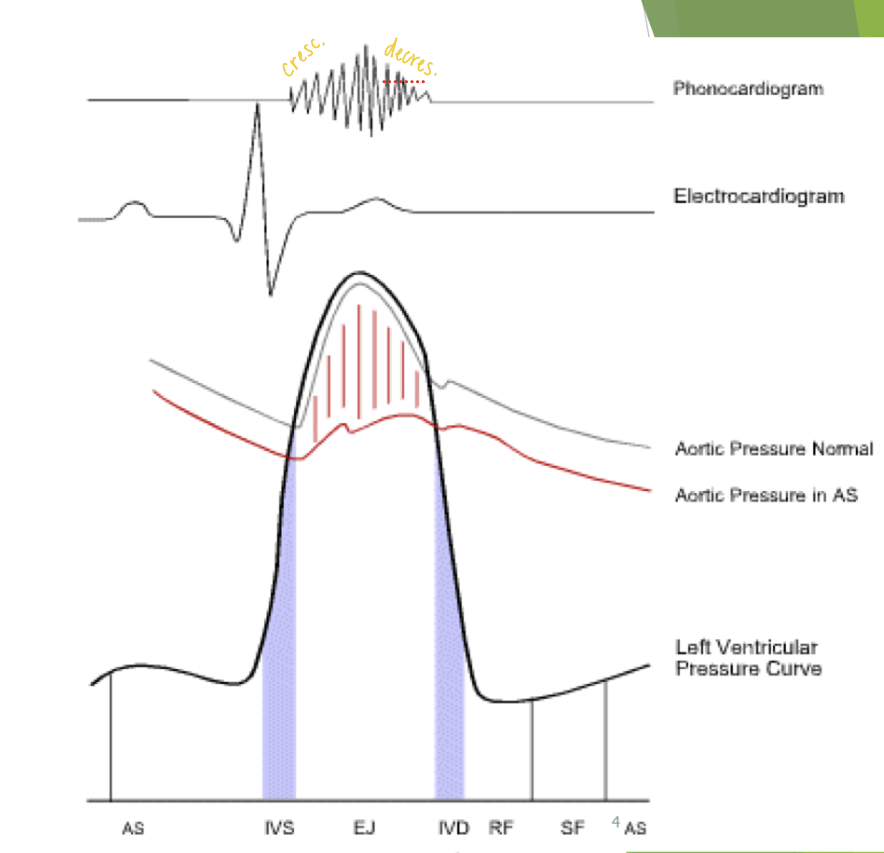
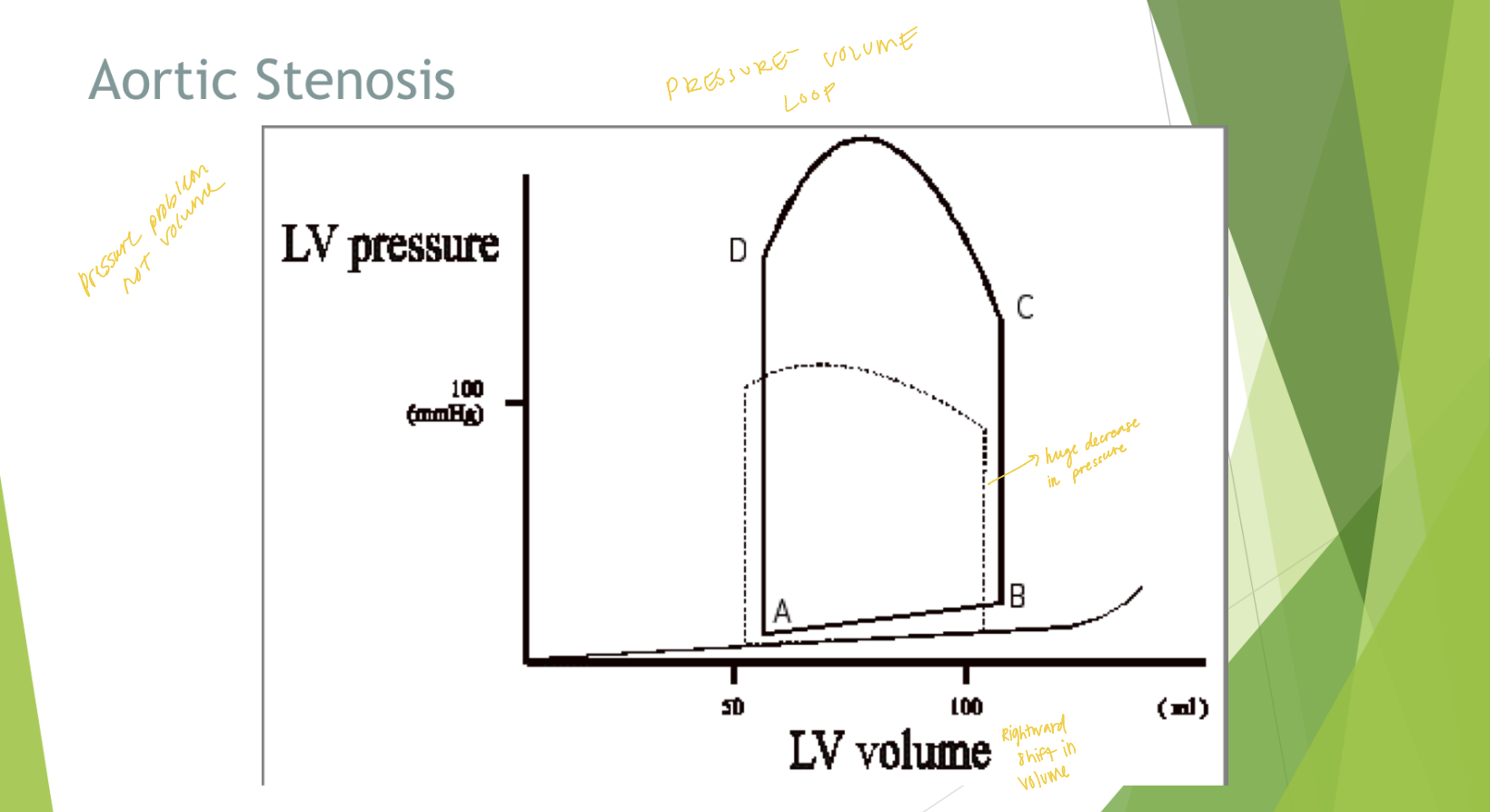
In aortic stenosis there is a _______ shift in LV volume
rightward
Is aortic stenosis a pressure or volume problem?
Pressure
There is a huge ____ in LV pressure in aortic stenosis
increase
Describe a pressure volume loop for aortic stenosis
What are the four things a sonographer uses to evaluate aortic stenosis?
2D, M-Mode, Aortic valve area (AVA), spectral Doppler
What does the sonographer evaluate in 2D when determining aortic stenosis?
Abnormal morphology (bicuspid AV?)
Systolic doming in PLAX
Football or cat eye shaped opening PSAX
Eccentric diastolic closure line (what can lead to doming appearance)
Degenerative disease (presence of calcification)
Decreased systolic excursion
Thickened leaflet appearance
What does the sonographer evaluate in M-Mode when determining aortic stenosis?
Aortic cusp separation of <11mm correlates with AVA of <.75 cm² (SEVERE)
What does the sonographer evaluate in AVA when determining aortic stenosis?
Planimetry (tracing opening in parasternal)
Continuity equation
What does the sonographer evaluate in spectral Doppler when determining aortic stenosis?
Peak aortic valve velocity
Peak instantaneous pressure gradient
Mean trans-valvar pressure gradient
Velocity ratio LVOTVTI / AoVVTI
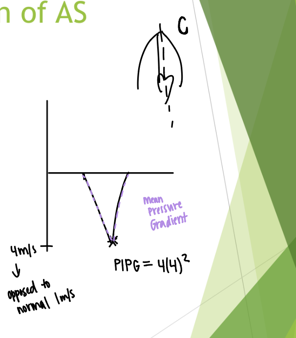
What does spectral Doppler of aortic stenosis look like?
What are some secondary finding on echocardiogram of AS (after years of stenotic condition)?
LV function
LV hypertrophy/mass
Post stenotic dilation of ascending aorta
The effective valve orifice of the stenotic flow area is what?
Very small (a pedoff probe can sometimes be used)
What are some peak aortic valve velocity windows?
Apical, suprasternal (aim towards ascending aorta), right parasternal (at end of study), supraclavicular
Multiple angles from multiple Doppler windows using imaging and non-imaging continuous wave transducers
Describe mean gradient for aortic stenosis

To determine AVA we need the SV of what two locations?
LVOT and AV

Where should the PW Doppler be placed for evaluation aortic stenosis?
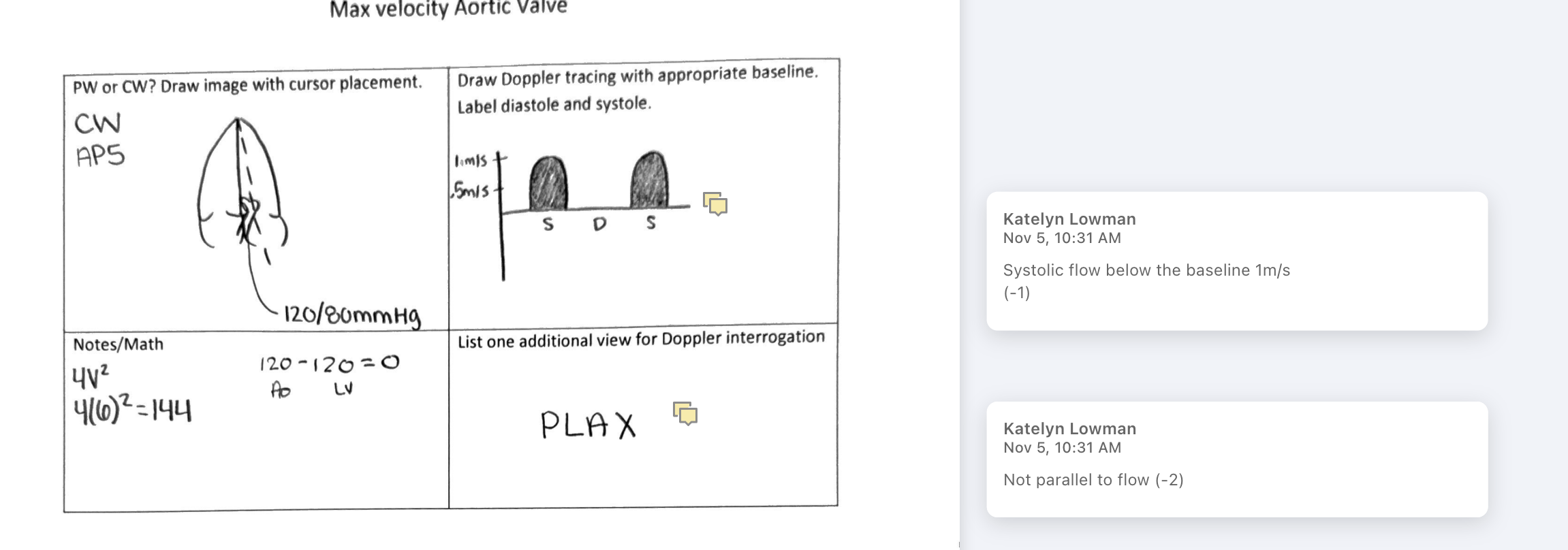
In the LVOT, right above the AV (in AP5)
Not on the valve itself

What are the pitfalls for an inaccurate LVOT diameter measurement?
Oblique image
Extensive annular/sub-valvular calcium
Sigmoid septum (basal portion of IVS is thick)
Error is squared
What are the pitfalls for inaccurate LVOT velocity (V1)?
Poor Doppler angle
Non-laminar flow if too close to valve (pre-stenotic flow acceleration)
What are some pitfalls for trans-valvar velocity (V2 or Vmax)?
Underestimation if angle too great
Confusion with MR jet (eccentric volume curves of mitral can get into aortic window)
Confusion with TR jet

What are some pitfalls for dynamic subaortic obstruction (obstruction in series)?
Inaccurate LVOT velocity (V1)
Use alternate method for measuring AV area
What is a pitfall related to irregular rhythm?
Atrial fibrillation, average 8-10 beats
What are some pitfalls for low output states?
Falsely low effective valve area
Consider inotropic challenge (low flow state)
Poor EF = not much squeeze = pressure gradient can’t be created
Stress echo can increase work load of the heart
Reduced ventricular function
AVA divided by what = AVA index
BSA
Ava index < 0.6 = severe AS
What do you think of when you think of aortic insufficiency?
Leaking Ao valve, retrograde/backflow problems
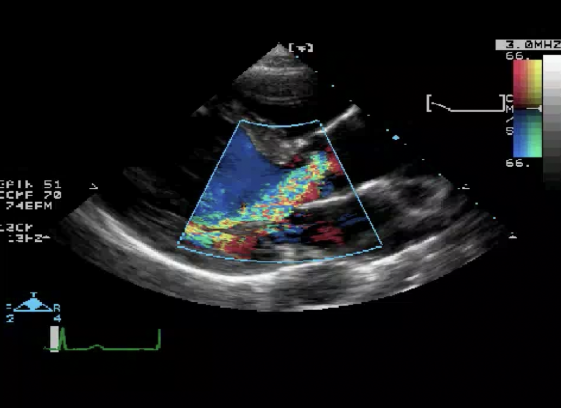
What is this showing?

What is aortic insufficiency?
Retrograde flow across the aortic valve into the LV
Not physiological in majority of population (normal) = most likely a reason
What is the etiology of aortic insufficiency?
Aortic valve pathology (abnormal morphology): no good closure lines = potential for leak
Marfan’s or Ehlers-Danlos Syndrome
Endocarditis
What is the murmur like in aortic insufficiency?
High pitched, diastolic decrescendo left sternal border
Possible Austin Flint murmur (i.e. abnormal mitral valve sound due to AI flow hitting anterior MV leaflet)
What does a Wigger’s diagram look like aortic insufficiency?

What does a pressure volume loop look like for aortic insufficiency?

What does the sonographer evaluate in M-Mode when determining aortic insufficiency?
Fine diastolic flutter of AMVL
Premature closing of MV
What does the sonographer evaluate in 2D when determining aortic insufficiency?
Incomplete closure of aortic valve (coaptation defect)
Dilation of LV
What does the sonographer evaluate in Doppler when determining aortic insufficiency? (Doppler ideally suited for detection of AI)
Color Doppler: jet visualization; assists with beam alignment
Spectral Doppler intensity
Regurgitant fraction
Deceleration time/pressure half time
Color Doppler area vs. LVOT area
Reversal of flow in abdominal aorta and descending aorta
What does mild versus severe AR look like in intensity of spectral Doppler (CW)?

What is the aortic insufficiency regurgitant fraction?

What is the effective regurgitant orifice (ERO)?
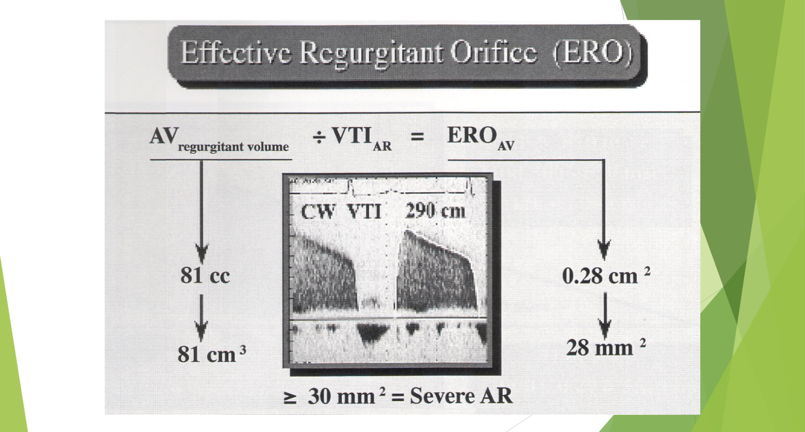
What are some pitfalls of aortic insufficiency?
Modal velocity not traced (inaccurate waveform used)
Inadequate beam alignment
Not well validated
Too few beats measured
Incorrect sample volume position
Significant mitral regurgitation (don’t use ERO if they have this)
Assumption of circular orifice
Incomplete Doppler spectrum
Describe the severity of AI
Deceleration slope of AI spectral envelope
Pressure gradient = 4V²
Fall in velocity during diastole related to decrease in pressure gradient
Flat slope indicates no change in gradient during diastole = mild AR
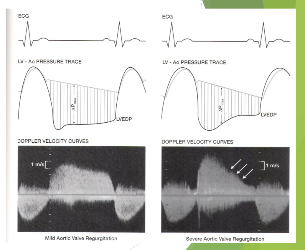
What is the difference between deceleration rate and half time (aortic insufficiency)


What is this?
Continuous wave Doppler of AI
Describe the grading of AI in deceleration slope
Mild <200 cm²/sec
Moderate 200 - 350 cm²/sec
Severe >350 cm²/sec
Dependent on ventricular compliance
What are some deceleration slope/half-time pitfalls
Eccentric jets may be difficult to assess
Poorly visualized Doppler signal
Too few beats measured
Width of jet compared to LVOT diameter helps determine the severity of what?
Aortic regurgitation
Measured in PLAX or in TEE longitudinal plane
Mild AR <30%
Moderate 30-50%
Severe >50%


What is the area ratio method for aortic insufficiency?
AR area short axis divided by (LVOT diameter x 0.785)
Make sure it is not at level of the cusps
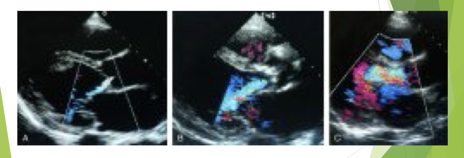
What is this?
Grading AI by regurgitant jet area/LVOT area (PLAX)
In diastolic reversal of flow where is the sample volume?
Sample volume in descending thoracic aorta from suprasternal notch
Sample volume in abdominal aorta from subcostal position
What does diastolic reversal of flow look in the descending aorta?

What does diastolic flow reversal look like in the abdominal aorta?

What are some pitfalls of aortic flow reversal?
Poor quality windows
Too few beats measured (don’t get rid of information)
High color and PW filters
Color Nyquist limit >50 cm/sec
Not well validated
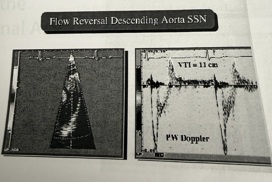
Is there holodiastolic flow reversal
There is a brief period of diastolic flow reversal, but it does not last all of diastole.
Based on the aortic regurgitation case study, how do I realize if it is moderate, mild, or severe AR?
Average the two percentages

Is there holodiastolic reversal of blood flow in the abdominal Aorta?
No
What is aortic valve area (AVA) using peak velocities and the continuity equation?

What is aortic valve area (AVA) using VTI and the continuity equation?

What does aortic insufficiency look like on Doppler?

In AP5
What does max velocity of aortic valve look like on Doppler?
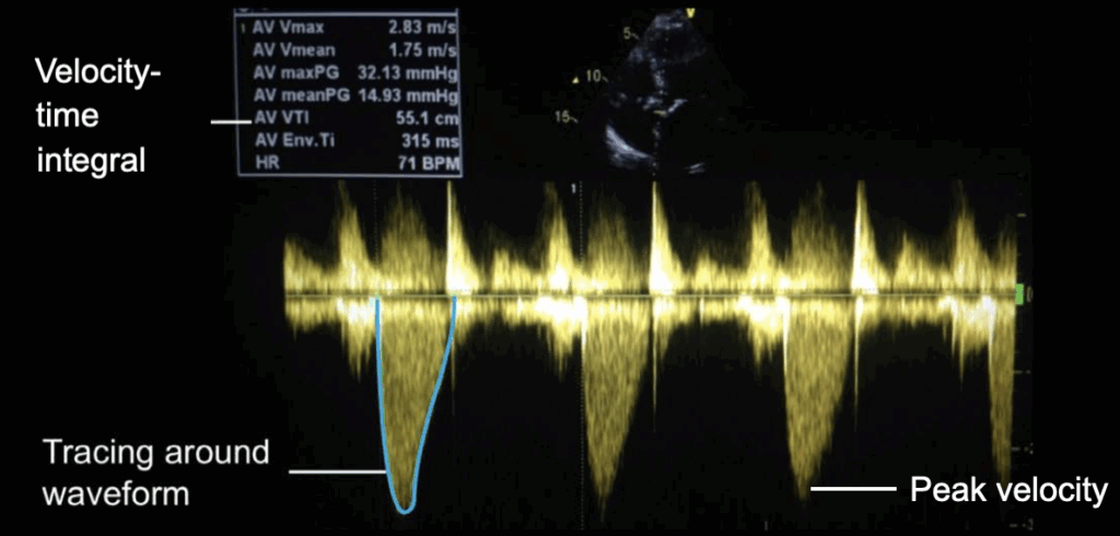
What is this?
Aortic stenosis