Structure and function of the oral cavity 1: The mouth - lips, cheeks, and palate
 Call Kai
 Learn
Learn Practice Test
 Spaced Repetition
 Match
Match Flashcards
 Knowt Play
1/41

There's no tags or description
Looks like no tags are added yet.
Name | Mastery | Learn | Test | Matching | Spaced | Call with Kai | Chat |
|---|
No analytics yet
Send a link to your students to track their progress
42 Terms
The Alimentary Tract/Canal
The passage (transport, physical processing and storage), from the Mouth to the Anus.
The Accessory Organs
the teeth, tongue, salivary glands, liver, gall bladder, pancreas and the anal sacs.
The mouth (Os/Oris): Rima oris /Oral fissure
the opening; Lips form the rim of the mouth
Ensures proper closure of the rima oris/oral fissure
Sucking milk in the young, Prevents loss of saliva and food while chewing
Angle of the mouth/oral commissure
Point where the upper and lower lips meet…
The Lips (labia oris)
Skin - Keratinization
Supported by Orbicularis oris m. a circular muscle and connective Tissue
Mucosa - Labial glands; minor salivary glands on the oral surface
The cheeks (buccae)
Form the caudo-lateral boundary of the oral cavity
Buccinator m. forms the major bulk
Inner mucosa and submucosa; Buccal glands – Minor salivary glands
Cheeks differences in species

Nerve supply
Sensory innervation – maxillary and mandibular branches of the trigeminal nerve (CN V)
Motor Innervation – Facial nerve (muscles of fascial expression)
Facial nerve supply

Blood Supply to the lips and cheeks
Facial artery: Superior labial and Angularis oris
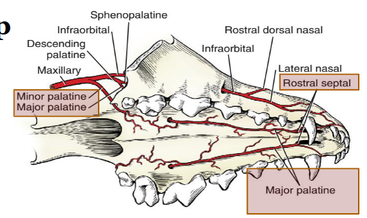
Maxillary artery: Inferior alveolar – mental branches, Infraorbital – lateral nasal a.
Blood supply in the face

The oral cavity boundaries
Hard and soft palate – Roof
The Mylohyoideus muscles – floor
Lips and Cheeks – Walls
Rostral limit – Rima oris
Caudal limit – Palatoglossal arch
The oral cavity
The vestibule (of the mouth); Space outside the dental rows (arcades)
The oral cavity proper; Space inside the dental rows (arcades)
Organs: The tongue and teeth, Openings of the Salivary glands
Hard palate (palatum durum)
Rostral bony portion
Incisive, maxillary, and palatine bones
Rugae -Transverse ridges (Palatine rugae)
Incisive papilla
opening of the incisive ducts on both sides – ducts of the vomeronasal organ
Hard palate of the horse
The mucosa of the hard palate is thick, particularly in its most rostral part, and incorporates a very generous venous plexus,
may become engorged(lampas) at the time of tooth replacement when it may project above the occlusal surfaces of the neighboring teeth
Anatomy of the hard palate of the horse

Soft palate (palatum molle or velum palatinum)
Caudal membranous portion formed by muscles and connective tissue
Relation with epiglottis/epiglottic cartilage - Normally lies slightly over the epiglottis -
allowing continuous respiration
Soft palate in horses
Below the epiglottic cartilage in the horse – Obligatory nasal breather
Parts of the soft palate
Palatoglossal arch
Palatopharyngeal arch
Palatine tonsils
Palatine (Salivary) glands
Functions of the soft palate
Respiration – relaxed
Swallowing/vomiting – must be lifted
Anatomy of the soft palate

Continued anatomy of the soft palate

Muscles of the soft palate
Palatine muscles
Tensor veli palatini muscles
Levator veli palatini muscles
Palatopharyngeus muscles
Palatinus m.
Anatomy of soft palate muscles

Motor innervation of the soft palate
Branches of the Vagus nerve- CN X via Pharyngeal plexus
Tensor veli palatini – Mandibular br. Trigeminal (CN V)
Sensory innervation of the soft palate
Major and Minor Palatine (Maxillary division of the trigeminal nerve)
Glossopharyngeal nerve CN –IX
Cleft Palate & / Cleft lip

Blood vessels to consider while cleft palate repair
General features of the oral mucosa
Stratified squamous epithelium (SSE)
Dog, Cat and Pig – Non keratinized
Ruminant and horse – Keratinized
Lamina propria and submucosa is usually intermixed in the oral region(Muscularis mucosae is absent)
Mixed salivary glands and skeletal muscles
The Lip
Outer – Cutaneous/Skin
Middle - Orbicularis oris muscle
Inner – Oral, Stratified squamous epithelium
Ruminant and Horses – Keratinized
Seromucous (Mixed) labial (salivary) glands
TS through the lip, Dog/Cat

1. Mucocutaneous junction
2. Stratifies squamous epithelium (SSE) - keratinized
3. Keratin layer of SSE, 4. hair follicle(cutaneous side)
5. sebaceous gland
6. Skeletal muscle (orbicularis oris)
7, Seromucous labial salivary glands
8. duct of the glands

1. Sinus Hair,
2. Hair follicles and glands in the outer skin,
3. Subcutis
4. Stratified Squamous Epith.
5. Lamina propria and Submucosa
6. Buccinator Muscle

The buccal papillae of the sheep and Cross-section of the cheek (Sheep). The buccal papillae: Keratinized stratified squamous epithelium

The Hard palate tissues
Keratinized Stratified squamous epithelium
Abundant connective tissue in the submucosa
CT Papillae projecting into the epithelial layer
Salivary gland in the caudal portion only
The mucosa of the hard palate has several ridges that are covered by keratinized
stratified squamous epithelium (e). Bone of the hard palate (not shown) is located
deep to the connective tissue (ct) of the propria- submucosa that underlies the
epithelium. Hard palate, cat

Soft palate histology species differences

Soft palate of cat showing oropharyngeal part lined by stratified squamous epithelial non keratinized (A) rested on thick layer of submucosa (B) with heavy layer of salivary glands (Blue arrow)

Buprenorphine
Pain relief/sedative, Effective in cats, oral spray/drops
Administration of allergy shots in Small animals
Sub-lingual Immunotherapy(SLIT)
Detomidine
pain relief/sedation
It has been described in horses and calves