Gen Bio 103 (UNIT 2) - DNA Replication and Gene Expression
 Call Kai
 Learn
 Practice Test
 Spaced Repetition
 Match
 Flashcards
 Knowt Play
1/94

There's no tags or description
Looks like no tags are added yet.
95 Terms
Semiconservative model
Each DNA serves as a template during replication—every DNA strand synthesized consists of an entirely new strand (daughter strand) and an old strand (parent strand)
DNA
a double-stranded molecule shaped like a twisted ladder. The "rails" of the ladder are made of an alternating sugar-phosphate backbone, while the "rungs" consist of nitrogenous bases pairing across the center.
Antiparallel strands
The two strands run in opposite directions. One strand runs from 5' to 3' (five-prime to three-prime), and its partner runs from 3' to 5'. The numbers refer to the carbon atoms on the sugar ring where the phosphate groups attach.
Proceeds in the amino-to-carboxyl direction, also known as the N-terminus to C-terminus direction.
The ribosome reads the mRNA strand from the 5′ to 3′ end and synthesizes the corresponding polypeptide starting with the very first amino acid (the N-terminus), which possesses a free amino group (—NH2)
What direction is a polypeptide synthesized in? What direction does ribosome read an mRNA strand?
Complementary strand
Due to specific base-pairing rules, the sequence of one strand strictly dictates the sequence of the other. If one strand reads 5'-ATCG-3', the complementary strand must read 3'-TAGC-5'.
Hydrogen bonds
Weak chemical bonds formed between opposing nitrogenous bases across the helix. A pairs with T via 2 ________, and G pairs with C via 3 _________.
Base stacking
Hydrophobic interactions and van der Waals forces that occur between adjacent, flat base pairs stacked on top of one another along the vertical length of the helix. Provides the vast majority of the double helix's thermodynamic stability.
Purines are larger with 2 rings, consist of adenosine and guanine. Pyrimidines are smaller with 1 ring, consist of thymine, cytosine, and uracil.
Purines vs Pyrimidines? (nitrogenous bases, structure)
mRNA
Type of RNA that carries the temporary genetic blueprint copied from DNA in the nucleus out to the cytoplasm. It serves as the direct template for protein synthesis during translation.
tRNA
Type of RNA that has a distinctive cloverleaf shape. It acts as an adapter molecule that matches specific amino acids to the corresponding codons on the mRNA strand using its unique anticodon loop—reads codons thru complementary base pairing with its anticodon.
rRNA
The structural and catalytic backbone/scaffolding of the ribosome. It aligns mRNA and tRNAs and catalyzes the formation of peptide bonds between amino acids to build proteins.
The sugar ribose has a reactive hydroxyl group at the 2’ position, which acts as a nucleophile that can attack the adjacent phosphodiester backbone, causing spontaneous self-cleavage and hydrolysis. RNA is flexible and single-stranded, exposing its chemical bonds to environmental and enzymatic attacks. Enzymes that break down RNA (RNases) are abundant in the environment and on human skin.
Why is RNA much less stable than DNA? (3)
Deoxyribose lacks an oxygen atom at the 2' carbon position, while has a hydroxyl -OH group at the 2' carbon position
Ribose vs deoxyribose sugar?
RNA does not have a lifelong partner strand to balance its hydrophobic bases. To shield its nonpolar bases from surrounding water, a single RNA strand will loop back onto itself. These architectural folds allow RNA molecules to transition from simple informational strings into structural machines or catalysts.
How and why does intramolecular base pairing occur in RNA?
To minimize contact with water, the partially hydrophobic nitrogenous bases twist inward and stack flatly on top of each other, pushing water out. Hydrogen bonds, while weak individually, collectively lock the matching bases into their native pairings.
Why is DNA structurally stable? (2)
Base stacking
the vertical alignment of hydrophobic, aromatic nitrogenous bases on top of one another within a nucleic acid strand. While hydrogen bonds lock base pairs together across the horizontal rungs of the double helix, ________ acts vertically along the length of the helix to hold the ladder together.
Nucleoside triphosphate (dNTP)
the generic term for the four molecular building blocks used to construct DNA. Each ______ consists of a deoxyribose sugar, a nitrogenous base (A, T, C, or G), and three phosphate groups that provide the energy needed to build the DNA chain. When DNA polymerase adds this to a growing DNA strand, it cleaves off two of these phosphates, releasing the necessary energy to form a covalent phosphodiester bond.
Phosphodiester bond
A highly stable covalent linkage that forms the structural backbone of nucleic acids. It is created when a phosphate group connects the 3’ carbon of one pentose sugar ring to the 5’ carbon of an adjacent sugar ring through a pair of ester bonds
Leading strand
the newly synthesized strand of DNA that is built continuously in the same direction that the replication fork is opening (the 5’ to 3’ direction). Only requires one single RNA primer at the very beginning (the origin of replication) to kick off synthesis
Lagging strand
The newly synthesized strand of DNA that must be built discontinuously in short, fragmented chunks because it is being replicated in the opposite direction of the unwinding replication fork. Requires multiple primers for DNA polymerase to synthesize in the 5’ to 3’ direction.
Replication fork
The dynamic, Y-shaped structure formed within the nucleus/cytoplasm when helical DNA is unwound by helicase. It represents the active site of DNA replication where the leading and lagging strands are simultaneously synthesized.
Replication bubble
The unwound, open region of a DNA double helix where active replication is occurring. It contains two replication forks moving symmetrically in opposite directions away from a central starting point.
Origin of replication
A specific, conserved sequence of nucleotides in a genome where DNA replication is initiated. These regions are typically rich in Adenine and Thymine (A-T) bases because their two hydrogen bonds are easier to melt than G-C pairs. (They have 2 hydrogen bonds instead of 3)
Origin recognition complex (ORC)
A multi-subunit DNA-binding protein complex found in eukaryotes. It binds directly to the origin of replication to initiate the assembly of the pre-replication complex and recruit helicases during the G1 phase of the cell cycle.
DNA polymerase
A massive family of enzymes responsible for synthesizing new DNA strands. They function by catalyzing the addition of incoming dNTPs to a pre-existing 3’ —OH group, matching the template strand with high fidelity in a strict 5’ to 3’ direction.
Primase
A specialized RNA polymerase that initiates DNA synthesis. Because DNA polymerases cannot build a strand from scratch, this enzyme synthesizes a short RNA primer (roughly 10–12 nucleotides) to provide the vital free 3’ —OH group required for DNA polymerase elongation.
DNA Polymerase I
A bacterial enzyme primarily involved in replication cleanup. It possesses 5’ to 3’ exonuclease activity, which allows it to chew away the temporary RNA primers on the lagging strand and replace them with standard DNA nucleotides
DNA Polymerase II
A bacterial DNA polymerase involved in inducible DNA repair and translesion synthesis. It acts as a backup system capable of bypassing damaged DNA templates when the main replication machinery stalls.
DNA Polymerase III
The primary, highly processive replicative enzyme in prokaryotes. It is responsible for the rapid, continuous synthesis of the leading strand and the discontinuous synthesis of Okazaki fragments on the lagging strand.
Helicase
A motor protein that uses energy from ATP hydrolysis to travel along a nucleic acid backbone and break the hydrogen bonds between complementary nitrogenous bases, forcibly unzipping the double helix.
Single stranded binding proteins (SSBP)
Specialized proteins that rapidly coat the single strands of DNA exposed by helicase. They prevent the unstable single strands from spontaneously re-annealing into a double helix or forming secondary hairpin structures, protecting them from nuclease degradation.
Okazaki fragments
Short, discontinuous stretches of newly synthesized DNA built backward on the lagging strand template. They are created because DNA polymerase must work away from the opening replication fork to maintain 5’ to 3’ synthesis.
DNA ligase
An essential molecular gluing enzyme. It seals structural nicks in the sugar-phosphate backbone by creating a covalent phosphodiester bond between an adjacent 3’ —OH group and a 5’ monophosphate, joining Okazaki fragments together.
Telomere
A repetitive, non-coding nucleotide sequence (such as TAGGG in humans) that caps the physical ends of linear eukaryotic chromosomes. They shield chromosome ends from degradation and prevent them from being mistaken for double-stranded DNA breaks.
Telomerase
A specialized ribonucleoprotein reverse transcriptase enzyme that carries its own internal RNA template. It functions to extend the 3’ ends of linear chromosomes, preventing telomere shortening primarily in germ cells, stem cells, and many cancer cells.
Mismatch Repair
A post-replication surveillance system. It identifies, excises, and replaces mispaired bases that escaped the proofreading activity of DNA polymerase, using structural clues (like parental strand methylation in bacteria) to distinguish the original template from the mutated new strand.
Polymerase Chain Reaction (PCR)
An in vitro laboratory technique used to exponentially amplify a specific target segment of DNA through repeated cycles of thermal denaturation, primer annealing, and enzymatic extension.
Plasmid
A small, circular, double-stranded DNA molecule that replicates independently of the host's chromosomal DNA. They are naturally abundant in bacteria, often carrying accessory genes like antibiotic resistance, and are widely used as vectors in genetic engineering.
PCR vs cellular DNA replication?
The Central Dogma of Molecular Biology
The foundational framework describing the directional flow of genetic information within a biological system. It states that information is stored in DNA, transcribed into temporary RNA messages, and ultimately translated into functional Proteins.
Transcription
The enzymatic process by which the genetic information encoded within a structural DNA sequence is enzymatically copied into a complementary single strand of RNA by RNA polymerase.
Coding strand
The strand of a DNA double helix whose nucleotide sequence exactly matches the newly synthesized RNA transcript (with Thymine instead of Uracil). It runs 5’ to 3’ parallel to the RNA copy.
Template strand
The structural DNA strand that runs 3’ to 5’ relative to the gene. It acts as the physical blueprint read by RNA polymerase via complementary base pairing to guide the assembly of the RNA molecule.
The template strand is the physical substrate that RNA polymerase binds to and reads; its bases directly dictate the sequence of the RNA via complementary base pairing. The coding strand is the non-template partner strand; it is not read by the enzyme, but its sequence is an exact structural mirror of the resulting RNA transcript.
Template vs Coding strand in transcription?
RNA polymerase
A complex enzyme that synthesizes RNA from a DNA template. Unlike DNA polymerase, it can initiate synthesis from scratch without a primer, unwinding the DNA ahead of itself and linking rNTPs together 5’ to 3’. Plays vital role in transcription/synthesizing mRNA.
Promoter
A specific regulatory sequence of DNA located upstream of a gene's coding region. It serves as the primary recognition and binding site for RNA polymerase and associated transcription factors to establish the correct initiation site and direction of transcription.
Transcription terminator sequence
A specialized genomic sequence at the end of a gene that signals RNA polymerase to cease synthesis. In prokaryotes, it can form structural elements like a hairpin loop or recruit specialized proteins to disrupt the transcription complex.
Prokaryotic core promoter regions
Region of a prokaryotic gene upstream of its transcription start site (+1) where the sigma factor binds to correctly position RNA polymerase and kick off transcription. Consists of the -10 element (Pribnow box) and the -35 element—both have specific sequences and positions relative to the starting site, which are conserved across several genome types
Sigma protein
A bacterial transcription initiation factor. It binds to the core bacterial RNA polymerase to form the active holoenzyme, granting it the structural specificity required to recognize and bind tightly to the -10 and -35 promoter elements. Ensures that RNA polymerase binds to DNA in the correct direction/orientation
Transcription factor
Any protein in eukaryotes that binds to specific DNA sequences (such as promoters or enhancers) to control the rate of transcription by either recruiting or blocking the binding of RNA polymerase.
Major groove
The wider, deeper structural indentation running along the outer surface of the DNA double helix. It exposes more of the chemical signatures of the nitrogenous bases, making it the primary site for sequence-specific binding by transcription factors.
Minor groove
The narrower, shallower structural indentation on the DNA double helix. It provides less distinct chemical accessibility to the base identities, generally hosting non-sequence-specific structural proteins.
TATA box
A conserved, core eukaryotic promoter sequence located roughly 25–30 base pairs upstream of the transcription start site. It is enriched with A-T pairs, serving as the primary landmark for the assembly of the transcription initiation complex.
TATA binding protein (TBP)
A highly specialized subunit of the general transcription factor TFIID. It recognizes and binds directly to the TATA box, bending the DNA double helix nearly 90 degrees to physically distort the strand and recruit RNA polymerase II.
RNA processing
The series of chemical modifications that a eukaryotic primary RNA transcript (pre-mRNA) must undergo in the nucleus to transform into a mature, stable messenger RNA (mRNA) ready for export to the cytoplasm. Consist of splicing, and the addition of the 5’ cap and poly-A tail
RNA splicing
The highly precise post-transcriptional process where non-coding sequences (introns) are cleaved out of a pre-mRNA transcript, and coding sequences (exons) are covalently joined back together.
Alternative splicing
A regulated regulatory process where different combinations of exons within a single pre-mRNA are selectively included or excluded. This allows a single gene to encode multiple distinct protein isoforms, vastly expanding eukaryotic proteomic complexity. In other words, exons of one protein can be treated as introns for another protein, and vice versa, depending on the target protein product.
5’ guanosine cap
A modified 7-methylguanosine nucleotide added backward via an unusual 5’ to 5’ triphosphate linkage to the front of a nascent pre-mRNA. It protects the transcript from 5’ exonuclease degradation (by preventing it from being recognized) and serves as a vital anchor for ribosome binding during translation.
Poly-A tail
A long stretch of adenine nucleotides (typically 100–250 bases) added enzymatically to the 3’ end of a cleaved pre-mRNA transcript. It regulates mRNA stability, governs export from the nucleus, and boosts translation efficiency.
Spliceosome
A massive molecular machine composed of small nuclear RNAs and over a hundred proteins. It coordinates the precise alignment, cutting, and pasting of exons during RNA splicing.
Splice junctions
The highly conserved nucleotide sequences at the borders of introns and exons that define where splicing cuts occur. They include the 5’ splice site (usually GU), the 3’ splice site (usually AG), and an internal branch point adenine.
Small Nuclear Ribonucleoprotein (snRNP)
Complexes made of small nuclear RNA (snRNA) molecules and proteins. Form the core catalytic and structural units of the spliceosome, directly recognizing splice junctions via complementary base pairing.
Prokaryotic vs Eukaryotic transcription?
Translation
The cellular process where the genetic code carried by a mature mRNA molecule is read by a ribosome to synthesize a polypeptide chain with a specific sequence of amino acids.
Codon
A specific sequence of three consecutive nucleotides in mRNA that specifies a single amino acid or acts as a start/stop signal during protein synthesis.
Anticodon
A triplet of nucleotides carried on the loop of a transfer RNA (tRNA) molecule that is perfectly complementary to an mRNA codon, enabling precise amino acid matching.
Untranslated regions (UTR)
Segments of mature mRNA located at both the 5’ end (upstream of the start codon) and the 3’ end (downstream of the stop codon). They do not code for amino acids but contain vital motifs regulating mRNA stability, localization, and translation rates.
Wobble position
The third 3’ nucleotide position in an mRNA codon and its corresponding 5’ partner position on a tRNA anticodon. The structural configuration of this site allows for non-standard hydrogen bonding, meaning a single tRNA species can pair with multiple different codons.
Ribosome
A complex molecular machine composed of ribosomal RNAs (rRNA) and structural proteins. It acts as the physical factory that stabilizes mRNA-tRNA interactions and catalyzes peptide bond formation.
Small ribosomal subunit (30S/40S)
The smaller section of the ribosome (30S in bacteria, 40S in eukaryotes) that binds directly to the mRNA transcript, scanning for the correct initiation sequence and ensuring accurate codon-anticodon pairing. This subunit first binds to the ribosome binding site, hold mRNA in place, and ensures that the proper codon is aligned with its corresponding amino acid in the aminoacyl site.
Large ribosomal subunit (50S/60S)
The larger section of the ribosome (50S in bacteria, 60S in eukaryotes) that binds atop the small subunit. It binds to RNA right after the small ribosomal subunit and initiator tRNA have located and aligned with the start codon (AUG) on the messenger RNA. It houses the catalytic peptidyl transferase center and the three structural tRNA binding pockets.
Kozak sequence
A conserved consensus sequence (ccGccAUGG) flanking the start codon in eukaryotic mRNAs. It acts as a structural signpost that helps the scanning small ribosomal subunit identify the correct functional AUG start codon.
Aminoacyl (A) site
The first structural pocket inside a ribosome. It serves as the physical landing pad for incoming, charged aminoacyl-tRNA molecules matching the next available mRNA codon.
Peptidyl (P) site
The middle pocket of the ribosome. It holds the tRNA molecule that is currently covalently linked to the growing polypeptide chain.
Exit (E) site
The final pocket inside the ribosome. It holds uncharged tRNAs that have surrendered their amino acids before they are ejected from the ribosome.
16S RNA
The essential catalytic structural RNA component of the prokaryotic small (30S) ribosomal subunit. It is responsible for recognizing the Shine-Dalgarno sequence on bacterial mRNAs to initiate translation.
23S RNA
The massive ribosomal RNA component forming the core of the prokaryotic large (50S) ribosomal subunit. It functions directly as a ribozyme to catalyze peptide bond synthesis.
Peptidyl transferase
The enzymatic activity located within the large ribosomal subunit that catalyzes the formation of a peptide bond between the amino acid in the A-site and the growing peptide chain attached to the tRNA in the P-site.
Missense mutation
A single nucleotide substitution in a DNA sequence that alters a codon so that it specifies a different amino acid than the original sequence.
Silent mutation
A nucleotide substitution that alters a codon but, due to the redundancy of the genetic code, still codes for the exact same amino acid, causing no structural change to the protein.
Frameshift mutation
The insertion or deletion of a number of nucleotides that is not a multiple of three. This physically shifts the reading frame of the ribosome, scrambling every downstream codon and completely altering the protein structure.
Nonsense mutation
A point mutation that changes a codon specifying an amino acid into a premature stop codon, leading to truncated, typically non-functional proteins.
Chromosome level mutation
Large-scale structural alterations affecting massive tracts of DNA, including deletions, duplications, inversions, or translocations of entire chromosomal regions.
Ribosome binding site (Shine-Dalgarno)
A purine-rich sequence (AGGAGG) found upstream of the start codon in prokaryotic mRNAs. It base-pairs directly with the 16S rRNA of the small ribosomal subunit to properly align the ribosome over the start codon.
Coding sequence
The actual continuous stretch of codons in an mRNA molecule that begins at the start codon (AUG) and terminates at the stop codon, explicitly dictating the final primary structure of the protein.
The structural configuration of the DNA polymerase active site favors geometrically stable, standard Watson-Crick base pairings (A-T and G-C). Mismatched incoming dNTPs distort this geometry and are sterically excluded from bonding easily. If an incorrect base is successfully linked, the structural misalignment stalls the polymerase.
The enzyme shifts gears, moving the frayed 3’ end of the new strand into its adjacent exonuclease pocket. It clips out the bad nucleotide, then slides the strand back into the polymerization site to continue forward.
Any errors that slip through the replication fork are identified by downstream MMR scanning complexes. The system cuts the newly synthesized strand, digests the region containing the mutation, and allows a new DNA polymerase to fill the gap with perfect accuracy
How is fidelity of DNA replication maintained via DNA polymerase fidelity, proofreading, and mismatch repair? (3)
DNA polymerases can only synthesize DNA 5’ to 3’, and they always require a pre-existing primer.
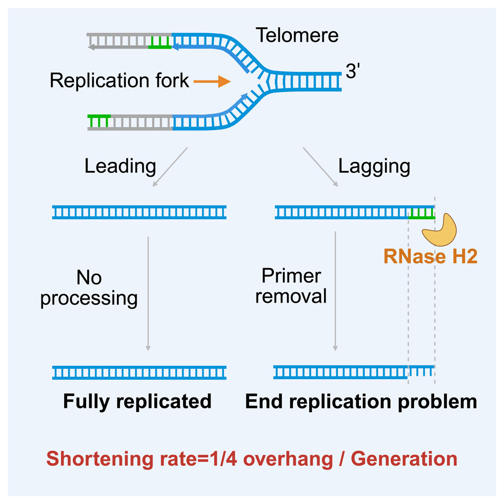
During lagging strand synthesis at the very tip of a linear chromosome, an RNA primer is laid down at the extreme 3’ edge of the template. Once DNA polymerase finishes extending the final fragment, the RNA primer is removed by nucleases. Because there is no upstream DNA to provide a 3'-OH group hook, DNA polymerase cannot fill the resulting gap. Consequently, a short single-stranded overhang is left behind at the tip, which is trimmed away by the cell, causing the chromosome to shorten slightly with every round of division.
Why is some DNA lost at chromosomal ends every time DNA is replicated? What does this stem from? (DNA poly. traits)
Intron
A non-coding segment of DNA or RNA located inside a gene that is transcribed into pre-mRNA but is excised during RNA splicing before the message is translated into protein.
Exon
A coding segment of a gene that contains the structural blueprint for a protein. Exions are retained in the mature mRNA molecule after splicing occurs.
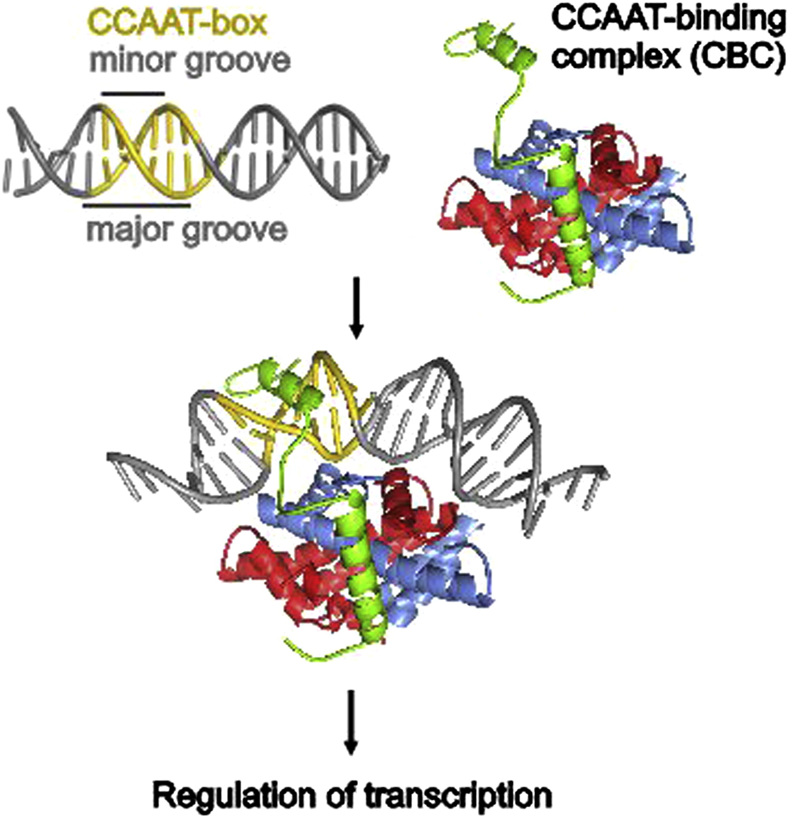
They use specialized structural arrangements called DNA Binding Domains (such as helix-turn-helix motifs) to read the code from the outside.
These protein motifs insert alpha-helices directly into the major groove of the DNA helix. Once inside, the specific amino acid side-chains of the transcription factor form a precise network of non-covalent interactions with the edge profiles of the base pairs exposed at the bottom of the groove. The protein binds tightly only when it finds the exact sequence that matches its chemical layout.
How do transcription factors use DNA binding domains to recognize specific DNA sequences?
Info Complexity: The specific order of these core promotors (-35 box, -10 box, and TATA box) represents a unique chemical landscape that can only be bound by transcription factors and sigma proteins in one exact spatial orientation.
Directionality: Because the binding proteins themselves are highly asymmetric, they can only land on the promoter facing one direction. When they recruit RNA polymerase, they lock the enzyme into a fixed physical orientation. This orientation determines which strand is treated as the template and forces the enzyme to march forward in the correct direction.
How does a core promoter provide sufficient information complexity for transcription factor binding/establishing RNA polymerase directionality? (2)
Prokaryotes:
Occurs entirely in the cytoplasm
Coupled; translation begins on the mRNA while transcription is still finishing.
Requires a Sigma protein to find promoters.
No processing; mRNA is raw and typically unmodified (no caps or tails)
Uses -35 and -10 core elements as core promoter region
Eukaryotes:
Occurs strictly within the nucleus.
Decoupled; transcription must completely finish and process before export to cytoplasm—spatially and temporally separated from translation
Requires an extensive array of General Transcription Factors and the TATA binding protein (TBD)
Processing required before mRNA leaves nucleus (5’ cap, poly-A tail, and splicing out introns)
Uses TATA box as core promoter region
Prokaryotic vs. Eukaryotic transcription? (5 differences) —> Where does it occur? What proteins are involved? What happens after transcription? What does it use as a promoter?
Unwinding: : Cellular replication uses the enzyme helicase and topoisomerase to unzip DNA at body temperature. PCR uses high heat (thermal denaturation at 95 C) to split the strands.
Priming: Cells use an RNA polymerase (primase) to synthesize temporary RNA primers. PCR relies on pre-designed, synthetic DNA primers added to the master mix.
Enzymes: Cells use a suite of complex polymerases (e.g., DNA Pol III). PCR uses a single, thermostable DNA polymerase that survives repeated boiling cycles.
Topology: Cellular replication duplicates entire chromosomes (both linear and circular) using leading and lagging strand coordination. PCR amplifies a short, isolated segment continuously across both strands.
Why does PCR only amplify a specific part of a template? (How is DNA unwound and primed, what enzymes are used, and what is the product?)
PCR product ends correspond exactly to the 5’ ends of the primers because the primers physically become the structural ends of the final DNA strands.
Why do PCR product ends correspond to the nucleotides at the 5’ end of each primer?

The two primers must be oriented in an opposing, "face-to-face" direction, pointing toward each other so that their 3’ ends extend inward across the target sequence.
DNA polymerase can only synthesize DNA by adding nucleotides to the 3’ end of a primer (moving in the 5’ to 3’ direction), the primers must be arranged like two runners starting on opposite sides of a track, running toward one another.
How must DNA primers be oriented and placed in order to form a PCR product? Why?