Development and Inheritance - Lecture Notes
 Call Kai
 Learn
Learn Practice Test
 Spaced Repetition
 Match
Match Flashcards
 Knowt Play
1/96

There's no tags or description
Looks like no tags are added yet.
Name | Mastery | Learn | Test | Matching | Spaced | Call with Kai | Chat |
|---|
No analytics yet
Send a link to your students to track their progress
97 Terms
Prenatal Period
Begins with fertilization
Secondary oocyte and sperm unite
Ends 38 weeks later with birth
Pre-embryonic period
Embryonic period
Fetal period

Pre-embryonic Period
First 2 weeks after fertilization
Zygote, cell produced by fertilization, becomes spherical multicellular structure blastocyst
Ends with blastocyst implants in uterine lining
Embryonic Period
3rd through 8th weeks of development
Rudimentary versions of major organs appears
Now called an embryo
Fetal Period
Remaining 30 weeks prior to birth (9-38 weeks)
Organism is now called a fetus
Continues to grow and increase in complexity
Embryogensis
Developmental period of pre-embryonic and embryonic periods
Fertilization
Fusion of sperm and secondary oocyte
Restores diploid number of chromosomes (46 total)
Determines the sex of the organism
Occurs in the ampulla of the uterine tube
Capacitation
Physiology condition undergone by sperm to become capable of fertilizing the secondary oocyte
Occurs in female reproductive tract
Glycoprotein coat and some proteins
Removed from sperm plasma membrane
Lasts several hours
Influx of Ca2+
Oocyte in surrounded by the cumulus oophorus (bound by pogesterone)
Necessary for capacitation, acrosome reaction, fertilization
Phase of Fertilization
Corona Radiata Penetration
Zona Pellucida Penetration
Fusion of Sperm and Oocyte

Phase of Fertilization: Corona Radiata Penetration
1st phase
Sperm reaching secondary oocyte
Initially prevented entry by corona radiata and zona pellucida
Can push through cell layers of corona radiata
Phase of Fertilization: Zona Pellucida Penetration
Acrosome Reaction:
Release pf digestive enzymes from acrosomes
Allows sperm to penetrate zona pellucida
After penetration of secondary oocyte:
Immediate hardening of zona pellucida, preventing other sperm from entering this layer
Ensures only one sperm fertilizes the oocyte
Polyspermy
If two sperm enter simultaneously
Immediately fatal with 23 triplets chromosomes
Phase of Fertilization: Fusion of Sperm & Oocyte
In the plasma membranes of sperm & oocyte
Immediate fuse; only sperm nucleus enters oocyte
Forms an ovum after second meiotic completion
Nucleus of sperm and ovum pronuclei
Zygote: single diploid cell form
Pronuclei
Each with haploid number of chromosomes
Fuse to become diploid nucleus
Cleavage
Rapid mitotic divisions increase cell number without increasing overall size
Before 8-cell stage
Become tightly compacted after 3rd cleavage divisions
Compaction: process by which contact between cells in increased to the max

Cleavage: Morula
16-cell stage; Pre-embryo continues division
Develops fluid-filled cavity, blastocyst cavity
Cleavage: Blastocyst
Fluid-filled cavity
Trophoblast: outer ring of cells surrounding cavity
Will form the chorion
Embryoblast: packed cells within one side of blastocyst
Will form embryo proper
Cells pluripotent, able to develop into any tissue
Transit of the Pre-embryo through the uterine tube: Fertilization through implantation

Implantation
Blastocyst embeds in uterine wall (day 7)
Burrows into the endometrium (implantation)
Human Chorionic Gonadotropin (hCG) is secreted, maintaining the corpus luteum for hormone production
Placenta beings to form by the 2nd week
Pregnancy detection

2 layers of Trophoblast
Cytotrophoblast: Inner layer
Syncytiotrophoblast: Outer layer
Produces hCG
Hormone levels during pregnancy

Clinical View: Infertility
Inability to conceive and maintain a pregnancy
Multiple causes:
Blocked uterine tubes
Caused by pelvic inflammatory disease or endometriosis
Ovulation disorders
Anti-sperm antibodies or low sperm count
Abnormal sperm, impaired sperm delivery
Clinical View: Infertility Treatments
Intrauterine insemination
Oral medications (Clomid)
In virto fertilization (IVF)
Donor oocytes
Clinical View: Chromosomal Abnormalities and Spontaneous Abortion
Occurs regularly during gametogenesis, fertilization, or cleavage
If severe, result in spontaneous abortion (miscarriage)
Many within 2 to 3 weeks after fertilization, before pregnancy known
Perhaps 50% of pregnancies terminated from spontaneous abortion
Half from chromosomal abnormalities
Changes to embryoblast
By day 8 (2nd week)
Cells start forming two layers:
Hypoblast layer adjacent to blastocyst cavity
Epiblast layer adjacent to amniotic cavity
Together form flat disc - bilaminar germinal disc
Extraembryonic membranes
Formed by bilaminar germinal disc and trophoblast
Mediate between them and environment
Protects embryo
Assist in nutrition, gas exchange, and removal of waste
Extraembryonic membranes: Yolk Sac
1st extraembryonic membrane to develop
Continuous with hypoblast layer
Does not store yolk (like birds)
Important site for early blood cell and blood vessel formation
Extraembryonic membranes: Amnion
Membrane continuous with epiblast layer
Eventually encloses entire embryo in fluid-filled sac - amniotic cavity
Protects membrane from drying out
Specialized to secrete amniotic fluid bathing embryo
Extraembryonic membranes: Chorion
Outermost extraembryonic membrane
Formed from cytotrophoblast cells and syncytiotrophoblast
Cells blend with functional layer of endometrium
Eventually form placenta
Site of nutrient exchange between embryo and mother
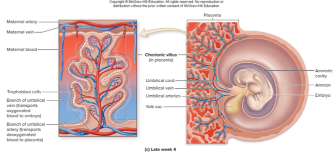
Placenta
High vascular structure
Functions:
Site exchange of nutrients, wastes, and respiratory gases between maternal and fetal blood
Transmits maternal antibodies to developing embryo or fetus
Produces estrogen, progesterone, and hCG
Maintains and builds the uterine
Placenta: Connecting Stalk
Connects early embryo to placenta
Eventually contains umbilical arteries and veins
Precursor to future umbilical cord
Placenta: Chorionic Villi
Fingerlike structures formed from chorion
Containing branches of umbilical vessels
Placenta: Gas and Nutrient exchange
Functional layer of endometrium with maternal blood vessels
Maternal blood does not mix with fetal blood
Bloodstreams so close that nutrients and gases mix
O2 diffusing from maternal blood to fetal blood
CO2 diffusing from fetal to maternal blood
Placenta: Selectively permeable
Example: respiratory gases passing freely
Microorganisms and certain maternal hormones prevented
Some harmful substances can cross:
Example: viruses, bacteria, drugs, alcohol, and toxins
May cause birth defects or death
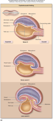
Formation of Extraembryonic Membranes: Week 3

Formation of Extraembryonic Membranes: Early Week 4

Formation of Extraembryonic Membranes: Late Week 4
Gastrulation1
Critical period of development; occurs during third week
Epiblast forms three primary germ layers
Cells from which all body tissues develop
Ectoderm, mesoderm, endoderm
Three-layered structure called an embryo
Gastrulation2
Begins with formation of primitive streak
Thin depression on surface of epiblast
Primitive node
Cephalic end of streak
Consists of elevated area surrounding small primitive pit
Invagination:
Cells detaching from epiblast layer
Migrate through primitive streak between epiblast and hypoblast layer

Formation of three primary germ layers:
Ectoderm: Becomes nervous system, epidermis
Mesoderm: Becomes muscles, bones, heart, kidneys
Endoderm: Becomes digestive, respiratory, and urinary linings

Embryonic Folding
Flattened, disc-shaped 3 week embryo
Starts to fold on itself during late 3rd and 4th week
Two types of folding:
Cephalocaudal and Transverse

Embryonic Folding: Cephalocaudal Folding
Occurs in cephalic and caudal regions of region
Rapid growth of embryonic disc and amnion
No growth of yolk sac
Causes head and tail to fold on themselves

Embryonic Folding: Transverse Folding
Left and right sides fold inward, creating a cylindrical body
Fusing of sides of embryonic disc in midline
Ectoderm solely along exterior of embryo
Endoderm confined to internal region of embryo
Yolk sac pinching off from most endoderm
Except vitelline duct
Creates torso region
Differentiation of ectoderm
On external surface of cylindrical embryo
Responsible for formng nervous system tissue: Neurulation
Forms:
Epidermis, sense organs, pituitary gland, adrenal medulla, enamel of teeth, lens of eye
Three Primary Germ Layers and Their Derivatives

Five categories of Mesoderm
Notochord
Paraxial mesoderm
Intermediate mesoderm
Lateral plate mesoderm
Head mesenchyme

Five categories of Mesoderm: Notochord
Formed by tightly packed midline group of mesodermal cells
Basis for central body axis and axial skeleton
Induces formation of neural tube
Five categories of Mesoderm: Paraxial Mesoderm
Found on both sides of neural tube
Forms somites, block-like masses
Forms axial skeleton, muscle, and cartilage, dermis, and connective tissue
Five categories of Mesoderm: Intermediate Mesoderm
Lateral to paraxial mesoderm
Forms most of kidneys, ureters, and reproductive system
Five categories of Mesoderm: Lateral Plate Mesoderm
Most lateral layers of mesoderm
Forms spleen, adrenal cortex, and cardiovascular system
Serous membranes and connective tissue of limbs
Five categories of Mesoderm: Head mesenchyme
Forms connective tissues and musculature of face
Differentiation of endoderm
Becomes innermost tissue after transverse folding
Forms:
Linings of GI, respiratory, urinary, and reproductive tracts
Tympanic cavity, auditory tube
Liver, gallbladder, pancreas, palatine tonsil, thyroid and parathyroid glands, thymus
Organogensis
Organ development when layers have formed and folding complete
By 8 weeks, upper/lower limbs have adult shape and most organ system have rudimentary form
Teratogens: substances causing birth defects or death (drugs or viruses)
Peak development period: time where most organization and construction of an organ occurs
Fetal period (Weeks 9-38)
Growth and organ maturation
Crown-rump length (CRL) and weight increase significantly
Movement begins (quickening)
Last two months: Rapid weight gain and lung maturation
Pregnancy: First Trimester
First 3 months of pregnancy
Zygote becoming embryo and then early fetus
Pregnancy: Second Trimester
4 to 6 months
Growth of fetus and expansion of maternal tissues
Pregnancy: Third Trimester
7 to 9 months
Fetus growing most rapidly
Mother’s body preparing for labor and delivery
Hormonal Changes: Estrogen & Progesterone
Produced by corpus luteum during 1st trimester. Mostly produced in placenta in 2nd & 3rd trimesters.
High levels suppressing FSH and LH secretion
Ovarian cycle and follicular development arrested
Facilitate:
Uterine & mammary gland enlargement, and fetal growth
Faster-growing nails & fuller hair
Relaxation of ligamentous joints
Uterus functional layer growth due to progesterone
Hormonal Changes: Relaxin
Secreted by corpus luteum and placenta
Promotes blood vessel growth ini uterus
Hormonal Changes: Corticotropin-releasing hormone (CRH)
Secreted from placenta in large amounts
Role in length of pregnancy and timing of childbirth
Responsible for aldosterone rise in mother
Promotes fluid retention and edema
Hormonal Changes: Human placental lactogen (HPL)
Secreted from placenta
Affects how pregnant woman metabolizes certain nutrients
Mother metabolizing more fatty acids instead of glucose
Inhibits effects of insulin; more glucose available for fetus
Hormonal Changes: Prolactin
Increased levels (10x) produced by anterior pituitary
Ensures lactation occurs after giving birth
Hormonal Changes: Oxytocin
Increased levels produced by hypothalamus
Involved in uterine contractions
Involved in milk expulsion from mammary glands
Increased in second and third trimester
In response to rising estrogen levels
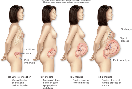
Uterine expansion
Begins once implantation occurs
By 12 weeks, uterus just superior to pubic symphysis
Most of enlargement due to:
Muscle hypertrophy, hyperplasia, placental growth, and amniotic fluid
By 16 weeks, fundus at midpoint between pubic symphysis and umbilicus
By ninth month fundus at xiphoid process of sternum
Melanocyte-stimulating hormoe
Secreted by placenta
Darkening of areola and nipples, linea alba, and now linea nigra
Growth of mammary glandular tissue
Development of additional acini
Increased insulin resistance in pregnancy can cause
Due to increased levels of corticosteroids, estrogen, progesterone, and HPL
Can lead to gestational diabetes in mother
Morning Sickness
Occurs in 1st trimester; not just in the morning
Some with a little nausea, others with severe symptoms (hyperemesis gravidarum [HG])
Cause unknown, possibly due to high hormones
Nutrition Changes
Weight gain due to fetus, fluid retention, adipose tissue, etc.
About 300 extra calories/day needed
Require adequate folic acid, calcium, protein, and iron
Clinical View: Gestational Diabetes
Diabetes that first develops during pregnancy
Increased insulin resistance and high blood glucose
Appearing in 2nd trimester
May develop high blood pressure and complications; risk of large baby
Increased risk of cesarean section and birth complications, hypoglycemia
Special diet to regulate blood glucose levels
At increased risk for type 2 diabetes later in life
Body System Changes:
Cardiovascular: Blood volume increases by 50%
Respiratory: Tidal volume increases
Urinary: Increased filtration due to fetal waste
Digestive: Increased nutrient absorption, but also reflux, nausea
Clinical View: Preeclampsia
High blood pressure occurring by second half of pregnancy
Risk factors: obesity, diabetes, older age, and previous preeclampsia
Cause is unknown but general risks of hypertension for mother
Only cure is giving birth, with medications or labor induction
Eclampsia:
High blood pressure causing seizures
Medical emergency
Stage of Labor: Dilation Stage
1st stage of labor
Longest stage; cervix dilates to 10 cm
Amniotic sac may rupture (water breaking)
Ends when cervix if effaced (thinned)

Dilation variability
Nulliparous women (who have not given birth) experience longer dilation stage, 8 to 24 hours
Parous women (who have given birth) may be in this stage for 4 to 12 hours
Stage of Labor: Expulsion Stage
Usually 30 mins to several house
Baby is pushed out of the birth canal
Crowning: When first part of baby’s calvarium distends vagina
Episiotomy: sometimes necessary
Perineal muscle surgically incised
Stage of Labor: Placental Stage
Placenta is expelled (afterbirth)
Uterus continuing to contract
Compresses uterine blood vessels; displaces placenta from uterine wall
Afterbirth:
Placenta and remaining fetal membranes; 30 minutes
Complications may occur if fragments are left
Clinical View: Fetal Positioning and the Dilation Stage
Fetus in vertex position - head down toward sacrum
Ideal position for pushing
Breech position
Buttocks first and may delay cervical dilation
Variant positions extraction
Forceps, vacuum may be needed
Cesarean section
Clinical View: Vaginal Bacteria and the Infant Microbiome
Vaginal delivery vs cesarean (C-section) delivery
Exposure to fecal bacteria during vaginal delivery
Infants have gut microbiome similar to the mother
Diverse, protective, supported by nutrients in breast milk
Exposure to surrounding environment during C-section
Hospital-acquired bacteria
Less diverse, prone to health issues later in life
True vs False Labor:
True Labor: Regular contractions, cervical dilation
False Labor (Braxton-Hicks): Irregular, non-progressive contractions
Postnatal Changes for the Newborn
Respiratory changes. in neonate
Fetus after being expelled from uterus
Fetal lungs not full inflated prior to birth
Neonatal Adjustment
Respiratory System: First breath expands lung
Circulatory System: Fetal shunts close
Thermoregulation: Brown fat helps generate heat
Digestive System: Meconium (first stool) is passed
Clinical View: Preterm (Premature) Birth
Baby born before start of 37th week pregnancy
Preterm births earlier than 24 weeks rarely survive, may have severe medical issues
Usually very low birth weight
May have jaundice, breathing issues, problems regulating body temp, and feeding difficulties
Postpartum
Time period after giving birth
Woman’s body undergoing further changes
Feed neonate
Return to pre-pregnancy form and function
Postpartum Hormonal Changes: Estrogen & Progesterone
Mood swings, postpartum depression risk - Baby Blues
Hair reverts back to normal hair loos cycle
Peak in loss about 3 to 4 months after delivery
Chemoreceptors less sensitive to CO2 due to low progesterone
Corticotropin-releasing hormone (CRH). Placenta stops producing.
Lochia
Portion of blood volume, mucus endometrial tissue
Expelled via the vagina
Heaviest first five days, continues for several weeks
Lactation: Prolactin
Produced by anterior pituitary
Responsible for milk production
Secretion inhibited by dopamine in nonpregnant women and in men
Increased by high levels of estrogen
Lactation: Colostrum (initial milk)
Produce by mammary glands
During late pregnancy and first few days after birth
Watery, yellowish, milk like substance and lower concentration of fat than true breast milk
Rich in immunoglobulins, especially IgA
Laxative effect
Lactation: Breast milk
Starts to be produced few days postpartum
Higher fat content than colostrum
Has essential fatty acids, enzymes for digestion, and immunoglobulins
More easily digestible than breast milk substitutes
Milk Letdown
Oxytocin stimulates release via positive feedback
With suckling, mechanoreceptors in breast stimulated
Send signals to hypothalamus
Hypothalamus is stimulated to produce oxytocin, released into blood by posterior pituitary
Targets myoepithelial cells in mammary acini
Cells contract, releasing breast milk from acini
As milk released, infant continuing to nurse
Facilitates further milk release
Genetics1
Heredity: Transmission of genetic characteristics from parent to child
Genetics: Field of biology studying heredity and transmission patterns
Karyotype: Display of chromosomes pairs, ordered and arranged by size and similar features
Homologous Chromosomes: Paired chromosomes with genes for equivalent biological characteristics
Genetics2
Sex Chromosomes: Last two chromosomes containing genes that specify sex
Genes: Units of DNA that code for a protein
Locus: Specific space where each gene is located on a chromosome
Alleles: Variants of one gene found at some locus on homologous chromsomes
Example: alleles determining type A or type O blood
Genetics3
Dominant allele
Expresses, or physically shows, the trait
Represented by capital letter
Recessive allele
Trait is masked, represented by lowercase letter
Expressed only if present on both homologous chromosomes
Punnet Square:
Square showing specific gene combinations resulting from two parents
Gives probability that a particular gene combination can occur

Genetics4
Homozygous: If identical alleles present
Heterozygous:
Both dominant and recessive allele present
But only dominant allele expressed
Expression of the receive allele may appear to skip generations
Its phenotype is masked by the dominant allele
Karyotype

Chromosomes & Heredity
Humans have 46 chromosomes (23 pairs)
Autosomes (22 pairs) vs. Sex Chromosomes (XX/XY)
Genotype: Genetic makeup
Phenotype: Physical trait
Patterns of Inhertitance:
Dominant-Recessive: Dominant allele expressed (e.g. eye color)
Incomplete Dominance: Blended traits (e.g. sickle cell trait)
Codominance: Both alleles expressed (e.g. ABO blood type)
Polygenic Inheritance: Multiple genes influence traits (e.g. height, skin color)
X-linked Inheritance: Traits carried on the X chromosome (e.g. color blindness, hemophilia)
Sex-linked traits
Traits expressed by genes on X or Y chromosomes
900 to 1400 genes on X chromosome
Most not involved in sex determination
70 to 200 genes on Y chromosome
Mostly for male development
Sex-linked traits most often involve X chromosome
X-linked recessive traits
