Chapter 6: The Skeletal System I Bone Tissue
 Call Kai
 Learn
Learn Practice Test
 Spaced Repetition
 Match
Match Flashcards
 Knowt Play
1/109

There's no tags or description
Looks like no tags are added yet.
Name | Mastery | Learn | Test | Matching | Spaced | Call with Kai | Chat |
|---|
No analytics yet
Send a link to your students to track their progress
110 Terms
tissue and organs of the skeletal system
bone, cartilage, and ligaments
cartilage
embryonic precursor of most bones and covers many joint surfaces
ligaments
hold bone to bone at joint
tendons
attach muscles to bone
functions of the skeleton
support
movement
protection of delicate organs
blood formation in marrow
electrolyte balance - calcium & phosphate
acid-base balance
detoxification
osteology
study of bone
bone
connective tissue with a hard matrix
mineralization (calcification)
process of hardening
other tissue present in bone
blood
bone marrow
cartilage
adipose tissue
nervous tissue
fibrous connective tissue
flat bone
thin, often curved
examples: ribs, skull
long bone
rigid levers for movement
examples: humerus, femur
short bone
glide within joints
example: carpals of wrists
irregular bones
complex shape
examples: vertebrae
sesamoid bone
oval, generally within tendon
example: patella
general features of long bones
compact and spongy bone tissues
two epiphyses (heads) at ends of diaphysis (shaft)
marrow (medullary) cavity
epiphysial line - remnant of growth plate
periosteum - covering
nutrient foramina - tubes/holes in bone for vessel
endosteum - lining
articular cartilage - covering joints
long bone anatomy

general features of short bones
composed of spongy bone sandwiched between plates of compact bone
flat bone anatomy

bone cells
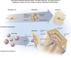
osteogenic (osteoprogenitor) cells, osteoblasts, osteocytes, osteoclasts
osteo- prefix
relating to bone
osteogenic (osteoprogenitor) cells
stem cells
osteoblasts
bone-forming cells
osteocytes
mature bone cells, contribute to bone maintenance/homeostasis
lacunae - cavities that house osteocytes
canaliculi - small canals that connect lacunae
lacunae
cavities that house osteocytes
cancliculi
small canals that connect lacunae
osteoclasts
bone-dissolving macrophages
"blasts build, clasts clean up"
bone cells and their development
bone matrix
one third organic and two thirds inorganic
organic bone matrix
one third of bone matrix
collagen and large protein-carbohydrate complexes
inorganic bone matrix
two thirds of bone matrix
85% hydroxyapatite (crystalized calcium phosphate salt), 10% calcium carbonate, and 5% other inorganic material
compact bone
osteon - basic unit - concentric lamellae, central (haversian) canal
perforating canals
circumferential lamellae

spongy bone
spicules - rods and spines of bone
trabeculae - thin plates of bones
porous appearance - spaces filled with bone marrow
lightweight but strong

bone marrow
soft tissue located in medullary cavities of long bones, spaces within spongy bone, and large central canals with osteons
two types: red marrow (myeloid tissue) and yellow marrow
red marrow (myeloid tissue)
hematopoietic tissue (blood-forming tissue) - both red and white blood cells are made here
yellow marrow
mainly fat
location of red marrow in adults
skull, vertebrae, sternum, ribs, parts of pelvic girdle, and proximal heads of humerus and femur

location of yellow marrow in adults
long bones of limbs

bone development
ossification (osteogenesis)
ossification (osteogenesis)
bone formation
begins within mesenchyme (an embryonic connective tissue)
two methods of ossification - intramembranous and endochondral
intramembranous ossification
produces flat bones of skull and most clavicle
bone develops in fibrous sheet resembling the dermis
endochondral ossification
*most bones develop this way
bone develops from hyaline cartilage model
stages of intramembranous ossification
1. deposition of osteoid tissue into embryonic mesenchyme
2. calcification of osteoid tissue and entrapment of osteocytes
3. honeycomb of spongy bone with developing periosteum
4. filing of space to form compact bone at surface, leaving spongy bone in middle

simple stages of intramembranous ossification
mesenchymal cells line up along vessels, become osteoblasts, make bone
stages of endochondral ossification
1. early cartilage model
2. formation of primary ossification center, bony collar, and periosteum
3. vascular invasion, formation of primary marrow cavity, and appearance of secondary ossification center
4. bone at birth, with enlarged primary marrow cavity and appearance of secondary marrow cavity in one epiphysis
5. bone of child, with epiphysial plate at distal end
6. adult bone with a single marrow cavity and close epiphysial plate

bone elongation
bones grow longer at epiphysial plates
metaphyses
plates made of hyaline cartilage
metaphysis
area of transition from cartilage (epiphyseal plate) to bone (diaphysis) at each end of marrow cavity
zones of metaphysis
think of it as cartilage providing a scaffold for bone to be formed
via endochondral ossification

bone widening and thickening
appositional and intramembranous ossification
apositional growth
growth in diameter and thickness
intramembranous growth
at surface
osteoblasts in periosteum deposit matrix
once matrix hardens, cells become osteocytes
circumferential lamellae are formed (not concentric lamellae)
osteoclasts widen medullary cavity
bone remodeling
absorption of old bone and deposition of new bone
bone is a living and dynamic tissue!
wolff's law of bone
wolff's law of bone
bone shape is determined by mechanical stress
bone adapts to withstand stress
form follows function
nutritional factors
calcium and phosphate
vitamin a
vitamin c (ascorbic acid)
vitamin d (calcitriol)
calcium and phosphate
raw materials for calcified ground substance
vitamin a
promotes formation of gylcosaminogylcans (protein-carb) fibers
vitamin c (ascorbic acid)
promotes collagen cross-linking-adding stiffness
vitamin d (calcitriol)
necessary for calcium absorption by small intestine and reduces urinary calcium loss
hormonal factors
calcitonin
growth hormone
estrogen and testosterone (sex steroids)
parathyroid hormone (PTH)
calcitonin
secreted by thyroid gland
stimulates osteoblasts in children and pregnant women
growth hormone
promotes intestinal absorption of calcium
stimulates growth plates and bone elongation
estrogen and testosterone (sex steroids)
stimulate long bone growth during adolescence
parathyroid hormone (PTH)
secreted by parathyroid glands
stimulates bone reabsorption to boost level of calcium in blood
the aging skeletal system
osteopenia
osteopenia
loss of bone (when severe it develops into osteoporosis)
resorption faster than deposition
after age 35 - osteoblasts less active than osteoclasts
after age 40 - females lose 8% of bone mass per decade and males lose 3% of bone mass per decade
fractures more common, heal slowly
fracture types
stress and pathological
stress fracture
caused by abnormal trauma
pathological fracture
occurs in bone weakened by a disease such as osteoporosis
fractures are classified according to
breaking of skin
direction of fracture
separation of bone pieces
treatment of fractures
closed reduction
open reduction
cast (fiberglass)
closed reduction treatment of fractures
nonsurgical manipulation of fragments
open reduction treatment of fractures
surgical setting involving plates, screws, or pins
how long does it take a fracture to heal
8-12 weeks
closed fracture
skin is not broke
(formerly called a simple fracture)
open fracture
skin is broken
bone protrudes through skin or wound extends to fractured bone
(formerly called a compound fracture)
complete fracture
bone is broken into two or more pieces
incomplete fracture
partial fracture that extends only partway across bone
pieces remained joined
nondisplaced fracture
the portions of bone are still in correct anatomical alignment
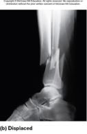
displaced fracture
the portions of bone are out of anatomical alignment
comminuted fracture
bone is broken into three or more pieces
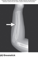
greenstick fracture
bone is bent toward one side and has incomplete fracture on opposite side
hairline fracture
fine crack in which sections of bone remain aligned
common in skull
impacted fracture
one bone fragment is drive into the marrow cavity or spongy bone of the other
depressed fracture
broken portions of bone forms a concavity
as in skull fractures
linear fractures
fracture parallel to long axis of bone
transverse fracture
fracture perpendicular to long axis of bone
oblique fracture
diagonal fracture
between linear and transverse
spiral fracture
fracture spirals around axis of long bone as the result of a twisting stress such as a skiing accident
x-ray of nondisplaced fracture

x-ray of displaced fracture
x-ray of comminuted fracture

x-ray of greenstick fracture
stages of the healing of a bone fracture
1. hematoma formation - the hematoma is converted to granulation tissue by invasion of cells and blood capillaries
2. soft callus formation - deposition of collagen and fibrocartilage converts granulation tissue into soft callus
3. hard callus formation - osteoblasts deposit a temporary bony collar around the fracture to unite the broken pieces while ossification occurs
4. bone remodeling - small bone fragments are removed by osteoclasts while osteoblasts deposit spongy bone and then convert it to compact bone

osteoporosis
bones lose mass and become subject to pathological fractures
hip, wrist, vertebrae are especially vulnerable
kyphosis ("dowager's hump")
postmenopausal white light built women are at greatest risk
kyphosis ("dowager's hump")
exaggerated thoracic curvature

why are postmenopausal white light build women at greatest risk of osteoporosis
decline of estrogen results in less inhibition of osteoclasts
by age 70 have typically lost 30-50% of bone mass
osteoporosis treatments
bis-phosphonates and parathyroid hormone
osteoporosis prevention
weight bearing exercise
adequate calcium and protein
healthy versus osteoporotic bone

orthopedics
prevention and correction of injuries and disorders of bones, joints, and muscles