BSCI202 LAB PRACTICAL 1
 Call Kai
 Learn
 Practice Test
 Spaced Repetition
 Match
 Flashcards
 Knowt Play
1/51

There's no tags or description
Looks like no tags are added yet.
52 Terms
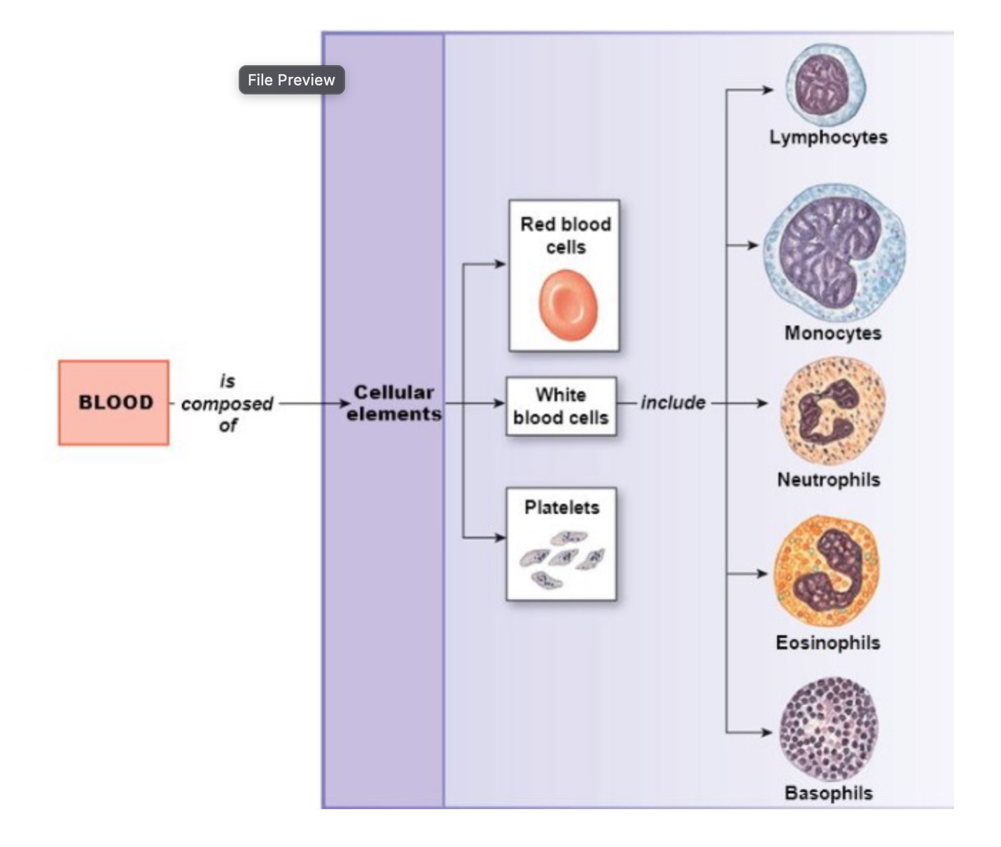
Blood & Hematocrit - Formed Elements (cells)
Erythrocytes use hemoglobin to carry oxygen to tissues throughout the body
The most abundant formed element in the blood
Leukocytes have many different immune functions
Granulocytes: neutrophils, eosinophils, basophils
Agranulocytes: lymphocytes, monocytes
Can travel out of blood vessels into tissues by diapedesis
Platelets are critical in blood clotting
Not complete cells – cytoplasmic fragments
Originate from megakaryocytes that fragment

Blood & Hematocrit - How to perform a hematocrit
Place a capillary tube into blood sample to draw up blood
Centrifuge the capillary tube
Measure total length of the fluid in the tube from the bottom
Then also measure in cm: RBC, WBC and plasma from the bottom
Calculate percentage of RBC/total, WBC/total, and plasma/total
Detailed instructions are in your lab manual

Blood & Hematocrit - Understanding Hematocrit
When blood is centrifuged:
Erythrocytes sink to the bottom (~45% of blood, a percentage known as the hematocrit)
Buffy coat contains leukocytes and platelets (less than 1% of blood), the buffy coat is a thin, whitish layer between the erythrocytes and plasma
Plasma rises to the top (~55% of blood)
Ranges:
Normal Adult Female Range: 37 – 47%
Normal Adult Male Range: 42 – 52%
Normal Newborn Range: 49 – 61%

Blood & Hematocrit - Conditions Detected by Hematocrit
Leukocytosis → leukocyte count too high
Buffy coat constitutes > 1%
caused by infection and some types of leukemia
Anemia → erythrocyte count too low
hematocrit value is below average
aplastic anemia → bone marrow doesn’t produce enough new erythrocytes
iron-deficiency anemia → erythrocytes are smaller
hemolytic anemia → erythrocytes are destroyed too quickly
sickle cell anemia → sickle-shaped erythrocytes
hemorrhagic anemia (caused by blood loss) NOT DETECTED by hematocrit
Individuals that live at high altitude tend to have higher hematocrit due to lower level of oxygen in the air
to compensate, these individuals produce more erythrocytes to increase blood oxygen levels
Dehydration: plasma percentage decreases
loss of water = less plasma
Lymphatic System
consists of two parts
lymphatic vessels
lymphoid tissues and organs
functions
transports escaped fluids back to the blood
plays essential roles in body defense and resistance to disease
also has a role in digestion
Lymphatic Characteristics
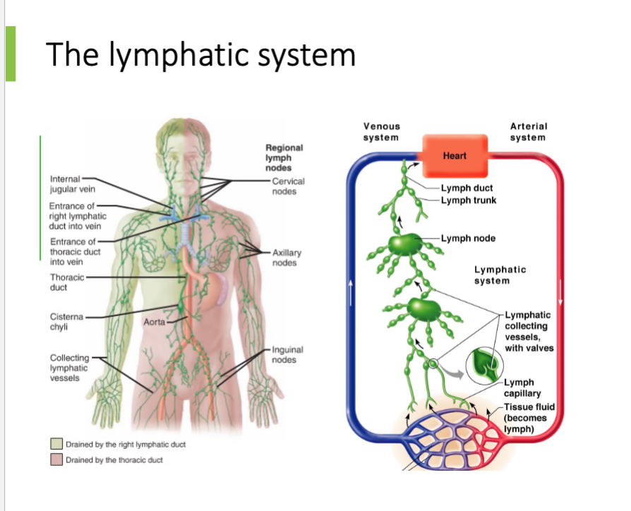
lymph fluid is carried by lymphatic vessels
fluid within bodily tissues (leaks out of the capillaries of the cardiovascular system)
fluid picked up by the lymphatic vessels
properties of lymphatic vessels
lymphatic vessels are one-way system that moves lymph back towards the heart
there is NO heart-like pump that moves lymph through lymphatic vessels
the “skeletal muscle pump” moves lymph through lymphatic vessels
squeezing motion of skeletal muscles
valves prevent back flow of lymph
Lymph Nodes
filter lymph (fluid) before its returned to the blood
defense cells within lymph nodes
macrophages engulf and destroy foreign substances
lymphocytes provide immune response to antigens
harmful materials that may enter lymph vessels
bacteria
viruses
cancer cells
cell debris


Lymph Node Filtration
filter lymph before its returned to the blood
typically kidney-shaped and less than one inch long
anatomy
cortex (outer part) → contains follicles that house collections of lymphocytes
medulla (inner part) → contains macrophages (phagocytosis cells)
lymph enters through afferent lymphatic vessels and exits through efferent lymphatic vessels
lymph enters the convex side through afferent lymphatic vessels
lymph flows through number of sinuses inside the node
lymph exits through efferent lymphatic vessels
fewer efferent than afferent vessels causes flow to be slowed
Lymphatic Vessels
collect lymph from lymph capillaries
carry lymph towards and away from lymph nodes
return fluid to circulatory veins near the heart
right lymphatic duct
thoracic duct

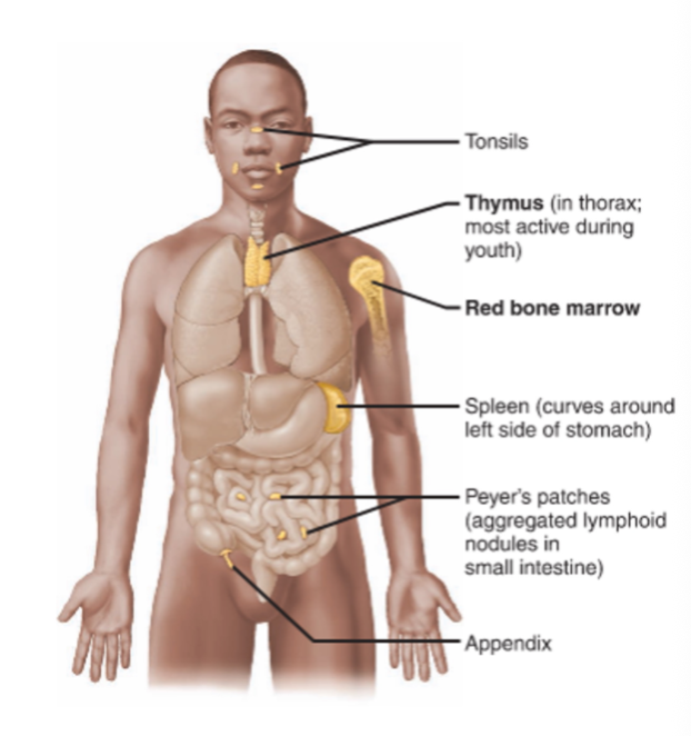
Lymphoid Organs
red bone marrow → site of lymphocyte production; B cells mature here
spleen → filters blood and destroys worn out blood cells
thymus → produces hormones to program lymphocytes
tonsils → trap and remove bacteria and other foreign material
peyer’s patch → capture and destroy bacteria in the intestine
appendix → reservoir for gut bacteria; may assist in B cell maturation
Primary Lymphoid Organs
Bone Marrow
Hematopoietic stem cells: precursors for all blood cells
Leukocytes except T lymphocytes full develop here
Thymus
T lymphocytes migrate from bone marrow to thymus
develop maturity in thymus
Secondary Lymphoid Organs + Tissues
spleen, tonsils, adenoids, appendix, peyer’s patches
collection of B cells, T cells, and macrophages
function to trap microorganisms and foreign particles
works to expose them to leukocytes in high concentrations
spleen and lymph nodes filter blood and lymph
Lymphoid Organs & Lymphatic Function
Lymphoid Organs that Contribute to Lymphatic Function:
Spleen → located on the left side of the abdomen
filters blood and destroys worn out blood cells
forms blood cells in the fetus
acts as blood reservoir
Thymus → located low in the throat, overlying the heart
functions at peak levels only during childhood
produces hormones (like thymosin) to program lymphocytes
Tonsils
small masses of lymphoid tissue around the pharynx
trap and remove bacteria and other foreign materials
tonsillitis is caused by congestion with bacteria
Peyer’s patches
found in the wall of the small intestine and capture and destroy bacteria in the intestine
resemble tonsils in structure
Lymphocytes Originate in Lymphoid Organs

Functions of Cells Involved in Immunity

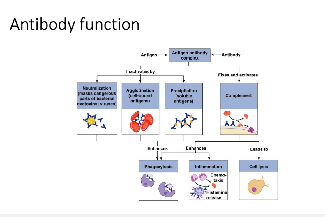
Antibodies
Overview
plasma cells (effector B cells) produce antibodies
antibodies are also called immunoglobulins
fiver major antibody classes: IgM, IgD, IgG, IgA, and IgE
antibodies are Y shaped and contain antigen-binding sites on their arms
each antibody binds to a specific antigen
Functions
binds to specific antigen
aids in inactivation or destruction of antigen
Structure
four amino acid chains linked by disulfide bonds
two identical amino acid chains are linked to form a heavy chain
other two identical chains are light chains
2 specific antigen-binding sites are present
constant region same within a class of antibodies
variable region gives specificity to antigen-binding site


Antibodies

Human Blood Groups
blood contains genetically determined proteins
antigens (a substance the body recognizes as foreign) may be attacked by the immune system
antibodies are “recognizers”
blood is typed by using antibodies that will cause blood with certain proteins to clump (agglutination or precipitation)
over 30 common red blood cell antigens
most vigorous transfusion reactions caused by ABO and Rh blood group antigens
Blood Groups
blood types are classified according to antigens found on the plasma membrane of erythrocytes
antigens are genetically determined proteins
blood plasma contains antibodies that can target antigens
blood is typically classified by two blood groups
ABO blood groups → based on the presence or absence of A and B antigens
Rh blood groups → based on presence of absence of one of the eight Rh antigens (agglutinogen D)
person’s blood is typically classified according to both groups (ex: B+)
blood transfusions from someone with antigens to which a patient has antibodies will result in a transfusion reaction
blood will agglutinate
Rh Blood Group
most people in the US are Rh-positive
denoted by a + or - added onto the ABO blood type
ex: O+, AB-, etc
Rh-negative individuals do not have Rh antibodies in their plasma
will become sensitized if they receive a blood transfusion from an Rh-positive individual
subsequent exposures will result in a transfusion reaction


How to Type & Read Blood
How to Type:
Collect a blood sample from a patient
Add a drop of blood from the sample to each well on a blood typing tray
Add antibody sera
To the A well → add anti-A serum
To B well → add anti-B serum
To Rh well → add anti-Rh serum
Mix contents of each well with a separate toothpick
How to Read Blood Type:
antibodies in sera will interact with corresponding antigens present within the blood
if antigens are present on the blood cell surface, interaction with the antibodies in the sera will cause agglutination
agglutination: clumping of particles in the blood; makes the sample appear clumpy/precipitated


Heart & Blood Vessel Anatomy
Location → Thoracic Cavity
size of a human fist, weighing less than a pound
located between the lungs in the inferior mediastinum (medial section of the thoracic cavity)


Gross Anatomy of Heart
Exterior
Apex is inferior, base is superior
Arteries carry blood away from the heart
Veins carry blood to the heart
Four chambers:
Two atria (singular: atrium) → receive blood from the vena cavae and pulmonary vein
Two ventricles → receive blood from atria, pumps blood into aorta and pulmonary artery
Interior
Septa (singular: septum) → separate the chambers of heart
intertrial septum separates the left and right atrium
interventricular septum separates the left and right ventricle
Four valves → prevent backflow of blood between heart chambers
semilunar (SL) valves → pulmonary and aortic
atrioventricular (AV) valves → tricuspid and mitral (bicuspid)

Valves
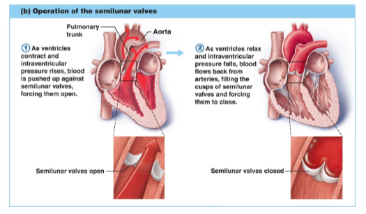
Semilunar Valves
located at the openings to the arteries
each semilunar valve consists of 3 moon-shaped cusps/flaps
pushed flat against artery wall during ventricular contraction
as ventricles relax, SL valves prevent the backflow of blood into the ventricles
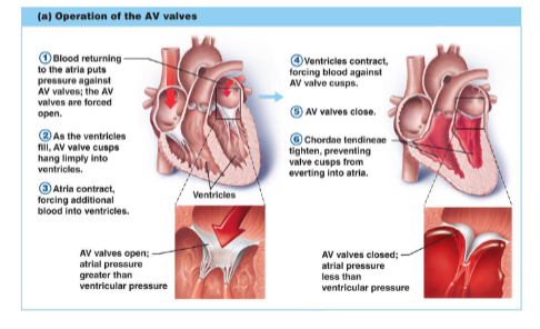
Atrioventricular Valves
located between atria and ventricles
during diastole, blood flows through the valves (from atria into ventricles)
right AV valve: tricuspid (3 cusps/flaps of endocardium)
left AV valve: bicuspid or mitral (2 cusps/flaps of endocardium)
prevent the backflow of blood into the atria
valves close passively as pressure builds in ventricles (during systole)
associated structures
chordae tendineae → cords prevent AV valve inversion
papillary muscle → anchors chordae tendineae

Features of the Heart
Pericardium
double-walled sac around the heart
outer wall → fibrous pericardium
inner wall → serous pericardium (double layered)
parietal pericardium → outer layer lining the fibrous pericardium
visceral pericardium → inner layer which forms the outermost layer of the heart wall called the epicardium

Heart Wall Layers
Epicardium → outermost layer of the heart (also called the visceral pericardium)
Myocardium → middle layer, made of cardiac muscle
striated, has intercalated discs, under involuntary control
fibrous skeleton composed of fibrous connective tissue that supports the cardiac muscle fibers and heart valves
Endocardium → innermost layer
composed of simple squamous epithelium


Circulatory Systems
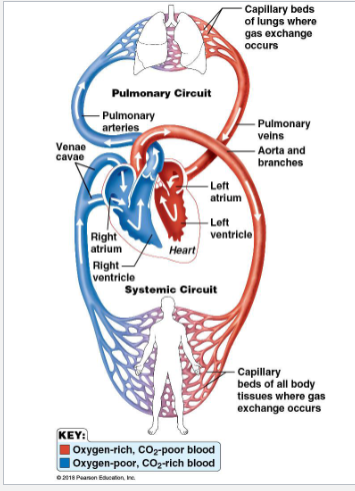
Systemic Circulation → carries blood to all tissues for nourishment, then transports deoxygenated blood back to heart
left ventricle → aorta → other arteries → arterioles → capillaries (within tissues) → venules → veins → venae cavae → right atrium
Pulmonary Circulation → carries deoxygenated blood back to the lungs for gas exchange, then brings oxygenated blood back to the heart
right ventricle → pulmonary trunk → pulmonary arteries → capillaries in lungs → pulmonary vein → left atrium
Circulatory System is Closed Transport System
path of blood flow in systemic circuit
heart → large arteries → medium arteries → arterioles → capillary beds → venules → medium veins → large veins → heart
substances diffuse across the capillary walls
from blood to tissues: O2 and nutrients
from tissues to blood: waste and CO2
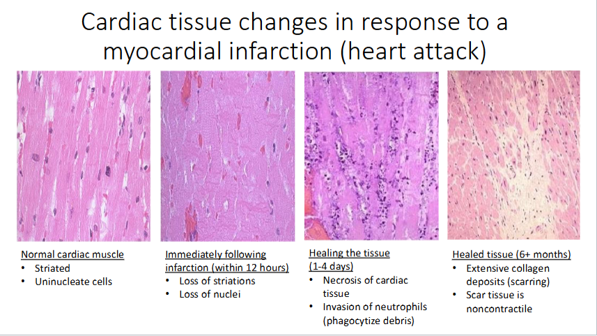
Cardiac Tissue Response to Myocardial Infarction
Types of Blood Vessels
Arteries → transport blood away from the heart
elastic arteries → large, highly expandable arteries close to the heart
muscular arteries → medium sized arteries that distribute blood around the body
arterioles → smallest arteries, with very thin tunica externa and tunica media
Capillaries → provide for exchange of materials in tissues
narrow vessels with very thin walls
Veins → transport blood toward the heart
low pressure vessels because of thing walls and large lumens
venules (smallest veins) drain capillary beds and then merge to form larger veins

Layers of Arteries and Veins
Tunica Intima → forms a friction-reducing lining to the lumen
composed of endothelium of simple squamous cells
innermost (deep)
Tunica Media → controls lumen diameter
composed of circulatory arranged smooth muscle and elastic CT
Tunica Externa → protects and anchors blood vessels
Composed of areolar or fibrous CT
outermost (superficial)

Venous Blood Return
blood pressure in veins is low, so three main mechanisms facilitate venous blood return to the heart
skeletal muscle pump → skeletal muscle contractions from normal movement squeeze veins and push blood through them
valves → prevent the backflow of blood
respiratory pump → during inspiration, a decrease in intra-thoracic pressure and increase in intra-abdominal pressure causes blood to flow from veins in the abdominal region to veins in the thoracic region

Five Special Circulations

Coronary Circulation
nourishes the myocardium tissue
blood in the heart chambers does NOT nourish the myocardium
heart has own nourishing circulatory system consisting of:
coronary arteries → branch from aorta to supply heart muscle with oxygenated blood
left → anterior interventricular artery and circumflex artery
right → posterior interventricular artery and marginal artery
cardiac veins → drain the myocardium of blood
deliver blood to the coronary sinus, a large vein on the posterior of the heart
blood empties into the right atrium via the coronary sinus

Pulmonary Circulation
oxygenates the blood supply
blood flow: right side of the heart → lungs → left side of the heart
pulmonary trunk and pulmonary arteries carry deoxygenated blood to the lungs
pulmonary veins carry oxygenated blood to the heart
capillary beds around alveoli permit gas exchange (exchange outgoing CO2 for incoming O2)
pulmonary circulation does NOT nourish lung tissue
blood supply for the lungs provided by bronchial arteries


Hepatic Portal Circulation
major vessels of hepatic portal circulation drain the digestive organs and carry the blood to the liver
superior mesenteric vein drains the small intestine, part of the large intestine, and stomach
inferior mesenteric vein drains the distal portion of the large intestine
splenic vein drains the spleen, pancreas, and stomach
gastric vein drains the stomach
Liver Function → helps maintain proper glucose, fat, and protein concentrations in the blood
capillary beds of the digestive system absorb nutrients and toxins
nutrient-rich blood drains into veins of the hepatic portal circulatory system
these veins empty into the hepatic portal vein, which carries the blood to the liver
liver helps maintain proper glucose, fat, and protein concentrations in the blood
liver also filters toxins from the blood
blood drains into the hepatic vein as it leaves the liver

Fetal Circulation
receives exchanges of gases, nutrients, and wastes through the placenta
umbilical cord contains three vessels
umbilical vein → carries blood rich in nutrients and O2 to the fetus
umbilical arteries (two of them) → carry CO2 and debris-laden blood from the fetus to the placenta
fetal blood flow bypasses:
the liver through the ductus venosus and enters the inferior vena cava
the lungs through the foramen ovale (blood entering right atrium shunted directly into the left atrium) and the ductus arteriosus (blood entering the pulmonary trunk passes through the aorta)

Cerebral Arterial Circle
major blood supply to the brain forms the cerebral arterial circle
also called the circle of willis
anterior and posterior blood supplies (internal carotid arteries and vertebral arteries) are united by small communicating arterial branches
result is a complete circle of connecting blood vessels

Atherosclerosis
condition where arteries are blocked by fat and cholesterol deposits


Major Arteries of Systemic Circulation
Aorta
largest artery in the body
leaves from the left ventricle of the heart
regions
ascending aorta → leaves the left ventricle
aortic arch → arches to the left
thoracic aorta → travels downward through the thorax
abdominal aorta → passes through the diaphragm into the abdominopelvic cavity

Arterial Branches of Ascending Aorta and Aortic Arch
ascending aorta
right and left coronary arteries serve the heart
aortic branch
brachiocephalic trunk
then splits into the: right common carotid artery and right subclavian artery
left common carotid artery
then splits into the left internal and external carotid arteries
left subclavian artery
then branches into the left vertebral artery and left subclavian artery
subclavian artery becomes axillary artery → brachial artery → radial and ulnar arteries

Arterial Branches of Descending Thoracic Aorta
intercostal arteries
supply the muscles of the thorax wall
bronchial arteries
supply the lungs
esophageal arteries
serve the esophagus
phrenic arteries
serve the diaphragm

Arterial Branches of the Abdominal Aorta
celiac trunk → first branch of the abdominal aorta
three branches:
left gastric artery → serves the stomach
splenic artery → serves the spleen
common hepatic artery → serves the liver
superior mesenteric artery → supplies most of the small intestine and first half of the large intestine
left and right renal arteries → serves the kidneys
left and right gonadal arteries → called ovarian in females, testicular in males
lumbar arteries serve muscles of abdomen and trunk
inferior mesenteric artery → supplies the second half of the large intestine
left and right common iliac arteries are the final branches of the aorta
internal iliac arteries serve the pelvic organs
external iliac arteries enter the thigh → femoral artery → popliteal artery → anterior and posterior tibial arteries

Major Veins of Systemic Circulation

Vena Cavae
two veins that return blood from the systemic circuit to the right atrium
superior vena cava → drains the head and arms
inferior vena cava → drains the lower body
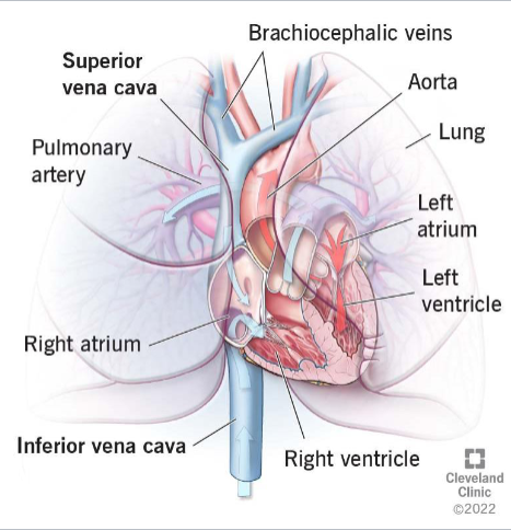
Veins Draining into Superior Vena Cava
draining the arm:
radial and ulnar veins → brachial vein → axillary vein
cephalic vein drains the lateral aspect of the arm and empties into the axillary vein
basilic vein drains the medial aspect of the arm and empties into the brachial vein
basilic and cephalic veins are jointed at the median cubital vein (elbow area)
subclavian vein receives:
venous blood from the axillary vei
venous blood from the skin and muscles of the head and neck via external jugular vein
vertebral vein drains the posterior part of the head
internal jugular vein drains the dural sinuses of the brain
left and right brachiocephalic veins receive venous blood from the:
subclavian veins
vertebral veins
internal jugular veins
brachiocephalic veins join to form the superior vena cava
azygous vein drains the thorax

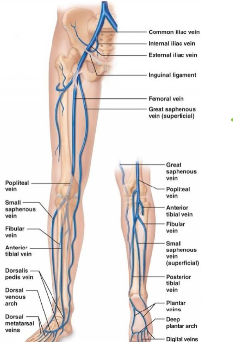
Lower Limb Veins Draining into Inferior Vena Cava
anterior and posterior tibial veins and fibial veins drain the legs
posterior tibial vein → popliteal vein → femoral vein → external iliac vein
great saphenous veins (longest veins of the body) receive superficial drainage of the legs
each common iliac vein (left and right) is formed by the union of the internal and external iliac vein on its own side
Abdominal Veins Draining into the Inferior Vena Cava
right gonadal vein drains the right ovary in females and right testicle in males
left gonadal vein empties into the left renal vein
left and right renal veins drain the kidneys
hepatic portal vein drains the digestive organs and travels through the liver before it enters systemic circulation
left and right hepatic veins drain the liver

Circulatory System Development in Fetus
a simple “tube heart” (without chambers) develops into the embryo and pumps by the fourth of the week post-fertilization
septa form between the atria and ventricles between 4-7 weeks post-fertilization
heart becomes a four-chambered organ by the end of seven weeks post-fertilization
fewer structural changes occur after the seventh week post-fertilization, the heart mostly just grows in size and strength for the rest of gestation
Lub Dub → Sounds of a Heartbeat
systole: contraction
diastole: relaxation
two sounds of heartbeat coincide with ventricular systole and diastole
heartbeat = sounds of blood hitting and closing the valves
lub (s1) → closure of the AV valves at the start of ventricular systole
dub (s2) → closure of the SL valves at the end of ventricular systole
Heart Rates and Beats
normal → 60-100 bpm at rest
average resting heart is 70-76 bpm
arrhythmia → irregular heartbeat
can describe bradycardia or tachycardia
or pattern of heartbeat is inconsistent
bradycardia → a heart rate (< 60 bpm)
considered abnormal, except in well-conditioned athletes
tachycardia → heart rate (>100 bpm)
abnormal when at rest
if untreated, may lead to fibrillation
fibrillation → rapid, uncoordinated contractions
renders heart useless as a pump
Intrinsic Conduction System
spontaneous contractions of the heart are coordinated by the intrinsic conduction system
cardiac muscle contractions occur in a regular and continuous way
atrial cells beat 60 times per minute
ventricular cells beat 20-40 times per minute
intrinsic conduction system ensures the heart beats as a coordinated unit
cardiac pacemaker cells cause orderly and sequential depolarization of regions of the heart
enforces a heart rate of 75 bpm
autonomic nervous system helps regulate heart rate (but does NOT initiate contractions)
sympathetic nervous system (SNS) accelerates heart rate
parasympathetic nervous system (PNS) decelerates heart rate
Composition
composed of specialized nervous tissue embedded in the myocardium
sinoatrial (SA) node → located in the right atrium and serves as the heart’s pacemaker
atrioventricular (AV) node → at junction of the atria and ventricles
atrioventricular (AV) bundle (bundle of His) and bundle branches → in the interventricular septum
purkinje fibers (or subendocardial conducting network) → spread within the ventricle wall muscles

Impulse through Intrinsic Conduction System
impulse that triggers contraction travels sequentially through the intrinsic conduction system
the SA node starts at each heartbeat
impulse spreads through the atria to the AV node
atria contract; blood pushed into ventricles
at the AV node, the impulse is delayed briefly
impulse travels through the AV bundle, bundle branches, and subendocardial network
ventricles contract, blood is ejected from the heart
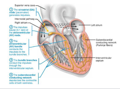
Conduction in the Heart
change in electric potential (depolarization) and the generation of action potentials result in muscle contraction
cardiac muscle cells are connected through gap junctions which allow for the spread of action potential
impulse starts at SA node (superior portion of the right atrium)
sets depolarization rate (it is the pacemaker)
stimulus travels through atria, causing atrial contraction to the AV node
impulse is delayed at the AV node (inferior atrial septum in right atrium)
allows for atrial contraction to be completed before ventricular depolarization begins → prevents premature closure of the AV valves
electrical conduction through the ventricles requires:
AV bundle (bundle of His) → located in interventricular septum
Bundle branches → located in interventricular septum
Subendocardial conducting network (purkinje fibers) → in muscles of ventricular walls
denser in left ventricle


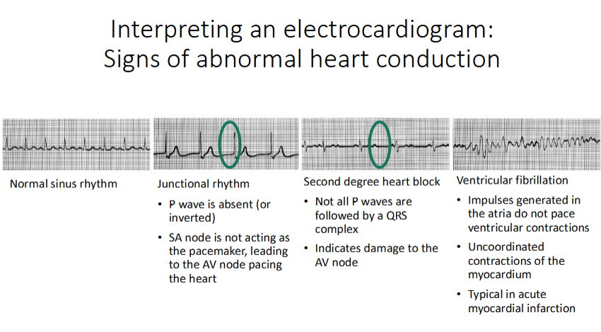
Electrocardiograms
measure the heart’s electrical activity from the body surface
P wave → depolarization of the atria
QRS complex → depolarization of the ventricles
atrial repolarization also happens here, concurrent with ventricular contraction
T wave → repolarization of ventricles

Interpreting Electrocardiogram