Lesson 16: Cell Signaling Overview and Gated Channels
 Call Kai
 Learn
 Practice Test
 Spaced Repetition
 Match
 Flashcards
 Knowt Play
1/36

There's no tags or description
Looks like no tags are added yet.
37 Terms
Signal Transduction
Is the process of translating information from outside of the cell into a specific & limited cellular response.
Receptor ligand binds causing a confirmational change to active state, ligand releases and returns receptor to inactive form.

Typical Signal Transduction Cascade
First messenger (signal)
Receptor protein interaction
Upstream signaling proteins
Second messengers
Relays info from ligand receptor complex
Downstream signaling proteins
Target proteins (cause a response)
First Messengers include…
Hormones
Ions
Small molecules
Metabolites
Gases
Second Messengers
Are molecules required by most signaling pathways
A point for amplification
Multiple 2nd messengers produced by one signal can give multiple responses
Protein kinase activity is central to many BLANK pathways
Signaling
Common method for amplifying the original signal
Can occur at any point in the pathway or multiple times
Generates diverse responses from one signal by phosphorylating multiple target proteins
Gated ion channels
Have a “gate” regulated by a biological signal
Eversion: Have both open and closed conformations
When gate is open, ions diffuse thorugh channel in direction dictated by electrochemical gradient.
Gated ion channels can be gated by:
Physical stimuli (mechanosensitive)
ligand binding
voltage (membrane potential)
pH
Voltage-gated membrane channels change conformation in response to BLANK across the membrane.
Voltage
Voltage-gated ion channels have positively charged voltage sensors. When the inside of the cell becomes more positive, the sensors move outward due to the change in electrical forces, causing the channel to open.

Membrane potential results from…
the unequal distribution of charges across a cell.
Nernst Equation:
Δψ = VEq = (RT/ZF) ln ([ion]out / [ion]in)
Δψ → potential across membrane, ΔV
Use when membrane permeable to a single ion
At room temp (20°C), the equation for equilibrium potential for a single monovalent cation (Z=1) is:
VEq = 58 mV log ([ion]out / [ion]in)
Name the subunits and what they do/form in voltage-gated channels
Each subunit has 6 transmembrane helices with an intracellular N-terminus
Helices 5 and 6 form the channel
Helix 4 is the voltage sensor
The intracellular N-terminus of the K+ channel forms an BLANK
inactivation domain
The ball and chain model…
Ball-and-chain model
Depolarization opens the channel → ions begin moving through it → positive charge on amino acids (the ball) move into the channel & plugs channel (keeps it open for a short time)
How quickly the ball plugs the channel is based off of chain length
Shorter chain = quicker inactivation
Most neurons have the same parts, which are?
Dendrite
Cell Body
Axon
Dendrites
They receive information from other neurons
The cell body (in neurons)
It contains the nucleus and most cell organelles
Axon
Conducts action potentials away from the cell body
Signal travels down axon
Axon terminals
They synapse with a target cell
Nerve impulses are propagated by changes in BLANK
membrane potential
Action potential
Its the rapid temporary change in membranes potential due to opening of ion channels
3 phases of action potential and what happens in each?
Depolarization - membrane potential becomes less negative and moves toward a positive charge
Repolarization - membrane potential changes back to negative charge
Hyperpolarization - membrane potential slightly more negative than the resting potential (also called the undershoot)
Action Potential cascade - starting at resting potential
membrane potential -60 mV
depolarization of membrane to approx -40 mV
opening of Na channels
membrane potential of +30 mV
closing of Na channels
opening of K channels
membrane potential -75 mV
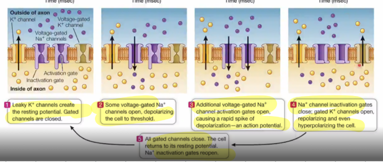
Action potenitals propagate rapidly along the length of axons, causing what?
electrical signaling as charge spreads down the membrane
Lots of positive charge at one spot gets attracted further down, so that charge moves down the axon
Where positive charge used to be, it resents itself because of potassium channels, so it has no movement backwards because of falling charge
this forces membrane potential all the way down the axon
Ligand-gated ion channels change conformation upon BLANK
ligand binding
can happen extracellularly or intracellularly
Ligand itself is not transported across membrane, only opens channel
3 classes of ligand-gated channels
Cys-loop
Glutamate (P pore-loop)
ATP-gated P2X channels
Cys-loop channels
consist of 5 subunits arranged around a pore (pentamer)
2 α-subunits
1 β-subunit
1 δ-subunit (delta)
1 γ-subunit (gamma)
Each subunit has has 3 domains
Extracellular (ligand binding)
Transmembrane
Intracellular (involved in opening and closing of the channel)
Acetylcholine Nicotinic Receptor
Is a Cys-loop channel
M2 helicies face the interior which make up the channel
acetylcholine binds to alpha subunit & causes confirmational change so Leu unblocks channel
Nicotinic Receptors transmit neuronal signals from BLANK cells to BLANK cells
nerve
muscle
Acetylcholine (ACh)
is a neurotransmitter synthesized from choline and acetlyCoA
Its released from vesicles in pre-synaptic neuron
Binding of it opens channel to Na and K ions
Acetylcholine is synthesized in, stored in, and released from BLANK
cholinergic neurons → those that respond to acytylcholine
Acetylcholine is released into the BLANK in response to an influx of Ca2+
synapse
Events in the transmission of nerve impulses in synapses
release of acetylcholine from the synaptic vesible into the synaptic cleft
increse of acetylcholine concentration in the synaptic cleft from ~10 nM to 500 nM
Binding of acetylcholine to the ACh receptor
increse in postsynaptic membrane permeability to Na+ and K+
depolarization of post-synaptic membrane
Muscles contract when…
they receive signals from motor neurons
Acetylcholinesterase
breaks down acetylcholine in the synapse cleft
Choline is transported back into the pre-synaptic cell to be used for ACh synthesis
Neuromuscular blocking agents (NMBAs)
are nicotinic receptor antagonists used in conjunction with anesthesia
Induces paralysis
Used for mechanical ventilation and traceal intubation
Used to relax muscles during surgery
No effet on the level of consiousness or ability to preceive pain
Nondepolarizing Agents
Competitive antagonist
Bind to the acetylcholine receptor but do not open the channel
Prevent depolarization of the muscle cell → inhibits muscle contraction
Effects of this can be reversed with acetylcholinesterase inhibitors