Hem quiz 1
 Call Kai
 Learn
 Practice Test
 Spaced Repetition
 Match
 Flashcards
 Knowt Play
1/110

There's no tags or description
Looks like no tags are added yet.
111 Terms
Hematology big picture
focuses on blood cells and the systems that influence them
blood transports oxygen to tissues and carries CO2 away
the liquid portion of blood is plasma
includes
RBCs
WBCs
PLTs
RBCs
erythrocytes
gas transport
WBCs
leukocytes
defense
PLTs
thrombocytes
hemostasis
erythrocytes
form: non-nucleated, biconcave discs
content: hemoglobin rich cytoplasm
size: approx. 6-8 micrometers
function: transport gases and support exchange
RBC evaluation
rbc count: # of erythrocytes
hgb: hemoglobin concentration
hct: packed cell volume
indices: MCV, MCH, MCHC, RDW (derived from previous values)
retics: marrow reponse
anemia
decreased red cell mass
polycythemia
increased red cell mass
which feature is most characteristic of mature erythrocyte?
biconcave shape
Leukocytes
neutrophils: segmented and band forms
basophils: granulocyte
monocytes: mononuclear
eosinophiles: granulocytes
lymphocytes: mononuclear
manual wbc differential
a sustained blood film is examined microscopically
cells are identified by morphology and counted
automated wbc differential
instrumentation classifies cells and generates flags that may require microscopic review
leukopenia
decreased wbc count
leukocytosis
increased wbc count
platelets
appearance:
2-4 micrometers
round or oval
anucleate
granular
function:
initiate vessel-wall repair and support clotting
hemostasis - plts
keeps blood within the vascular system while allowing repair of injury
hemostasis 4 interacting systems
platelets: primary plug
vascular: vessel response
coagulation: fibrin formation
fibrinolytic: clot breakdown
COMMON SCREENING AND MONITORING TESTS:
PT
PTT
D-DIMER
platelet counting methods
light microscopy
manual counting w a hemacytometer
platelets can be difficult to identify
phase microscopy
preferred manual method in the source presentation
automation
reports platelet count and may provide mean platelet volume (MPV)
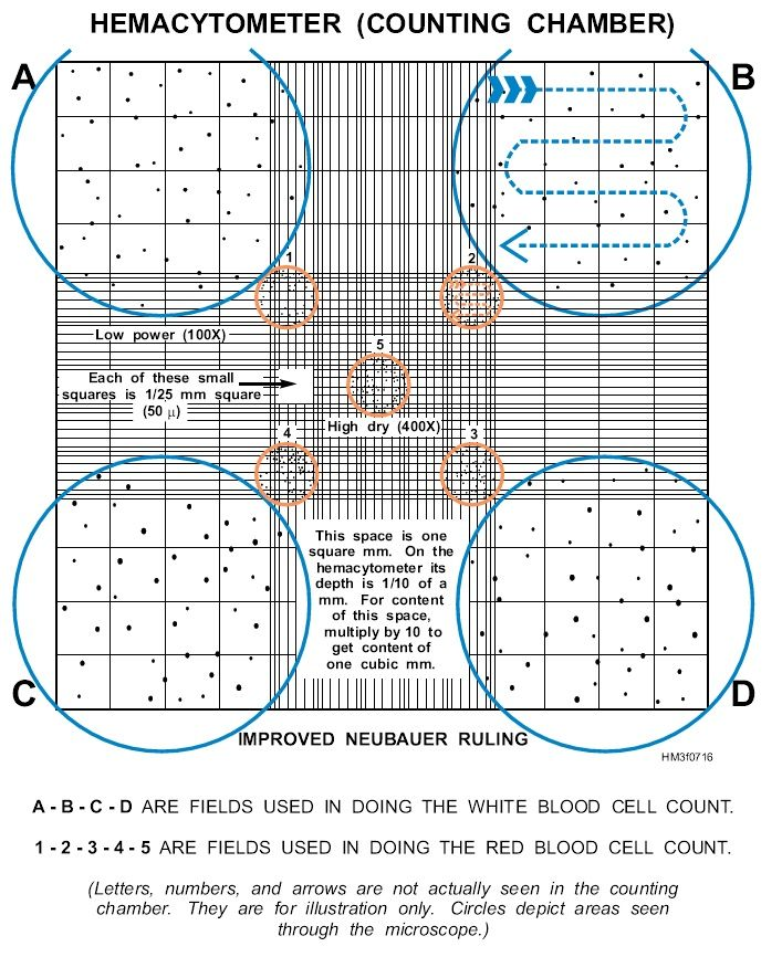
hemacytometer: manual cell counting
a ruled counting chamber allows cells to be counted within a known area and depth
the calculation incorporates the area counted and specimen dilution
the principle is similar for rbcs, wbcs, and platelets, but diluent and counting area vary
manual counts remain important for body fluids and selected situations
counting chamber: orientation matters
identify the correct counting area
apply a consistent boundary rule
record dilution and area before calculating
coulter principle (automation)
direct current electrical impedance remains a foundational method for counting cells in many analyzers
CBC includes…
counts
WBC
RBC
PLT
oxygen carriers
hemoglobin
hematocrit
RBC indices
MCV
MCH
MCHC
RDW
differential
distribution of WBC types
morphology
RBC
WBC
PLT abnormalities
blood film examination
prepare: create a wedge film on a glass slide
stain: use Wright stain
estimate: visually estimate WBCs and platelets
examine: assess size, shape, color, inclusions, and abnormalities
advanced hematology procedures
bone marrow: aspirates + biopsies evaluate immature blood cell precursors
cytochemistry: stains help differentiate abnormal myeloid, erythroid, and lymphoid cells
flow cytometry: provides quantitative and qualitative cellular information
molecular testing: detects selected genetic abnormalities, including the philadelphia chromosome
special tests: examples include osmotic fragility, g6pd, sickle solubility, and hemoglobin electrophoresis
which sequence best reflects professional review of an abnormal automated CBC?
review flags and correlate w smear
lavender tube
EDTA
anticoagulant
cbc/hematology
light blue tube
sodium citrate
anticoagulant
coagulation
red tube
no additive
serum after clotting
chemistry/serology
light green tube
lithium heparin
plasma separator
chemistry
EDTA and sodium citrate
EDTA - PURPLE top
commonly associated w hematology testing, including CBCs
additive PREVENTS CLOTTING so whole blood can be analyzed
SODIUM CITRATE - BLUE TOP
used for coagulation testing
serum path
red top
no additive → blood clots → serum is separated
common use: chemistry/serology
plasma path
light green top
lithium heparin + separator → plasma is obtained
common use: chemistry
“Phuck cLots” - anticoagulant
which color tube is associated with CBC testing?
purple top
preanalytical variables can affect results
posture
diurnal rhythm (varies by time of day)
exercise/stress
diet
smoking
timing
collection equipment
evacuated system: tube, needle, adapter
torniquet: disposable strap, velcro, or blood pressure cuff
sharps safety: require safe handling
skin prep: approved antiseptic prep
needles
different lengths and gauges are available
21 gauge, 1-1.5 inches is common
needle holders
use the holder intended for the manufacturer
do not interchange incompatible components
torniquet
do not exceed 1 minute
RBCs can lyse (hemolysis)
additional collection equipment
syringes: available in different sizes w different needle attachments
butterflies: winged infusion sets contain short needle, thin tubing, and plastic wings
skin prep: examples listed include alcohol, providone-iodine, 1-2% iodine, and benzalkonium chloride
venipuncture sites
cephalic
median cubital
basilic
blood smears
capillary or venous blood may be used for wedge or coverslip preparations
how long should a tourniquet remain applied at the most?
1 minute
when should a hematology specimen be rejected?
ID
unlabeled or incorrectly labeled
order/tube
request and tube do not match; wrong tube
specimen quality
hemolyzed, clotted when whole blood is needed, or lipemic
timing
collected at the wrong time
contamination
contaminated with IV fluid
specimen check sequence
identify: is it correctly labeled?
match: do request, tube, and timing agree?
inspect: is the specimen clotted, hemolyzed, lipemic, or contaminated?
decide: accept for testing or follow rejection policy
a CBC specimen contains a visible clot. what is your primary concern?
whole blood is required
order of draw
Yellow/ blood culture: SPS
Light blue: citrate
Red: serum/ no additive
Gold/tiger: SST
Green: heparin
Lavender/pink: EDTA
Gray: fluoride/oxalate
“You Look Really Good Girly, Live Great”
quality
accurate + reproducible results that provide clinically useful info
what % of clinical decision making is based on lab results?
70%
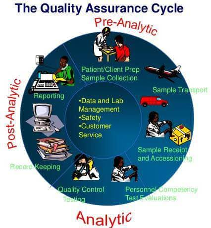
quality assurance cycle
PRE ANALYTIC
patient preparation
specimen collection
ANALYTIC
testing and QC
POST ANALYTIC
result verification
reporting and follow up
accuracy of assay
agreement btwn assay value and the theoretical “true value” of the analyte
primary standard of assay
reference material w fixed, known composition that can be prepared in essentially pure form
secondary standard calibrator
concentration established thru primary standard or reference assay
certified known concentration
incorporated during validation or revalidation
used to adjust instrumentation or develop a standard curve
precision
reproducibility or dispersion about the mean, often expressed as standard deviation (SD) or coefficient of variation (CV%)
relatively easy to measure and maintain
tightly grouped results
the validation goal
accurate: near the true value
precise: results cluster together
accurate + precise: near truth and consistent
STRONG ASSAY PERFORMANCE REQUIRES BOTH

reference interval
also called a reference range or normal range
adult intervals should reflect the demographics served by the laboratory
gaussian distribution
a symmetric, BELL SHAPED, distribution
the peak represents the mean
the width reflects variability
special populations
local intervals may be impractical for infants, children, and geriatric populations
published intervals may be selected by the lab director
standard deviations
a mathematical expression of how values are dispersed around the mean
confidence limits
common limits are described as 1 SD, 2 SD, and 3 SD from the mean
coefficient of variation (CV%)
a measure of variation relative to the mean, usually expressed as a percentage
CV% = (SD/mean) * 100
normal gaussian distribution
± 2 SD FOR HEMATOLOGY
includes approx. 95.46% of results from a healthy population
outside the interval
approx. 4.54% fall outside the interval, about 2.27% in each tail

which statement best describes quality in laboratory testing ?
accurate, reproducible results that provide clinically useful information
approximately what percentage of clinical decisions are based on lab results ?
70%
what are the 3 quality assurance variables?
preanalytical, analytical, and postanalytical
which statement correctly distinguishes accuracy from precision?
accuracy is closeness to the true value; precision is reproducibility around the mean
HGB control range: 13 ± 0.4 g/dL. RESULTS: 12.0, 12.3, 12.0, 12.2, 12.1 g/dL. How should the results be classified?
precise but not accurate
2 SD = 0.8 g/dL
range = 12.2 - 13.8
ONLY 2 VALUES WITHIN RANGE: 12.3, 12.2
ALL VALUES MUST BE WITHIN RANGE FOR RESULTS TO BE CLASSIFIED AS ACCURATE
internal quality control
controls and monitors and the testing process so results remain valid and reproducible
control samples are analyzed statistically to recognize unacceptable performance
internal QC is part of the analytical phase of quality assurance + includes corrective action when performance is unacceptable
controls in practice
MONITOR
analytical bias
test method precision
systematic error
calibration verification
RUN
treat like patient samples
run 2 controls per test
use one within and one above/below reference range
FREQ. AND LIMITS
once per shift
when new reagent is constituted
results must fall within ± 2 SD

the ideal control
affordable
stable (long term)
compatible (direct sampling; similar to blood)
reliable (easily suspended; does not agglutinate)
trackable (results displayed on a Levey-Jennings chart)
Levey - Jennings chart
distribution
control results are expected to distribute in a Gaussian manner, with limits at 1, 2, and 3 SD above+below the mean
analysis
used to recognize single-run errors and sequential errors that develop over time
westgard rules
internal QC rules developed by Dr. James Westgard and applied to deviations in control performance

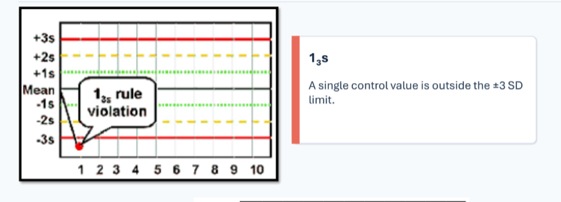
13s rule
one control exceeds ± 3 SD
22s rule
2 controls exceed the SAME ± 2 SD limit

R4s rule
2 controls in 1 run are more than 4 SD apart

41s rule
4 consecutive controls exceed the same ± 1 SD limit

10x rule
10 consecutive controls remain on one side of the mean

7T rule
at least seven controls trend in one direction

systematic error
occurs within the test system or method
causes may include incorrect calibration or instrument malfunction
associated patterns: 22s , 41s, 7T, 10x
random error
unpredictable irregular mistakes that create scattered performance
associated patterns: 13s, R4s
10x shift
SYSTEMATIC ERROR
calibration issue
reagent deterioration
pump fitting or light source deterioration
abrupt reagent or instrument fitting range

7T trendline
SYSTEMATIC ERROR

delta check
compares most current analyte result with the patient’s most recent prior result
20% deviation → investigate
external quality control
compares laboratory performance w/ a group mean used as the target value
supports proficiency testing requirements
assessments:
CAP - college of american pathologists
API - american proficiency institute
NASCOLA - north american specialized coagulation lab association

false positive
a positive test result generated from a specimen without disease
false negative
a negative test result generated from a patient with proven disease
diagnostic outcomes

sensitivity
among people WITH disease, what proportion tests positive?
Sensitivity (%) = TP / (TP + FN) * 100
ability to detect disease when disease is present
be WITH your SENSES
specificity
among people WITHOUT disease, what proportion tests negative?
specificity (%) = TN / (TN + FP) * 100
ability to correctly identify those without disease
which statement best explains external quality control?
It compares a lab’s performance w peer labs using proficiency testing specimens
which situation is a false positive result?
a specimen without disease produces a positive result
which situation is a false negative result
a patient with proven disease has a negative result
besides being valid, accurate, linear, and precise, assays must be diagnostically ________
effective
which term means the proportion of people with disease who test positive?
sensitivity
which term means the proportion of people without disease who test negative?
specificity
cell organization
plasma membrane: selective boundary
nucleus: genetic control center
cytoplasm: cytosol + organelles
organelles: specialized work places
translate micrograph into a model
find the boundary : trace plasma membrane
locate the nucleus: look for largest enclosed region
scan the cytoplasm: id organelles by shape and texture
ask what function fits: connect cell structure to cell work

plasma membrane
MANAGES EXCHANGE
PROTECTS: separates cell from extracellular environment
REGULATES: interchange of substances w/ the environment
ESTABLISHES: electrochemical gradients between the interior and exterior of the cell
COMMUNICATES: receptors enable signal transductio
nucleus
STORES AND MANAGES GENETIC INFO
membrane bound control center, except in mature RBCs and PLTs
CHROMATIN: dna + associated proteins
NUCLEAR ENVELOPE: double membrane boundary
NUCLEOLI: ribosome production regions
cytoplasm + organelles
provides the setting and machinery for specialized cellular functions
surface markers
CREATE CELLULAR “ID”
cd antigens: cluster of differentiation markers receive a cd number
unique repertoire: marker patterns change w blood cell type and stage of differentiation
flow cytometry: monoclonal antibodies identify cell surface antigens
marker pattern → cell identity → maturation stage
cytoplasm
THE CELL’S WORKING ENVIRONMENT
CYTOSOL: homogenous, continuous, aqueous, THE MEDIUM IN WHICH ORGANELLES EXIST AND FUNCTION
ribosomes, ER, golgi apparatus, mitochondria, lysosomes, cytoskeleton, microtubules, centrosomes