5. EPITHELIAL PATHOLOGY pt 1 ✅
 Call Kai
 Learn
 Practice Test
 Spaced Repetition
 Match
 Flashcards
 Knowt Play
1/36

There's no tags or description
Looks like no tags are added yet.
37 Terms
ORAL SQUAMOUS PAPILLOMA
Benign proliferation of squamous epithelium, HPV- 6, 11
Common between 30 - 50 years, Equal frequency among sexes
Soft, painless, pedunculated
Exophytic, finger-like projections, "cauliflower“ appearance
White, slightly red, or normal color
Tongue and soft palate most common
Solitary, enlarge rapidly to 0.5 cm
no malignant potential
ORAL SQUAMOUS PAPILLOMA: Histopathology
Keratinized stratified squamous epithelium with finger-like projections

ORAL SQUAMOUS PAPILLOMA: Treatment
Conservative surgical excision
CONDYLOMA ACUMINATUM (Venereal Wart)
Papillary proliferation of genitalia, anus, larynx, mouth
STD
HPV- 6, 11,16, 18
Develop at site of sexual contact, 20% of all STDs
Teenagers / young adults
Labial mucosa, soft palate, lingual frenum– Common sites
CONDYLOMA ACUMINATUM: appearance
Sessile, pink, well-demarcated, non tender, exophytic
Larger than papillomas
Multiple and clustered
Average size 1.0 to 1.5 cm

CONDYLOMA ACUMINATUM: Treatment and Prognosis
Conservative surgical excision
Should be removed - Contagious
VERRUCA VULGARIS
Benign, focal hyperplasia of squamous epithelium
HPV-2, 4, 40
Can spread by autoinoculation
Uncommon on oral mucosa
Extremely common on skin
Frequent in children, skin of hands usual site
Vermillion, labial mucosa, or anterior tongue
VERRUCA VULGARIS (Wart): appearance
Painless papules or nodules with papillary projections; occur in clusters
Usually surgically excised
2/3 disappear within 2 years, especially in children

in oral cavity this looks like papilloma
can only differentiate under microscope bc focus of the lesion is in the center
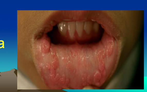
Heck’s Disease Multifocal Epithelial Hyperplasia
Mostly in Native Americans, Eskimos, esp. kids
Familial predisposition
Lower socioeconomic status, poor hygiene, malnutrition, crowded living conditions – Additional etiologies
HPV 13, 32
Most regress spontaneously
Multiple lesions- Buccal / labial mucosa
Sessile, pink or white
HPV - HISTOPATHOLOGY
Squamous epithelial proliferation
Koilocytes - Hyperchromatic nuclei, clear perinuclear halo

SEBORRHEIC KERATOSIS
Positive correlation with chronic sun exposure
Acquired, benign proliferation of epithelial basal cells
Sudden multiple Seb K associated with internal malignancy
Skin of face, trunk, extremities
Multiple, small tan to brown macules initially, become papillary raised, Usually less than 2 cm
Appear "stuck onto" the skin
Except for aesthetic purposes, seldom removed
KERATOACANTHOMA (Self-Healing Carcinoma)
Self-limiting
Extremely well-differentiated squamous cell carcinoma , called “SCC, “keratoacanthoma type”
MALIGNANT
Rarely before 45, male predilection
Does not occur intraorally
Rapid enlargement, diameter of 1 to 2 cm within 6 weeks
Regress spontaneously within 6 -12 months leaving depressed scar
KERATOACANTHOMA appearance + tx
Firm, non tender, dome-shaped nodule with central keratin plug
Epithelium at lateral edge of tumor normal
Epithelium exhibits a downward growth pattern
Treatment is surgical excision

well differentiated means very good prognosis, wont metastasize, likely to not return
ORAL MELANOTIC MACULE
Unknown etiology, focal increase in melanin, Unrelated to solar exposure
2:1 female predilection
Vermillion border (lower lip) >buccal mucosa >gingiva > palate
Solitary, uniform tan to dark-brown, round, oval macule, <7 mm, Lesions do not enlarge
flat
No treatment usually required

MELANOCYTIC NEVUS
“Nevus" - Congenital / Developmental
Skin, mucosa
Most common - Acquired nevus, or common mole
Benign, localized proliferation of neural crest cells
Sharply demarcated, brown / black macule
Intraoral nevi distinctly uncommon
Most arise on hard palate or gingiva
ACQUIRED MELANOCYTIC NEVUS (Mole)
White adults 10 - 40 cutaneous nevi
Head and neck region common site
Remains less than 6 mm
Melanoma can arise from nevi
The risk of transformation to melanoma is one in a million
ACQUIRED MELANOCYTIC NEVUS (Mole) Classified histopathologically
Junctional - Nevus cells nests within epithelium
Compound - Nevus cells within epithelium & connective tissue
Intramucosal– Nevus cells only within connective tissue (most common intraoral type)
No treatment for cutaneous nevi

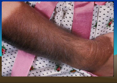
CONGENITAL MELANOCYTIC NEVI
1% of newborns, Small (<20 cm), large (>20 cm)
Large – Brown/ black plaque, rough multi-nodular surface
Hypertrichosis, more prominent with age, called bathing trunk or garment nevus
Congenital nevi excised for aesthetic purposes
5 -10% of large congenital nevi change into melanoma
BLUE NEVUS
Benign proliferation of melanocytes, deep in connective tissue - Dark, sometimes blue color
Intraoral - Most common on palate (and all pigmented lesions)
Mimics an early melanoma

you do biopsy
LEUKOPLAKIA
"white patch" / plaque that can’t be characterized clinically or pathologically as any other disease
Cannot be scraped off
Clinical term, does not imply specific histopathology
Surface keratin or thickened spinous layer masks vascularity of connective tissue
Precancerous, dysplasia / carcinoma in 5 to 25%
Malignant transformation rate 4%
Increases with age, esp for males, 8% of men 70+
causes of LEUKOPLAKIA
Tobacco smoking - 80+% of patients
Heavy smokers - More / bigger lesions
Alcohol strong synergistic effect with tobacco
Alcohol alone not associated with leukoplakia
UV radiation - Vermilion border
Candidal leukoplakia / candidal hyperplasia can → dysplasia
dysplasia
precancerous
LEUKOPLAKIA characteristics
70% - Lip vermilion, buccal mucosa, gingiva (low risk areas)
Tongue, oral floor, 90+% dysplasia / carcinoma (high risk areas)
Early lesions - Flat, fissured, wrinkled white plaques
2/3 of thin leukoplakia thicken, become distinctly white: homogeneous or thick leukoplakia
Exophytic / multiple projections:-verrucous leukoplakia
findings for leukoplakia biopsy
Hyperkeratosis - 80%
Dysplasia or ca-in-situ - 17%
Squamous cell carcinoma - 3%
you do incisional biopsy
it is a big lesion all over
you need to make sure it is not malignancy
LEUKOPLAKIA - DYSPLASIA stages
Mild - Limited to basal layer
Moderate - Basal layer to mid portion of spinous layer
Severe - Basal layer to level above midpoint of spinous layer
Ca-in-situ - Basal layer to surface ("top-to-bottom"), No invasion (high chance of SSC, frequent recall)

no histopathology on this but know the stage descriptions
LEUKOPLAKIA Histologic features of Dysplasia
(similar to squamous cell carcinoma)
• enlarged nuclei and cells
• increased nuclear/cytoplasmic ratio
• hyperchromatic nuclei
• pleomorphic nuclei and cells
• dyskeratosis (abnormal cellular keratinization)
• increased mitotic activity
• abnormal mitotic figures
TREATMENT for leukoplakia
Biopsy mandatory- Guides Tx *golden standard
Tissue sample from clinically most "severe" areas
Dysplasia or worse - Complete removal
Long-term follow-up, recurrences / additional lesions may develop
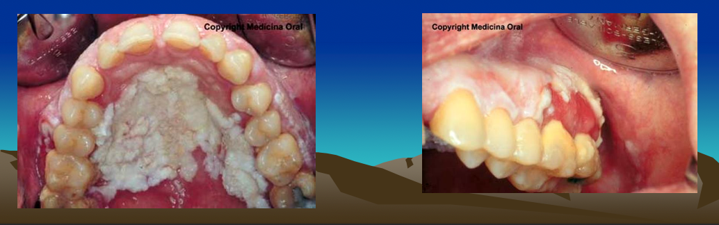
PROLIFERATIVE VERRUCOUS LEUKOPLAKIA
Extensive white plaques, Multiple sites, Invariable progression
Female 4:1, Usually nonsmokers, nondrinkers, HPV 16
Transform into carcinoma (verrucous carcinoma or SCC)
Recur after excision
main etiology for leukoplakia
smoking!!!
if not smoking, then likely hpv 16
ERYTHROPLAKIA
Red patch that can’t be clinically or pathologically diagnosed as any other condition
Related to alcohol and tobacco use
Older men, peak 65 – 74 years
Well-demarcated red macule or plaque, soft, velvety
Differential diagnosis: Inflammation, candidiasis, vascular lesions
ERYTHROPLAKIA; Histologic spectrum
Benign keratosis – 0%
Mild dysplasia - 10%
Severe dysplasia OR carcinoma-in-situ - 40%
Invasive carcinoma - 50%
Suspicious may be observed for 10-14 days, if persists biopsy
Recurrence & multifocal involvement common, long-term follow-up needed
TOBACCO POUCH KERATOSIS (Snuff Pouch)
Chewing tobacco / finely ground tobacco (snuff) : Smokeless tobacco or spit tobacco
Painless gingival "recession" & facial bone destruction
Correlates with quantity and duration
Thin gray-white, translucent plaque
1-5 years to become white
Fissured or rippled with pouch
Later becomes thickened leathery or nodular
TOBACCO POUCH KERATOSIS (Snuff Pouch) outcomes
Hyperkeratosis, acanthosis, edema
Dysplasia uncommon, when present, is typically mild
Biopsy intensely white, granular/verruciform, ulcerated, mass forming, indurated, or hemorrhagic lesions
Cancer risk 4 times greater than non-users
verrucous carcinoma is most common but SCC possible
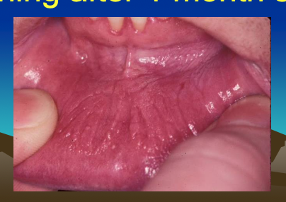
Habit cessation normal mucosa within 1 to 2 weeks
Lesions remaining after 1 month should be biopsied
NICOTINE STOMATITIS
Not premalignant, response to heat, not chemicals in tobacco, slightly elevated papules with punctate red centers
Inflamed & metaplastic salivary gland ducts
Completely reversible if habit discontinued
White lesion persisting after a month of habit cessation -True leukoplakia and biopsied

actinic
refers to sun exposure
ACTINIC CHEILITIS
Diffuse, premalignant alteration of lower lip vermilion, from excessive UV light
Light-complexioned, Strong male(45+)predilection(10:1)
Blurring of margin between vermilion & cutaneous portion of lip
Rough, scaly areas on drier portions of vermilion
can be reversable before its too bad
Ulcerations - Progression to squamous cell carcinoma
shown in pic
do incisional biopsy, NOT SHAVE

ACTINIC CHEILITIS outcomes
Marked hyperkeratosis, varying degrees of dysplasia
Degeneration of collagen fibers (solar elastosis)
Sunscreens prevent further degeneration
If severe – Vermilionectomy - Shave procedure
SCC- 6-10%
Enlarge slowly and metastasize late