blood
 Call Kai
 Learn
Learn Practice Test
 Spaced Repetition
 Match
Match Flashcards
 Knowt Play
1/46

There's no tags or description
Looks like no tags are added yet.
Name | Mastery | Learn | Test | Matching | Spaced | Call with Kai | Chat |
|---|
No analytics yet
Send a link to your students to track their progress
47 Terms
blood
connective tissue
transportation: delivers O2/CO2 + nutrients, removes waste (urea, creatinine, lactic acid)
immune defense
homeostasis (heat, pH, H2O)
clotting
plasma
extracellular matrix containing dissolved substances
suspends + circulates elements of blood
92% water
50%> of total blood volume
blood characteristics
bright/dark red based on O2
viscosity 5x than water
avg pH = 7.4
avg volume: 5
avg temp: 38 degrees C
8% of body weight, inverse to body fat
hematocrit
percentage of RBCs in a blood sample
erythrocyte: 42-52% (M), 37-47% (F)
buffy coat: >1% WBCS + platelets
require 38% to donate

albumin
most abundant plasma protein
transport vehicle for fatty acids + steroid hormones
osmotic pressure
globulin
plasma protein responsible for transport of iron, lipids, + fat-soluble vitamins
immunity
antibodies
fibrinogen
least abundant plasma protein
blood clotting
antigen
erythrocyte
element of blood responsible for transporting O2 to body tissue
no organelles to save space for hemoglobin
biconcave disk w/ thick edge + thin center → greater surface area for gas exchange + fit through capillaries
anaerobic respiration to prevent O2 consumption

hemoglobin
four folded chains of protein (globin)
each globin is bound to a red pigment molecule heme, which contains an ion of iron
each iron in the heme can bind to one O2 molecule

oxyhemoglobin
hemoglobin + oxygen
deoxyhemoglobin
reduced hemoglobin; release of some of the O2 molecules
carbaminohemoglobin
hemoglobin + CO2
can bind about 23-24% of the total CO2 transported through blood
hematopoeisis
formation of all blood cells
hemocytoblasts + EPO
controlled by several growth factors + hormones

hemocytoblast
stem cell found in red bone marrow used in hematopoiesis
erythropoietin
main hormone in hematopoiesis, produced by kidneys in response to low RBC count, hemoglobin, + O2 levels
targets red bone marrow to raise levels

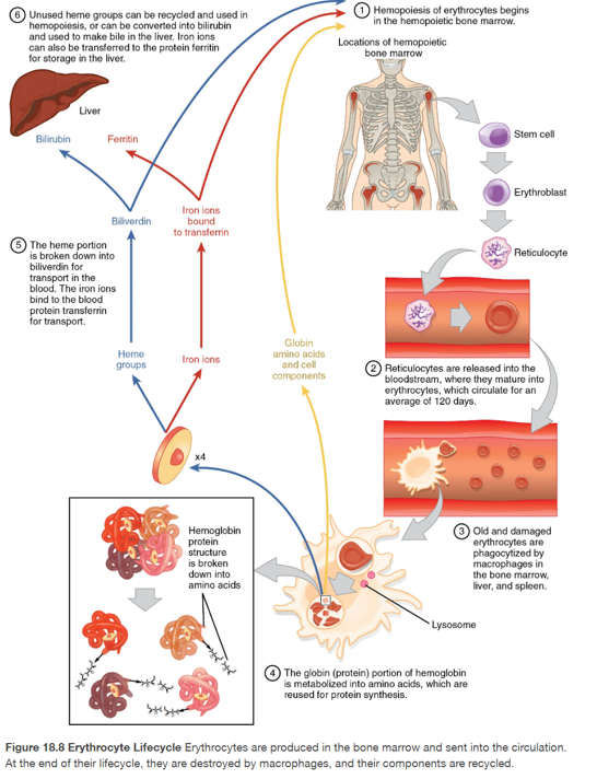
erythrocyte breakdown
cells replaced by macrophages, located w/in bone marrow, liver spleen
span of 120 days
globin → amino acids for reuse
iron stored in liver/spleen
non-iron degraded into the waste bilirubin
bilirubin → liver to use for bile
bilirubin → stercobilin in feces in LI
anemia
low oxygen carrying capacity
low RBCS/hemoglobin
blood loss, fault/decreased RBC, excessive destruction of RBC
sickle cell anemia
genetic disorder caused by mutated hemoglobin
crescent shaped RBC
polycythemia
disorder caused by elevated RBC count + detected in hematocrit
may occur transiently in dehydration: plasma volume falls from inadequate water intake or excessive loss
normal in high altitudes
insufficient anemia
hemorrhagic: excessive blood loss
hemolytic: RBCs are destroyed prematurely
aplastic: red bone marrow is destroyed/faulty
leukocyte
WBCs responsible for immune function
granular: lobed nucleus, phagocytic, stain
agranular: less-visible granules, simple nucleus
neutrophil
granular, multilobed, phagocytic WBC
most common: 50-70%
bacterial infection
eosinophil
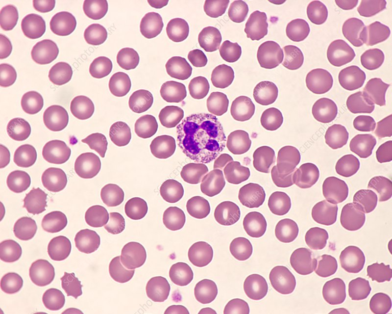
granular WBC, 2-3 lobes w/ red to orange granules containing antihistamine molecules
consume inflammatory chemicals + active immune complexes
phagocytic against parasitic worms
2-4% of WBC count

basophil
granular, two-lobed WBC that releases histamine, which contributes to inflammation, and heparin to oppose blood clotting
large dark granules, S-shaped nuclei
>1% of WBC

lymphocyte
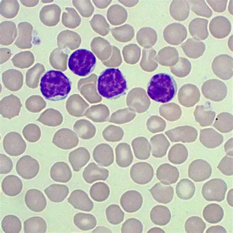
agranular, second most common WBC
20-30%
immune response
NK, T, + B cells
NK cell
lymphocyte responsible for recognizing cells that do not express “self” proteins
T cells
lymphocyte responsible for defense against specific pathogens like viruses and cancer
B cells
lymphocyte responsible for producing antibodies
monocyte
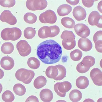
largest WBC with kidney-shaped nuclei
2-8%
mature into macrophages that release antimicrobial chemicals that attract other leukocytes to site of infection
phagocytosis of debris, pathogens, worn-out cells
leukemia
cancer involving abundance of leukocytes
chronic: mature WBCS accumulate + fail to die
acute: overproduction of young WBCs
cells do not function properly
platelet
fragment of cytoplasm of megakaryocyte
small, numerous
stop blood flow following damage to a vessel

hemostasis
process of repairing a damaged blood vessel to stop the bleeding and prevent hemorrhage
vascular spasms
platelet plug
coagulation

vascular spasms
first step in hemostasis, when blood vessels is damaged and loses blood
smooth muscles constrict to lower blood loss + withdraws from injury site
platelet plug
second step of hemostasis
platelets stick to injured vessel and transform to become spiky and sticky, forming a seal
secrete chemicals to attract more platelets
coagulation
third step of hemostasis
platelets + fibrin clump together to form clot
fibrin
form mesh-like structure that traps platelets to form blood clot
antigen
glycoprotein body identification marker on the surface of RBCs
neutralized by antibodies
antibody
neutralize antigens
A-
anti-B
A antigen
receive from: A-, O-
donate to: A+, A-, AB+, AB-
A+
anti-B
antigen A, Rh
receive from: A+, A-, O-, O+
donate to: AB+, A+
B-
anti-A
B antigen
receive from: B-, O-
donate to: AB-, AB+, B-, B+
B+
anti- A
B, Rh antigen
receive from: B+, B-, O+, O-
donate to: AB+, B+
AB-
no antibodies
A, B antigens
receive from: A-, B-, AB-, O-
donate to: AB-, AB+
AB+
no antibodies
A, B, Rh antigens (universal recipient)
receive from: All
donate to: AB+
O-
A, B antibodies
no antigens (universal donor)
receive from: O-
donate to: All
O+
A, B antibodies
Rh antigen
receive from: O+, O-
donate to: A+, B+, AB+, O+
HDN
hemolytic disease of newborn
occurs during transfusion of Rh incompatible blood
mother is exposed to baby’s Rh+ cells and generates anti-Rh that can attack
RhoGAM prevents development of anti-Rh