Chapter 8 skeletal system
 Call Kai
 Learn
 Practice Test
 Spaced Repetition
 Match
 Flashcards
 Knowt Play
1/28

There's no tags or description
Looks like no tags are added yet.
29 Terms
Bones in the adult and child body
Adult: 206 bones (some variation within bones that form within tendons (sesamoid), and bones that deveelop in skull suture lines (sutural bones).
At birth 270 bones but number decreases with fusion.
ex- ilium, ischium, and pubis fuse to form a hip bone.
Paranasal cavities
Frontal, sphenoid, ethmoid, and maxiallary
air filled cavities connected to nasal cavity and lined by mucous membranes.

cranial bones
There are eight cranial bones:
1 frontal
2 parietal
2 temporal
1 occipital
1 sphenoid
1 ethmoid
Old people from texas eat spiders.

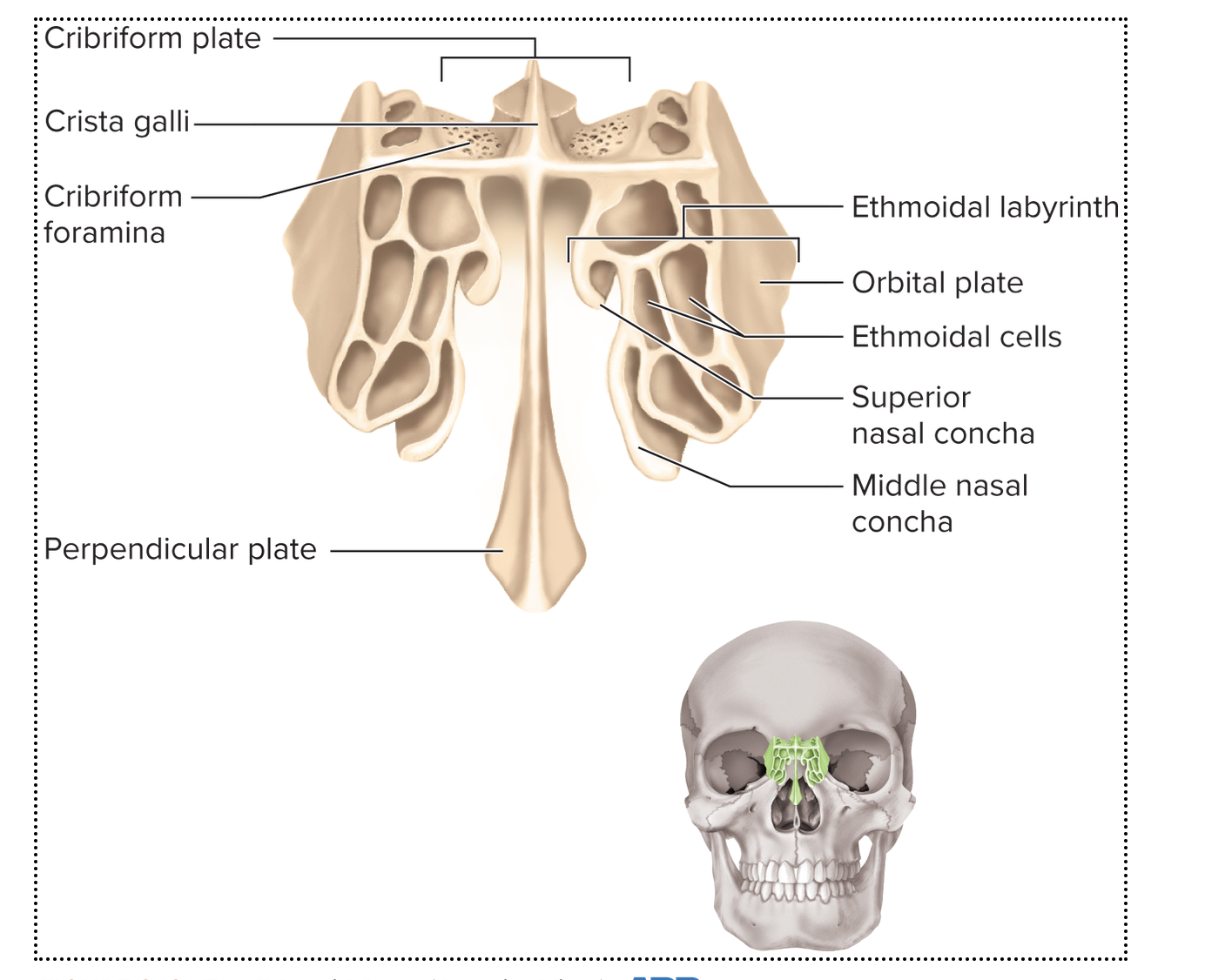
ehtmoid bone
The ethmoid bone is located between the eyes
Contributes to medial wall of orbit, walls and roof of nasal
cavity, and nasal septum

The Mandible
Structure of each Y-shaped ramus:
Posterior branch is condylar process containing mandibular
condyle for articulation with temporal bone to form
temporomandibular joint (TMJ)
Anterior branch called the coronoid process2attachment site for
temporalis muscle involved in mastication
Mandibular notch between the two processes
Mandibular foramen2opening below notch on medial surface of
ramus; allows passage of nerves and vessels that supply lower
teeth

Hyoid bone
Slender U-shaped bone between chin and larynx
Does not articulate with any other bone
Suspended from styloid processes of skull
fractured hyoid is sign of strangulation
vertebral column
The vertebral column (spine) is a flexible chain of 33
vertebrae with fibrocartilaginous intervertebral discs
between them
protects spinal cord
Supports skull and trunk and allows for their movement
Protects spinal cord
Absorbs stresses of movements
Provides attachments for limbs, thoracic cage, and postural
muscles
vertebral column
7 cervical vertebrae in the neck
12 thoracic vertebrae in the chest
5 lumbar vertebrae in lower back
5 sacral vertebrae at base of spine
4 coccygeal vertebrae (tail bone_

reason for most variations in adult bone number?
Variations in number of vertebrae occur in 1 in 20 people.
Generally due to differences in the number of fused
vertebrae in inferior part of spine.
Kyphosis
concave in anterior direction (curved outward
toward back)

Lordosis
concave in posterior direction (curved outward
toward front)
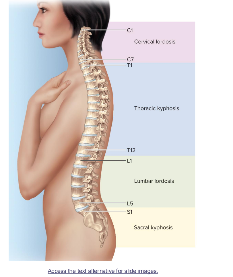
Beyond 3 years of age, the vertebral column has four bends
Cervical lordosis, thoracic kyphosis, lumbar lordosis, pelvic
kyphosis
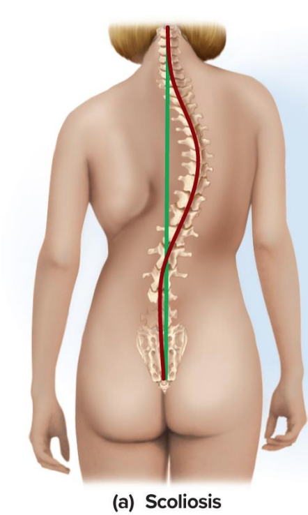
scoliosis
most common in teenagers.
curves laterally/ deviates from midsaggital plane
hyperkyphosis
most common in older women
cervical goes from lordosis to kyphosis
Hyperkyphosis—an excessive forward curvature of the thoracic (mid-to-upper) spine—is most common in older adults, particularly postmenopausal women. [1, 2]

hyperlordosis
common in pregnant women
Hyperlordosis is an excessive inward curvature of the lower back (the lumbar spine) or, less commonly, the neck (the cervical spine)
. While a healthy spine naturally curves inward in these regions to absorb shock and support weight, an exaggerated curve creates a distinct "swayback" appearance.

superior view vertebra

posterior view vertebrae

left lateral view vertebrae

Intervertebral disc-
cartilaginous pad located between
bodies of adjacent vertebrae
Consists of two parts:
Nucleus pulposus-inner gelatinous mass
Anulus fibrosus-outer ring of fibrocartilage
Functions: bind vertebrae together, support weight of the
body, absorb shock
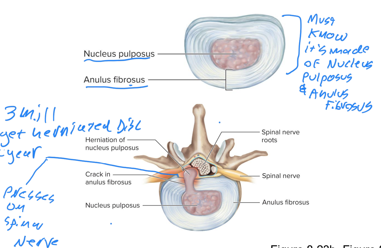
Herniated disc
causes pinched nerve: The spinal canal and the tiny exit holes for nerves (the foramina) are already highly cramped, narrow spaces. When the disc material bulges into this narrow pathway, it physically collides with and squishes the nearby spinal nerve root. we get chemical burn too- molecules body doesnt recognize leads to inflammatory response
ruptured or slipped sic puts painful pressure on spinal nerve or spunal cord
True ribs
(vertebrosternal) ribs (ribs 1 to 7)- each rib has its
own costal cartilage connecting it to the sternum
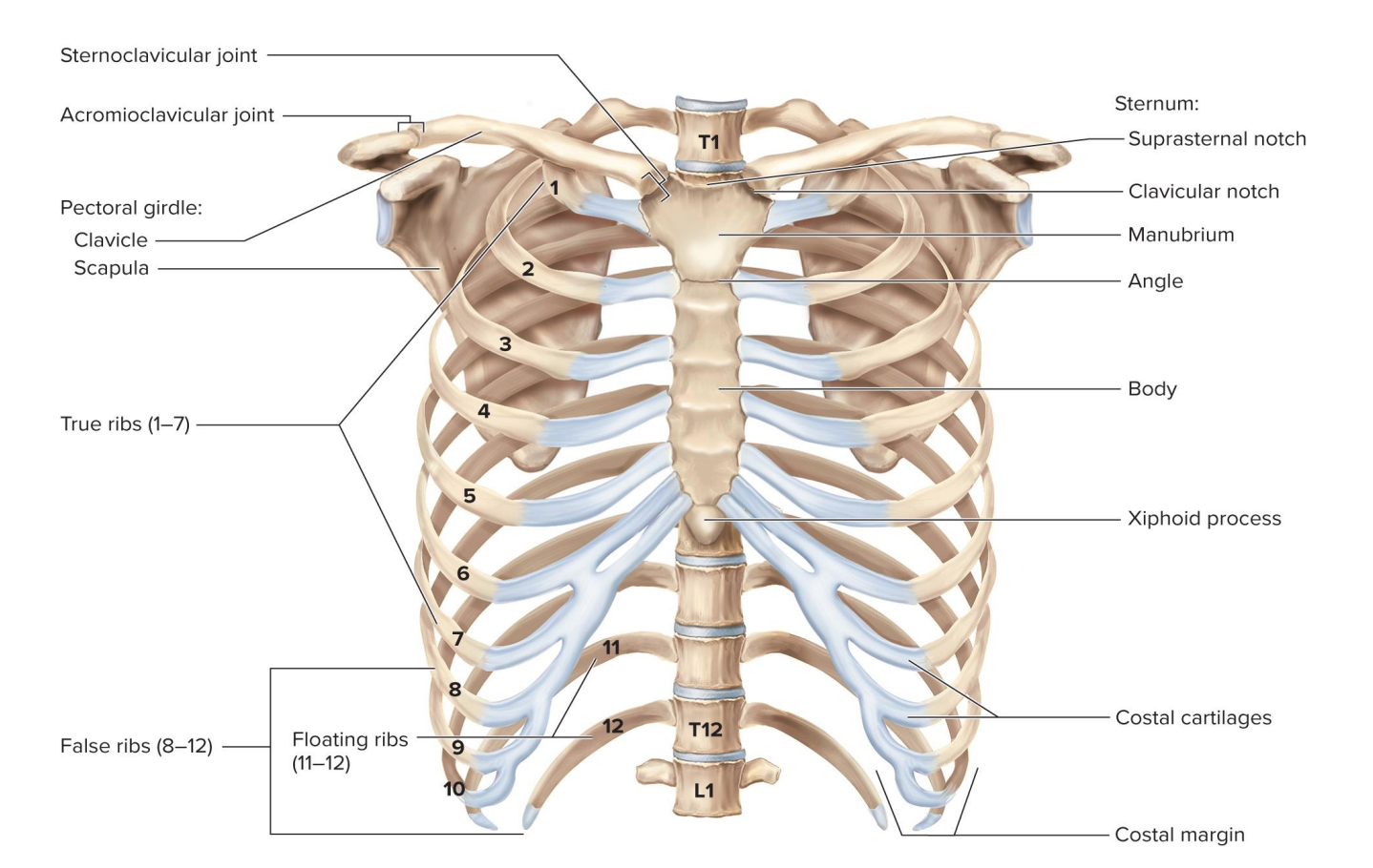
False ribs
(ribs 8 to 12)-each rib lacks an independent
cartilaginous connection to sternum

vertebrochondral ribs
Ribs 8 to 10 have associated cartilages that merge with that of rib 7;
also called ?

floating (vertebral) ribs
Ribs 11 to 12 do not connect to cartilage at all, so they are also
called

humerus
The humerus is the only bone in the brachium
Features of the proximal end:
Head-hemispherical projection that articulates with the glenoid
cavity of scapula
Anatomical neck-groove bordering head
Greater and lesser tubercles-bumps for muscle attachments
Intertubercular sulcus-groove between tubercles accommodates
tendon of biceps muscle
Surgical neck-narrowing just distal to the tubercles; common
fracture site
Deltoid tuberosity-rough area on lateral surface of shaft;
attachment site for deltoid muscle
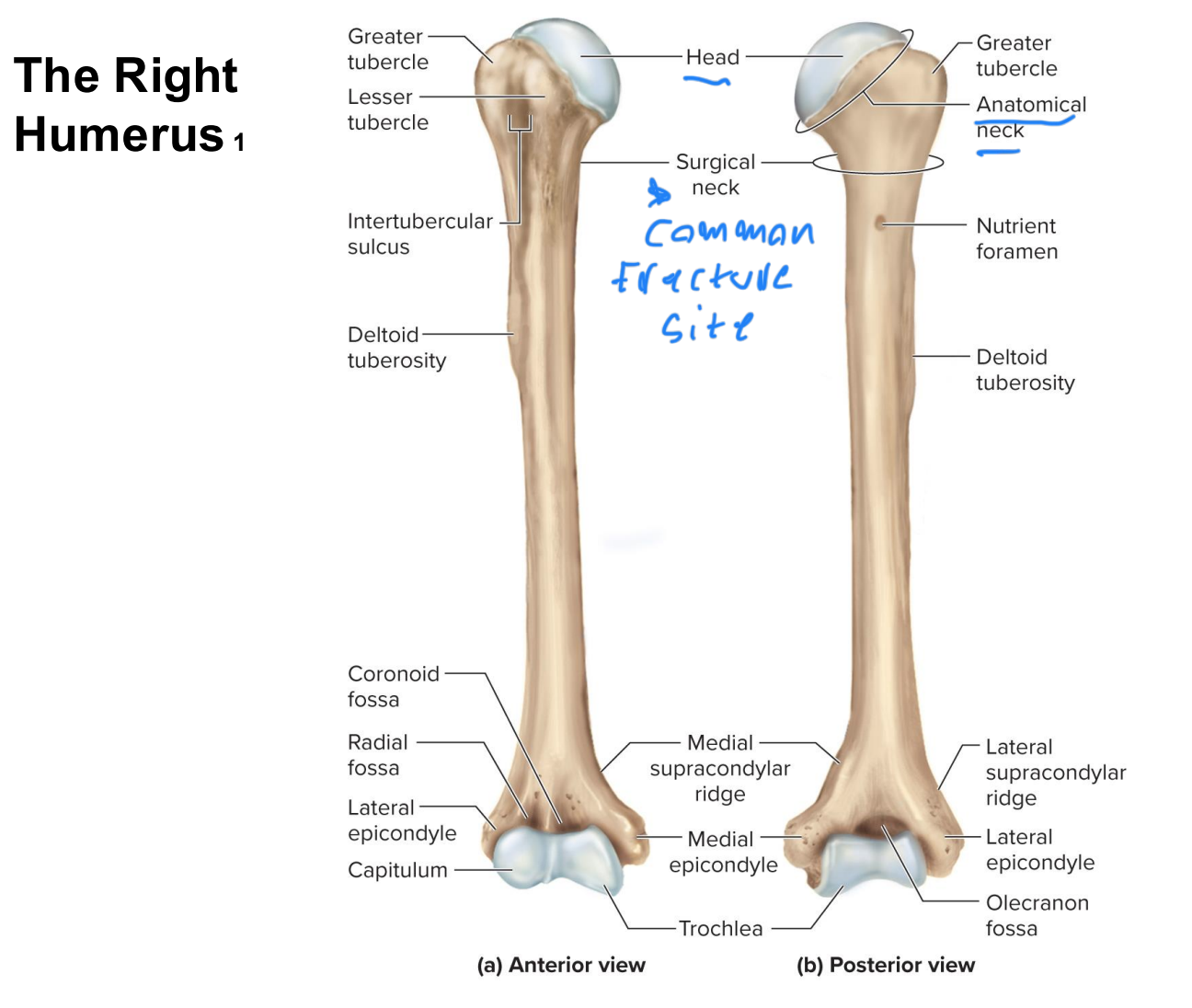
superior/proximal end of right humerus

distal/inferior view of right humerus

The Carpal Bones
8 carpal bones: Allow movements of flexion, extension, abduction, and
adduction
Arranged in two rows of four bones each
Proximal row: scaphoid, lunate, triquetrum, and pisiform
Pisiform is a sesamoid developed by age 9 to 12 in tendon of flexor carpi
ulnaris muscle
Distal row: trapezium, trapezoid, capitate, and hamate
peter told lisa she has curly toes today
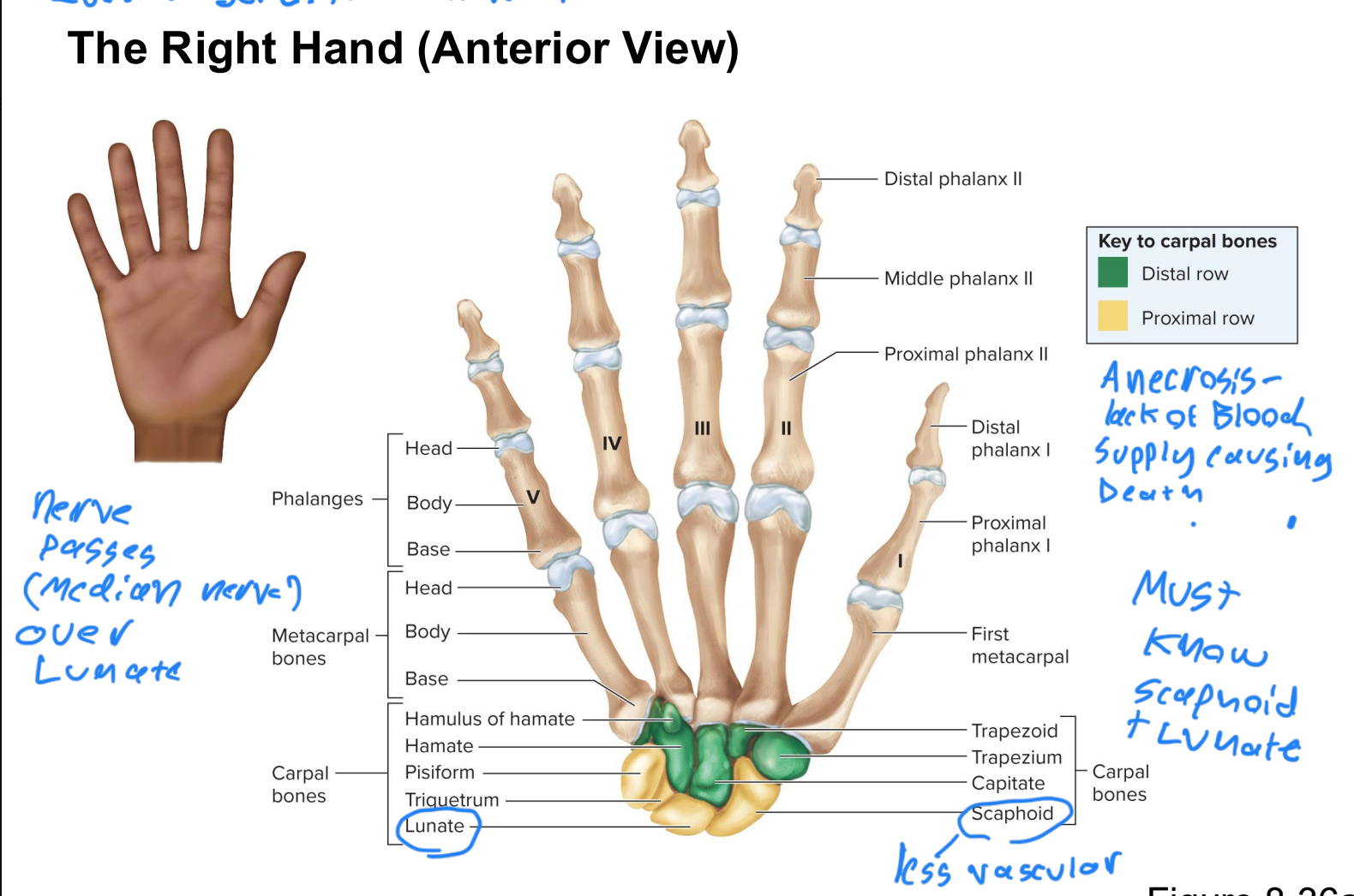
hamate
