[5] Comitancy Testing
 Call Kai
 Learn
 Practice Test
 Spaced Repetition
 Match
 Flashcards
 Knowt Play
1/121

There's no tags or description
Looks like no tags are added yet.
122 Terms
Why is a recent-onset noncomitant deviation considered a red flag?
Recent-onset noncomitant strabismus may indicate a serious neurologic or systemic disorder and can represent a health- or life-threatening condition. These patients may require immediate referral.
Why is it important to determine whether a deviation is longstanding?
In longstanding deviations, identifying the characteristics and pattern of the deviation helps guide treatment decisions and provides prognostic information.
What are the major causes of noncomitant (incomitant) deviations?
Common etiologies include:
Obstetric trauma (e.g., forceps delivery)
Idiopathic causes
CNS dysfunction
Extraocular muscle anomalies (abnormal insertion, development, or length)
Trauma
Vascular disease
Neoplasms
Viral disease
Chronic systemic disease
Which causes of noncomitant deviations are especially associated with certain age groups?
Vascular disease: more common in older patients
Viral disease: more common in younger patients
Neoplasms and chronic diseases: can occur in both younger and older patients
What defines a comitant deviation?
A comitant deviation has approximately the same angle of deviation in all positions of gaze and with either eye fixating.
What numerical criteria are used to classify a horizontal deviation as comitant vs. noncomitant?
Comitant: within 5Δ across positions of gaze/fixation
Noncomitant: differs by >5Δ
What numerical criteria are used to classify a vertical deviation as comitant vs. noncomitant? (sometimes)
Comitant: within 2Δ
Noncomitant: differs by >2Δ
How is a noncomitant (incomitant) deviation defined?
The magnitude of the deviation changes in different positions of gaze and/or differs depending on which eye is fixating.
What common misconception exists regarding noncomitancy?
Noncomitancy is not the same thing as having different deviations at distance and near. Noncomitancy specifically refers to changes with gaze position and/or fixation eye.
What are the most common causes of noncomitant deviations in adults?
The major causes are:
Microvascular disease
Trauma
Neoplasm
When an adult presents with a recent-onset noncomitant deviation, what etiologies should be high on the differential?
Consider:
Microvascular cranial nerve palsy
Traumatic injury affecting extraocular muscles or cranial nerves
Intracranial or orbital neoplasm
What are the two broad categories of noncomitant deviations?
Paretic (Neurogenic) deviations
Mechanical Restriction (Myogenic) deviations
What causes a paretic (neurogenic) noncomitant deviation?
An innervational problem causing weakness or paralysis of an extraocular muscle due to dysfunction of the nerve supply.
What is the hallmark mechanism of a paretic deviation?
Reduced neural input to an extraocular muscle → impaired muscle action → gaze-dependent deviation.
What causes a mechanical restriction (myogenic) noncomitant deviation?
Physical limitation of eye movement due to:
An abnormal extraocular muscle
Structural/orbital restriction
What is the hallmark mechanism of a restrictive deviation?
The muscle may receive normal innervation, but the eye cannot move normally because a muscle or surrounding orbital structure mechanically limits movement.
How do paretic and restrictive noncomitant deviations differ?
Paretic (Neurogenic) | Restrictive (Mechanical/Myogenic) |
|---|---|
Problem of innervation | Problem of movement restriction |
Weak muscle action | Physically limited movement |
Cranial nerve dysfunction common | Abnormal muscle or orbital structure common |
Eye cannot move because signal is weak | Eye cannot move despite normal signal |
What symptoms are commonly reported by adults with a recent-onset noncomitant deviation?
Adults typically present with:
Recent-onset diplopia
Ocular discomfort or strain
Why do adults with recent-onset noncomitant deviations commonly complain of diplopia?
Adults have established binocular vision and normal retinal correspondence, so a sudden ocular misalignment often results in symptomatic double vision.
How may children with recent-onset noncomitant deviations present differently from adults?
Children may:
Have no complaints
Present with an abnormal head posture (head turn, chin-up/down posture, or head tilt)
What clinical sign may be the only clue to a noncomitant deviation in a child?
A compensatory head turn, head tilt, or head tip may be present even when the child reports no symptoms.
What are the most common causes of noncomitant deviations in pediatric patients?
Common causes include:
Congenital/developmental abnormalities
Trauma
Neoplasm
Infection/inflammation
What should be included in the differential diagnosis for a child with a recent-onset noncomitant deviation?
Consider:
Congenital/developmental abnormalities
Trauma
Infection or inflammation
Intracranial or orbital neoplasm
What is the difference between congenital/nonocular torticollis and ocular torticollis?
Congenital/nonocular torticollis: caused by fibrosis, shortening, or tightening of the sternocleidomastoid (SCM) muscle.
Ocular torticollis: a compensatory head posture adopted to maintain binocular single vision, usually due to a noncomitant strabismus, often a cyclovertical deviation.
What is the typical cause of congenital torticollis?
Fibrosis or shortening of the sternocleidomastoid muscle causing persistent head tilt and abnormal neck posture.
What type of ocular misalignment most commonly causes ocular torticollis?
A noncomitant cyclovertical deviation that improves binocular vision when the head is tilted or turned.
How can the patch (occlusion) test differentiate ocular from congenital torticollis?
Congenital torticollis: head tilt remains despite patching.
Ocular torticollis: head tilt improves or straightens when one eye is patched.
What historical and examination findings suggest congenital torticollis rather than ocular torticollis?
Present at birth or shortly thereafter
Difficulty passively straightening the head
SCM muscle hardening/tightening
No diplopia
No change with occlusion
What findings suggest ocular torticollis rather than congenital torticollis?
Usually develops after infancy
Head can be easily straightened
No SCM fibrosis on palpation
Associated vertical deviation/cyclovertical strabismus
Diplopia may occur when the head is forced straight
Head straightens with patching
What type of extraocular muscle dysfunction is commonly associated with a compensatory head turn?
Dysfunction of the horizontal recti (medial rectus or lateral rectus) often produces a compensatory head turn.
What type of abnormal head posture is associated with vertically acting muscles?
A chin elevation or chin depression is commonly associated with dysfunction of the vertically acting muscles.
What pattern strabismus may cause chin-up or chin-down posture?
Significant differences in horizontal deviation between upgaze and downgaze, such as A-pattern or V-pattern strabismus.
What type of diplopia is most associated with a compensatory head tilt?
Vertical and torsional diplopia, classically seen in cyclovertical muscle palsies (e.g., superior oblique palsy).
How do congenital and acquired noncomitant deviations differ in terms of diplopia?
Congenital: Diplopia is uncommon due to suppression/adaptation (unless decompensation occurs).
Acquired: Diplopia is common, often worse in a particular field of gaze.
How does abnormal head posture differ between congenital and acquired noncomitant deviations?
Congenital: Abnormal head posture is often present, and the patient may be unaware of it.
Acquired: Usually absent initially but may develop as a compensatory mechanism to eliminate diplopia.
How do primary and secondary deviations differ in congenital versus acquired noncomitant deviations?
Acquired: Difference between primary and secondary deviation is typically present.
Congenital: May not show this difference in paretic cases; mechanical congenital deviations may show a difference.
Which sensory adaptations are more likely in congenital noncomitant deviations?
Amblyopia may be present.
Suppression may be present.
These adaptations are generally absent in acquired deviations because binocular vision was already established.
How does ocular motility limitation differ between congenital and acquired noncomitant deviations?
Congenital paretic: motility limitation often mild.
Congenital mechanical: motility limitation may be marked.
Acquired: motility limitation is often severe.
How does the likelihood of systemic or neurologic disease differ between congenital and acquired noncomitant deviations?
Congenital: neurologic/systemic disease is relatively rare.
Acquired: neurologic/systemic disease is much more common and should be considered in the workup.
What are the three major causes of an underacting extraocular muscle?
Trauma → direct injury or weakness of the muscle
Mechanical causes → faulty insertions, tendon abnormalities, or adjacent structural abnormalities
Innervational causes → impairment of CN III, IV, or VI
How can trauma cause an underacting extraocular muscle?
Trauma can directly damage the muscle, resulting in weakness and reduced ocular motility.
What mechanical abnormalities can cause an extraocular muscle to underact?
Faulty muscle insertions
Tendon abnormalities
Orbital or adjacent structural abnormalities that restrict normal muscle action
Which cranial nerves are involved in innervational causes of underacting extraocular muscles?
CN III (Oculomotor)
CN IV (Trochlear)
CN VI (Abducens)
What are the three major causes of an overacting extraocular muscle?
Mechanical
Idiopathic
Hering's Law of Equal Innervation
How can a mechanical abnormality cause an overacting muscle?
Abnormal muscle insertions or tendon/ligament abnormalities can alter muscle mechanics and create apparent overaction.
What role does Hering's Law play in muscle overaction?
Hering's Law states that yoked muscles receive equal neural input. When one muscle is weak, increased innervation is sent to overcome the weakness, causing the contralateral yoke muscle to appear overacting.
Which concept explains apparent overaction of a yoke muscle in the fellow eye when its partner is paretic?
Hering's Law of Equal Innervation.
What is the first step when evaluating a patient with a deviation?
Determine whether the deviation is comitant or noncomitant.
Once a deviation is identified as noncomitant, what are the next two steps?
Identify the underacting muscle.
Determine whether the underaction is paretic (neurogenic).
After identifying a paretic muscle, what important historical question should be asked?
Is the onset recent?
Recent-onset paresis may indicate a serious neurologic or systemic condition requiring prompt evaluation.
What are the three primary tests used to evaluate ocular motility?
Versions (binocular eye movements)
Ductions (monocular eye movements)
Forced duction testing (assesses mechanical restriction)
What are versions and what do they evaluate?
Versions are binocular movements of the eyes and are used to evaluate how the eyes move together in the cardinal positions of gaze.
What are ductions and what do they evaluate?
Ductions are monocular movements and assess the movement capability of each eye individually.
What is the purpose of forced duction testing?
Forced duction testing helps determine whether limited eye movement is due to a mechanical restriction rather than a paretic muscle.
What objective tests can be used to evaluate comitancy?
Observation
Version testing
Duction testing
Forced duction testing
Cover testing in all diagnostic positions of gaze
Park's 3-Step Test
What subjective tests can be used to evaluate comitancy?
Red Lens Test
Maddox Rod Test
Hess-Lancaster Test
Double Maddox Rod Test
Which subjective test is particularly useful for evaluating torsion?
Double Maddox Rod Test
What should you look for during observation when evaluating a noncomitant deviation?
Head turn
Head tilt
Head tip/chin posture
Eye closure or covering one eye
Consistency of abnormal head posture
Why is the duration of an abnormal head posture important?
Longstanding abnormal head posture suggests a chronic or congenital condition, whereas a recent onset may suggest an acquired process.
If a patient is unsure how long an abnormal head posture has been present, what can help determine chronicity?
Review old photographs to see whether the head posture has been present over time.
Why is it important to determine whether the head is always tilted or turned in the same direction?
A consistent abnormal head posture often represents a compensatory position used to minimize diplopia or maximize binocular vision.
How is version testing performed?
Have the patient follow a target binocularly through all Diagnostic Action Fields (DAFs) while observing for underactions or overactions.
How can the Hirschberg test be incorporated into version testing?
Move both the penlight and yourself into each DAF while observing corneal light reflexes. This increases sensitivity and can make subtle ocular asymmetries or misalignments more apparent.
What is an alternative way to evaluate eye movements if keeping the target moving is difficult?
Keep the target stationary and move the patient's head into different positions, or move yourself into the diagnostic fields.
Why is Hirschberg assessment useful during version testing?
It enhances detection of subtle ocular misalignment and asymmetrical eye movements that may be harder to appreciate with gross observation alone.
What grading scale is used to quantify overaction (OA) and underaction (UA) during version testing?
A scale from −4 to +4:
+4 = severe overaction
−4 = severe underaction
How much change in muscle action does each grade represent during version assessment?
Each grade corresponds to approximately 25% of normal function.
Examples:
−1 = 25% underaction
+2 = 50% overaction
What does a grading of −4 indicate on version testing?
Severe underaction of a muscle, representing near-complete loss of expected movement in that diagnostic field.
What does a grading of +4 indicate on version testing?
Severe overaction of a muscle, representing markedly excessive movement in that diagnostic field.
During version testing, what findings indicate underaction versus overaction?
Observe the non-fixating eye:
Lags behind expected position → Underaction (UA)
Moves excessively beyond expected position → Overaction (OA)
Why is version testing performed binocularly?
Binocular viewing allows comparison of yoked muscle actions and identification of relative underactions and overactions in different diagnostic fields of gaze.
When should duction testing be performed?
Duction testing is performed when underaction (UA) is noted during version testing.
What is the primary purpose of duction testing?
To determine the extent of true ocular motor limitation by evaluating movement of each eye individually.
How is duction testing performed?
Test monocularly (one eye at a time)
Move the target into the diagnostic action field (DAF)
Observe the eye's ability to move into that field
What does improvement on duction testing compared with version testing suggest?
The apparent limitation may be due to a binocular/innervational issue rather than a true restriction of eye movement.
What does persistent limitation on duction testing suggest?
It suggests a true motility deficit caused by either:
Paresis (innervational)
Mechanical restriction
Additional testing is needed to differentiate the two.
What is the purpose of forced duction testing?
To determine whether an underacting muscle is due to an innervational (paretic) cause or a mechanical restriction.
How is forced duction testing performed?
Instill local anesthesia.
Grasp the eye with forceps or a cotton-tipped applicator.
Attempt to passively move the eye in the direction opposite the suspected restriction.
What does a positive forced duction test indicate?
Mechanical restriction.
If the examiner cannot move the eye passively, a restrictive process is preventing movement.
What does a negative forced duction test indicate?
No significant mechanical restriction, making an innervational (paretic) cause more likely.
What is the purpose of Park's 3-Step Test?
To identify an isolated cyclovertical muscle palsy in a patient with a noncomitant vertical deviation.
What are the three steps of Park's 3-Step Test?
Which eye is hyper?
Is the hyperdeviation greater in right or left gaze?
Is the hyperdeviation greater with right or left head tilt?
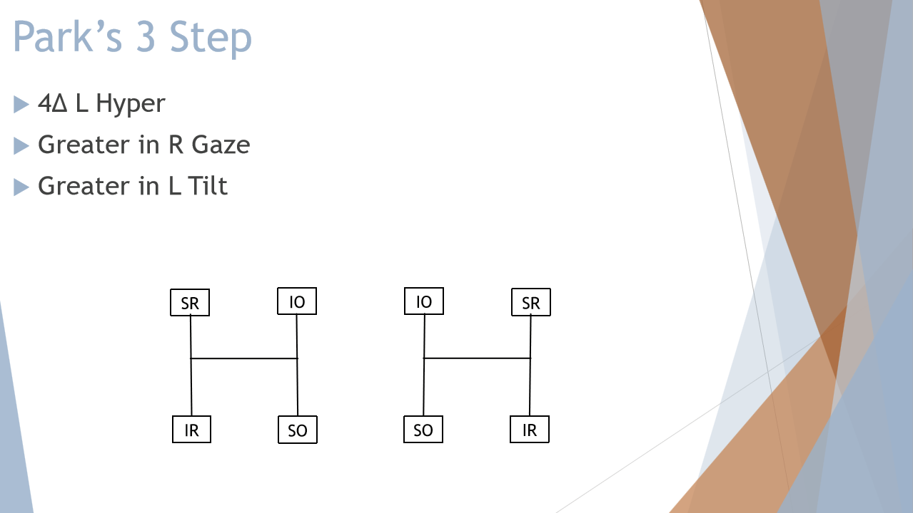
Which extraocular muscles are evaluated by Park's 3-Step Test?
The cyclovertical muscles:
Superior rectus (SR)
Inferior rectus (IR)
Superior oblique (SO)
Inferior oblique (IO)
What torsional actions are important to remember when performing Park's 3-Step Test?
Superior muscles (SR, SO) → intort
Inferior muscles (IR, IO) → extort
Mnemonic:
"Superiors Spin In, Inferiors Exit Out."
In what clinical situation is Park's 3-Step Test most useful?
An isolated vertical strabismus where a single cyclovertical muscle palsy is suspected (classically a superior oblique palsy).
How is the alternate cover test used when evaluating comitancy?
Measure and neutralize the deviation in all 9 diagnostic action fields (DAFs) to determine whether the magnitude changes with gaze position.
Why is the alternate cover test valuable in detecting noncomitancy?
It is highly sensitive to small changes in deviation size between different gaze positions.
Why should alternate cover testing be performed with each eye fixating?
Comparing measurements with right versus left eye fixation can reveal differences between primary and secondary deviations and help identify paretic deviations.
What is the primary purpose of Maddox Rod/Red Lens testing in a noncomitant deviation workup?
To evaluate diplopia in all 9 positions of gaze and help confirm the underacting (paretic) muscle identified on version testing.
How is Maddox Rod/Red Lens testing performed?
Place the Maddox rod or red lens over one eye
The fixating eye is the eye NOT behind the red lens
Test all 9 diagnostic action fields (DAFs)
Repeat with each eye fixing
Patient maintains fixation on a penlight (typically ~40 cm)
Patient should not move their head during testing
What is the most important finding to look for during Maddox Rod/Red Lens testing?
Find the position of gaze with the greatest separation of the two images.
How can Maddox Rod/Red Lens testing help identify the paretic eye?
The eye that sees the leading image (the image projected the farthest away) contains the paretic muscle.
What type of diplopia is seen with exodeviations versus esodeviations?
Exotropia (Exo) → crossed diplopia
Esotropia (Eso) → uncrossed diplopia
How is image location related to the direction of the deviation?
The perceived position of the image is opposite the direction of the deviation.
When is Maddox Rod/Red Lens testing unreliable or unusable?
When the patient has:
Abnormal correspondence (AC)
Deep suppression
These sensory adaptations may prevent reliable diplopia responses.
What is the purpose of the Hess-Lancaster screen test?
To:
Identify the affected (paretic) extraocular muscle
Quantify the magnitude of deviation in each field of gaze
Map ocular motility defects across the diagnostic positions of gaze
Is the Hess-Lancaster test a direct or indirect projection test?
It is a direct projection test. The image from the deviated eye is projected in the same direction as the deviation.
How does image projection differ between Hess-Lancaster and Maddox Rod/Red Lens testing?
Test | Projection Direction |
|---|---|
Maddox Rod / Red Lens | Image projected opposite the deviation |
Hess-Lancaster | Image projected in the same direction as the deviation |
In Hess-Lancaster testing, what type of projection occurs with exo vs eso deviations?
Exodeviation → uncrossed projection
Esodeviation → crossed projection
Which eye contains the paretic muscle during Hess-Lancaster testing?
The eye that sees the lagging image contains the paretic muscle.
Maddox = Leading image = Paretic eye
Hess-Lancaster = Lagging image = Paretic eye