a&p lab exam unit 2
 Call Kai
 Learn
Learn Practice Test
 Spaced Repetition
 Match
Match Flashcards
 Knowt Play
1/88

Earn XP
Description and Tags
Name | Mastery | Learn | Test | Matching | Spaced | Call with Kai | Chat |
|---|
No analytics yet
Send a link to your students to track their progress
89 Terms
head of bone
prominent, expanded, rounded end of a bone
closed (simple) fracture
bone break beneath the skin
open (compound) fracture
broken bone pierces the skin
greenstick fracture
partial breaks in a bone that occur as a result of bending
comminuted fracture
bone crushed into 3 or more pieces
impacted fracture
one end of a broken bone is driven into the other end
condyle
rounded end of a bone
pivot point
often comes in pairs
canal
tubular passage through bone
crest
raised edge of bone that forms a narrow ridge
epicondyle
rounded prominence above condyle of bone
facet
flat articular surface of bone that allows for flexion, extension, and rotation
foramen
passageway/hole that allows for the passage of blood vessels
fossa
shallow, broad depression within bone
supporter for joint
sulcus
narrow channel within bone that allows a vessel or nerve to pass while being protected from compression
process
outward growth from a larger portion of the bone
meatus
funnel shape thats an opening to a canal
ramus
curved edge to bone that gives it structure
sinus
air filled cavity in bone
spine
small outward growth from bone
tubercle
small rounded and raised area where connective tissues attach
tuberosity
raised rough area that serves as an attachment site for muscles and ligaments
foramen magnum
large opening at bottom of occipital bone
passage for spinal cord and vertebral arteries
occipital condules
two bumps at the bottom of the occipital bone next to the foramen magnum
external occipital protubrence
small piece of bone that pokes out of the middle of the occipital bone
zygomatic process
long bone pointing towards the front of the face
styloid process
sharp/pointy bone coming out of the bottom of the temporal bone, pointing downwards
mastoid process
Big bump towards the bottom side of the temporal bone, behind the ear
external acoustic meatus
small cave/indentation in middle bottom of temporal bone, behind zygomatic process
mandibular fossa
wider cave next to acoustic meatus, under the zygomatic process
on bottom side of the bone
frontal bone
forehead
mandible
jaw bone
coronoid process
anterior points a the top of the mandible
mandibular condyle
posterior points at the top of the mandible, bigger than coronoid process
mental foramen
two lateral divots in lower part of mandible ( snake bites)
mandibular foramen
two divots on the inside of the “arms“ of the mandible
squamous suture
parietal and temporal bones connect, on the side of the skull
lamboid suture
parietal and occipital bones connect, back of the skull
saggital suture
parietal and parietal bones connect, on the top middle of skull
coronal suture
frontal and parietal bones connect, on the top of skull going side to side

sphenoid

mastoid process
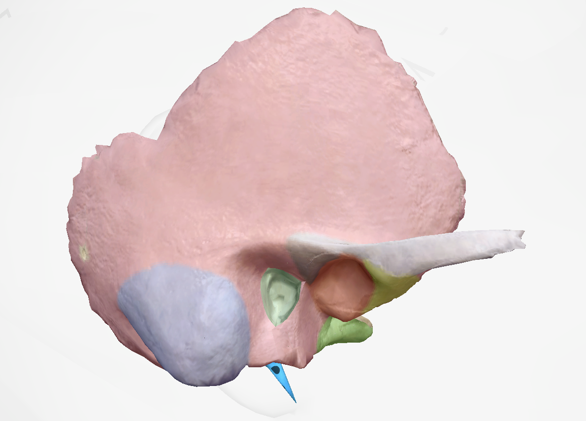
styloid process

zygomatic process
gliding joint
allows gliding movement between 2 articular surfaces, moves along the same plane
Gliding joint example
carpal and tarsal joints, intervertebral joints
Hinge joints
flexion and extension, moves along one axis hinge
hinge joint example
elbow and femur joints, finger joints
Pivot joint
rotational movement around an axis
Pivot joint example
atlas and axis
condyliod joint
allows flexion, extension and circumduction
can rotate within the joint
condyloid joint example
carpals on the radius
saddle joint
bones wrapped inside each other
high degree of movement with stability
saddle joint example
first metacarpal and trapezium
ball and socket joint
ball in concave structure
highest degree of motion
ball and socket joint example
shoulder and hip
the most mobile a joint is ______________
the less stable it is
Ligaments
Dense connective tissue that helps hold bone to bone
Joints of the shoulder
Sternoclavicular, Acromioclavicular, Glenohumeral
Sternoclavicular joint
Fibrous tissue attaches clavicle to manubrium/sternum
Sternoclavicular movement
elevation, depression, rotation, some protraction and retraction
Acromioclavicular (AC)
attaches clavicle and acromion/scapula
Acromioclavicular movement
not much movement, but allows very little gliding movement
Glenohumeral joint
ball and socket joint of shoulder
Glenohumeral joint movement
a lot of movement
rotation, flex, extend, abduction, adduction, circumduction
ligaments of hips
Iliofemoral, pubofemoral, ischiofemoral
Purpose of meniscus
cushion of knee for stress, shock absorber
increases surface are of condyle on tibia
What is the role of cartilage in the joint
protects bone from breaking down, protective membrane
Compact bone
Dense bone tissue on exterior of bone
Spongy bone
Interior of bone, looks like sponge
Medullary cavity
Cylindrical cavity in the center of long bones
Bone marrow stored here
Yellow bone marrow
Mostly fat
Red bone marrow
Makes red blood cells
Endosteum
Inner membrane
Separates compact and spongy bone
Periosteum
Fibrous outer layer
On the outside of the bone
Epiphyseal line
Growth plate
Long bone grows here
Turns into a scar when you’re done growing
Osteoporosis
Bone tissue loss is greater than bone tissue generated
Results in bone pain, tenderness and brittle bones
Parts of the sternum
Manubruim, body, xiphoid process
How are the atlas and axis different to other vertebrae
Axis has a notch to connect to atlas, used to pivot moving head
What is the role of synovial fluid in a joint
Lubrication
Role of cartilage in a joint
Protects bone from breaking down
Protective membrane that stops synovial fluid from entering the bone
Fibrous joints/Syntharoses
Joints with no movement
Gomphosis
Articulation between the teeth and the mandible or maxilla
Where the teeth go into the skull
Synchodrosis
Immobile cartilaginous joint between the first pair of ribs and the sternum
Syndesmosis
Ligament connects two bones, allows a little bit of movement
Symphyses
Fibrocartilatinous pad that connects two bones allowing for some movement
Syndesmosis example
Tibiofibular ligament
Symphyses
Pubic symphysis- between pubic bones
Intervertebral disks
Synovial joints
6 types that allow for different ranges of motion
Basic structure of a synovial joint
Membrane around joints that create synovial fluid