5. Jaw Relation – Part-1 Vertical Dimension of Occlusion ✅
 Call Kai
 Learn
Learn Practice Test
 Spaced Repetition
 Match
Match Flashcards
 Knowt Play
1/41

There's no tags or description
Looks like no tags are added yet.
Name | Mastery | Learn | Test | Matching | Spaced | Call with Kai | Chat |
|---|
No analytics yet
Send a link to your students to track their progress
42 Terms
Review of Clinical /Lab Procedures in Complete Dentures
Primary Impression
Custom Tray
Final Impression
Pour and Trim Casts to Ideal
Record base, wax occlusal rims
Records taking (bite registrations/facebow) to mount on articulator, tooth shade and mold selection.
Distinct differences between natural dentition & Complete denture occlusion.
Natural teeth are suspended in the bone by the periodontal
Ligament which acts as a shock absorber.
Denture teeth are part of the denture base which rests on movable or displaceable tissues.
Premature deflective contacts between the artificial teeth cause movement of the denture resulting in damage to the supporting tissues
Goals of Complete Denture Occlusion
Limit trauma to the supporting structures
Preserve remaining structures
Enhance stability of the dentures
Restore esthetics, speech and mastication
Recording Jaw Relations
To achieve this goal, the recording must include an approximate vertical dimension
of occlusion, stable occlusal contacts in harmony with existing TMJ and masticatory
muscle functions
In other words, we are transferring our patient’s maxillo-mandibular relationship to the articulator.
After preparing the record base and occlusal rims, we “customize” the rims to fit each patient’s esthetics and function using anatomical landmarks.
The maxillary occlusal wax rim is ALWAYS adjusted prior to adjusting the mandibular wax occlusion rim.
Adjustments are done to attain the following based on averages and observations mentioned in studies: Lip support Incisal level and midline Occlusal plane
Record Base
The average distance between the upper and lower labial sulci adjacent to the labial frenula when teeth are in occlusion is considered to be 40 mm
Maxillary Rim
22 mm measured from the highest point of the sulcus adjacent to the labial frenum to the occlusal surface of the wax.
Mandibular rim
18 mm from deepest point of sulcus adjacent to labial frenum
Maxillary record base
22 mm measured from the highest point of the sulcus adjacent to the labial frenum to the occlusal surface of the wax.
18 mm measured from the highest point of the sulcus adjacent to the buccal frenum to the occlusal surface of the wax

Mandibular record base-
18 mm from deepest point of sulcus adjacent to labial frenum
why follow the averages if we are doing refinements and changing contours later?
to save time
The measurements provide a guide in fabricating the record bases without the
patient being present.
This saves appointment time where only minor adjustments are done during record base try-in, instead of fabricating the wax rim while the patient waits in the chair
Advantages of Occlusal rims need to be refined to assume final teeth positioning
By doing it at this stage, a trial-and-error arrangement of teeth is minimized or eliminated.
In private practice, the lab will rely on your formed and adjusted wax rim to set the teeth
Anterior Rim for Lip Support
Restore the labial contour by shaping the wax to the proper width, position and
angulation
This determines the final position of the denture teeth helping in esthetics and
phonetics.
Maxillary wax rim –anterior Rim Guidelines for anterior teeth arrangement
1. Upper lip should be everted and not fallen/sunken in
2. Tooth support of the lip is by 2/3 of the incisal labial surface of the anterior teeth.
3. Incisal level of the central incisors are 1-2 mm below resting or low lip line.
This may change with age. Due to physiologic attrition, older patients may not show incisor edges at all.
4. Incisal edges of anterior teeth should meet the wet- dry line of the lower lip when pronouncing “fricative” sounds, i.e. “ F ” and “V”
5. Schiffman (1964) has shown that the maxillary central incisors fall approximately 8-10 mm anterior to the point of a line that bisects the midline of the palate perpendicularly through the incisive papilla.

Adjustment of the maxillary wax rim – Posterior rim
Follows the occlusal plane established through the use of extra-oral landmarks

The occlusal plane should be parallel to the interpupillary line
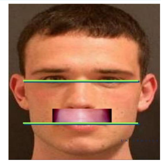
Sagittal plane
the occlusal plane should be parallel to the ala-tragus line (a.k.a. Camper’s Line)
Measure from mid- to superior edge of the tragus to the inferior border of the ala of the nose.

At this point, you can try the maxillary record base and wax rim in
the patient and adjust accordingly to meet these requirements
The Fox occlusal plane, designed by Dr. Frank Fox, is a very useful tool for establishing these requirements
Try-in of record base and wax rim
Adjust the plane of the wax rim so that it is parallel with the ala-tragus line (Camper’s line).
Adjust the plane of the wax rim so that it is parallel with the interpupillary line
With the Fox plane in, the frontal plane has met the requirement, but not the sagittal plane.
How would you fix this to make it parallel?
Reduce wax rim height posteriorly OR increase wax rim height anteriorly assuming you lack incisal edge visibility (1-2 mm)

After the maxillary occlusal wax rim has been adjusted, it is time to adjust the mandibular rim
Dimensions are dictated by the vertical dimension of occlusion (VDO)
Anterior Rim Adjustment
With the lips at rest, the wax rim should project 1-2 mm below the rested or low lip line. Patients 60 and older may not show

Vertical Dimensions
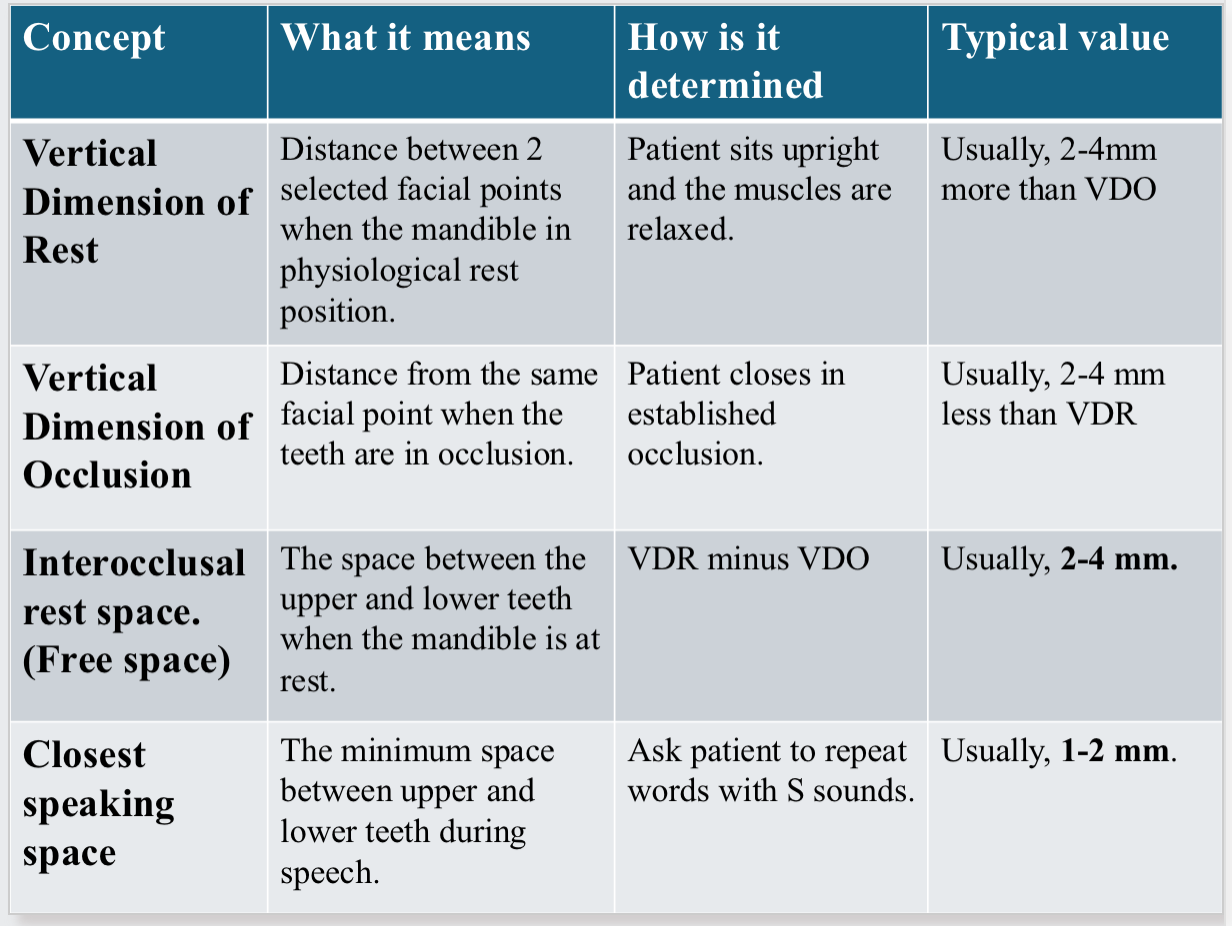
The distance between two selected anatomic or marked points (usually one on the tip of the nose and the other upon the chin), one on a fixed and one on a movable member
Simply put, it is the length of the face
Vertical Dimension of Occlusion (VDO)/Occlusal Vertical Dimension (OVD)
the distance measured between two points when the occluding members are in contact
Simply put: VD when teeth are in contact
Vertical Dimension of Rest (VDR)/ Physiologic Rest position, Rest Vertical Dimension (RVD)
the postural position of the mandible when an individual is resting comfortably in an upright position and the associated muscles are in a state of minimal contractual activity
Simply put: VD with no tooth contact and jaw is in rest position
Vertical Dimension of Rest (VDR)
The vertical separation of the jaws when the opening & closing muscles of the mandible are at rest in tonic contraction.
It is the length of the face when the mandible is in physiologic rest position.
In the absence of pathosis, the relation is constant throughout life.
The position can be accurately recorded and measured within acceptable limits.
It is used as an aid in the determination of the vertical dimension of occlusion
VDR + VDO

Interocclusal Rest Space (IRS)
Formerly known as “freeway space”, it is the difference between the vertical
dimension of rest and the vertical dimension of occlusion.
The distance between the occluding surfaces of the maxillary and mandibular teeth when the mandible is in its physiologic rest position.

Determining Vertical Dimensions
A. Mechanical Methods
Pre-extraction records, old photos.
Profile Radiograph.
Casts teeth in occlusion.
Facial Measurements.
B. Ridge Relations
Incisive Papilla to mandibular incisors.
Parallelism of the ridges
Measurement of the former dentures
C. Physiological Methods
Physiologic rest position
Phonetics
Esthetics
Swallowing threshold
Tactile sense and patient- perceived comfort
Mechanical Determination of VDO: profile radiographs
Very much used in research of VDO but not adequate for routine use due to radiation risks
Mechanical Determination of VDO: Casts of teeth in occlusion
gives an indication of the amount of space required between the ridges for teeth
Mechanical Determination of VDO: Facial measurements
one of the most popular methods; recording the distance from the chin to the base of the nose using calipers or a simple ruler
Mechanical Determination of VDO: Ridge relations
Incisive papilla to mandibular incisors – distance of papilla from the incisal edges of the mandibular anterior teeth on diagnostic casts averages approximately 4 mm in the natural dentition.

Parallelism of the ridges – removal of the teeth tend to leave the residual alveolar
ridges nearly parallel to each other (assuming extraction of all teeth was simultaneously done)
Measurement of former dentures; technique: Measure between the ridge crests of both dentures with a Boley gauge and determine the need for change in VDO through observations of the patient’s face.
Physiological determination of VDO: Physiological rest position
One of the most used methods
Ask patient to relax in an upright position with the head unsupported.
With the wax rims in place, the patient is asked to swallow, let the jaw relax and the
lips are parted to reveal the amount of space present between the occlusion rims.
Adjustments (addition or reduction) to the rims are made accordingly, to achieve a
good interocclusal rest space.
Factors affecting the Physiological rest position
the presence or absence of dentures or wax rims these can affect the rest position.
rapid adaptation may take place after changing the VD, leading to another rest position and creation of a new IRS.
The use of the rest position alone is not a reliable basis in determining maxillomandibular relationships.
Physiological determination of VDO: Phonetics
Another widely used method preferred most by clinicians
Tests include listening to speech sound production and observing the relationships of teeth, tongue, denture base and lips
The production of ”Ch" , "S" , and "J” sounds brings the patients anterior teeth close together
Physiological determination of VDO: Phonetics- Closest Speaking Space
Is an important concept used to assess the VD and the position of anterior teeth.
Closest Speaking space is the smallest distance between the upper and lower teeth when a person is speaking, especially during pronunciation of sounds such as S, Z, and Sh.
When the patient says words containing S sounds, the mandibular incisors move very close to the maxillary incisors but normally should not touch .
Typical space approximately 1-2 mm between the incisal edges of the of the upper and lower anterior teeth
This relationship must be taken in context to a constant or fixed position when speaking the sibilant sounds. It is believed that the ” S “ position remains constant throughout a person's life because it is a functional muscular position not influenced by hard structures like teeth in contact. Whether a patient is dentate or edentulous, the position should not change.
However, it has been reported and demonstrated that a person's oral muscle memory can be reprogrammed into a new VDO. Nevertheless, to prevent iatrogenic TMD, we should not change someone's VDO but instead restore it which brings us back to hopefully the same position that the patient originally had as a dentate patient.
Needless to say, that you should always satisfy other factors like esthetics, profile, physiologic rest position, etc
If you get a patient that you know has reprogrammed oral musculature, but you definitely see esthetic or facial profile issues, you can have the patient wear your rims for a certain amount of time to see if they can reprogram themselves back to where esthetics and phonetics would harmonize.
With a fixed "S" position in mind, if you were to make short occlusal rims resulting in a larger speaking space, then the patient would close farther into a decreased VDO. On the other hand, if you built tall rims resulting in rim contact even before the patient says "S", then you can expect your VDO to be too much
Why the Closet speaking space is important in complete dentures?
It help to evaluate whether the vertical dimension of occlusion (VDO) and anterior tooth position are correct.
It is not the same as the Interocclusal rest space!!
Interocclusal rest space = static position
Closest speaking space = functional position
Technique (Silverman technique)
during the pronunciation of the “s” sound, the interincisal separation, vertical distance, should average 1 to 2 mm.
1. Seat both maxillary and mandibular wax rims
2. Have patient say “yes”, “Mississippi”, or count quickly from 1 to 10 or 60 to 70
3. Observe approximation of wax rims at the “S” part:
a. If there is 1.0 to 1.5 mm of space between rims, VDO is correct – this is IDEAL
b. If space is greater than ideal amount, speech will sound altered.
c. If space is non-existent, teeth may contact during speech/clicking of denture, lisping or discomfort
Esthetics
Vertical relation of the mandible to the maxilla affects esthetics
The contour of the lips depends on their intrinsic structure and the support behind them
Technique: Labial surfaces of wax rims must be contoured to closely simulate the anteroposterior tooth positions and the contour of the base of the denture.
Problem: Recent evidence suggest this method to be unreliable thus should be used only in combination with other methods
Swallowing threshold
The position of the mandible at the beginning of the swallowing act used as a guide to determine VDO
Theory: teeth come together with very light contact at this phase of the cycle
Technique: Build soft cones of wax on mandibular record base to an excessive VDO and ask patient to swallow reducing the height of the cones.
Problem: inconsistent results.
Tactile sense and patient-perceived comfort
Technique: An adjustable central bearing screw is attached to one of the
occlusion rims, and a central bearing plate is attached to the other.
Technique: The central bearing screw is adjusted to an excessive VDO and slowly brought down until the patient indicates that the jaws are closing too far. Adjustments are reversed alternately until the height is comfortable to the patient
Problem: Having foreign objects in the palate and tongue space and inconsistent
results.
overall
No one method for determining rest position can be accepted as being valid for all patients.
Therefore, it is advisable to use several methods and compare the results.
Which technique do we use at UDMSOD?
Usually, a combination of the physiologic rest position, verified by Silverman’s “closest speaking space” technique and a check on the patient’s esthetic profile.
Clinical procedure for obtaining VDO
Establishing the occlusal vertical dimension (Remember), you are ready for this phase only after the maxillary wax rim has been refined according to your landmarks
Establishing the vertical dimension
Place the patient in an upright position looking straight ahead (or not).
Place marks on the tip of nose and the tip of the chin, on the greatest height of
curvature.
Make sure the chin is unstrained
Insert maxillary record base ONLY.
Instruct patient to lick lips and swallow.
Mandible comes to rest position.
Measure the distance between reference points.
Soften mandibular wax rim with hot spatula
Temper in water bath
Insert mandibular record base
Have patient bite down on the softened wax rim
Repeat until patient is at previously determined VDO position.
VDR - (2-4mm) = VDO
Let wax cool down.
Smooth the wax rims making sure both are flush with each other (right photo).
Confirm with closest speaking space technique to look for that 1.0-1.5 mm space on the letter “S“.
If there is a discrepancy in VDO, adjust accordingly!!
adjust the maxillary wax rim bc you have fixed points
If satisfied, proceed with determining centric relation.
Problems with insufficient IRS results in:
Clicking of the teeth.
Facial distortion, tense strained. appearance.
Difficulty closing lips.
Difficulty swallowing.
Soreness and discomfort under the denture.
Increased ridge resorption due to Trauma.
Problems with excessive IRS results in:
Establishing the vertical dimension
Reduced inter-arch distance when the teeth are in occlusion
Overclosure is potentially damaging to the TMJ
Normal tongue space is limited
Facial distortion, chin is closer to nose, commissure of the lips turns down, the lips lose their fullness
Muscles of facial expression lose their tonicity
Face appears flabby
Angular cheilitis is sometimes attributed to overclosure
overview chart
Summary of registering Vertical Dimension of Occlusion
Customize maxillary wax rim first to follow natural tooth positions.
Proper lip support.
Appropriate incisal edge position.
Occlusal plane parallelism to interpupillary line and ala-tragus line using Fox Plane.
Adjust mandibular rim flush with maxillary rim.
Test for proper VDO using different techniques (physiologic rest space, closest speaking space, external profile, etc.) and adjust mandibular rim height accordingly.