Session 1: Cell structure
 Call Kai
 Learn
 Practice Test
 Spaced Repetition
 Match
 Flashcards
 Knowt Play
1/15

There's no tags or description
Looks like no tags are added yet.
16 Terms
TEM (Transmission Electron microscope )
Beam of electrons transmitted through specimen to form an image
Specimen is very thin, 100nm
Electrons interact with specimen and image captured
High resolution
Interpreting E.M.
Light areas: attract fewer electrons
Dark areas: Electron dense
Phospholipid Bilayer:
Amphipathic: A molecule with both hydrophobic and hydrophillic parts
To form a bilayer
Proteins on membranes:
All membranes contain proteins that can move in the bilayer => Fluid Mosaic Model
Some proteins are attached to cytoskeleton
Others are glycosylated (have short polysaccharide chains)
KEY: Eukaryotic cells contain many types of membranes (RER is single convoluted layer, plasma membrane contains triple layer)
About the cell membrane:
Glycocalyx:
Sugar coat formed by glycoproteins and glycolipids.
Involved in protection, cell recognition, adhesion, and immune responses (the glycoproteins and glycolipids are recognised by the bodys immune system => immunogenicity)
Functions of the plasma membrane:
Exocytosis => Exit so the vesicle forms a membrane with the cell and leaves
Endocytosis +> Entering so the vesicle fuses its membrane with the cell and enters

Nucleus
Contains DNA, RNA and nucleoproteins
TEM reveals electron dense heterochromatin (DNA and associated nucleoproteins not active in RNA synthesis) => coiled structure // Electron-lucent (light) euchromatin (active in RNA synthesis) => uncoiled
Inactive cells = small nuclei containing heterochromatin
Active cells = large nuclei containing euchromatin
Nuclei not present in differentiated cells such as erythrocytes (most common RBCs), lens fibre cells
Nuclear envelope:
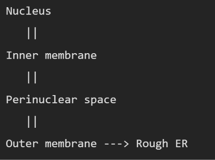
Has inner and outer membrane
The membrane of nucleus connects directly to the membrane of the ER
Nuclear pores that allow transport of molecules
RER (Rough endoplasmic reticulum)
Contain interconnecting set of membranes, vesicles and cisternae (flattened sacs)
Ribosomes on surface, undergoing protein synthesis
RER = Flattened + Ribosomes + protein production
SER (Smooth endoplasmic reticulum)
SER = Tubular + no Ribosomes + lipid production
Site of steroid production (Cortisol, Oestrogen, progesterone), cells that make a lot of steroid hormones = lots of SER
Golgi apparatus:
Proteins move from nucleus to the glogi at the cis face (smaller face) and travel to the trans face (larger face), the proteins then transport out to the lysosomes.
CIS (COMES IN) TRANS (TRANSPORT OUT)

Mitrocondria:
Large numbers in liver and skeletal muscles
Mitrocondria can divide => have their own genetic information

Lysosomes:
Produced by the Golgi apparatus
They digest and re-use carbohydrates
Contain acid hydrolases at pH 5 (nucleases, proteases…)
Lysosomes membranes are highly glycosylated (lots of carbohydrate chains)
Wide variety of shapes => spherical or oval
How do Lysosomes digest ?
Where does intracellular digestion occur?
Secondary lysosomes => Where the lysosomal enzymes meet the material that needs to be digested

Peroxisomes:
Small membrane-bound organelles found free in the cytoplasm
They react with oxygen, and form Hydrogen peroxide
H2O2 is a reactive and harmful chemical
However, catalase uses H2O2 to detoxify other substances
Abundant in liver and kidney, to remove toxins from body.
Neutrophils (white blood cell) + H2O2 = to kill bacteria
Cytoskeleton:
The skeleton of the cell
allows to maintain, and allow flexibility of cell

Microfilaments:
Microfilaments: Thin and flexible fibres involved in shape and movement, made from double stranded helix of 2 actin proteins joined together.
They maintain the cells’ shape
Allow movement (assemble = actin molecules join the filament) // (disassemble = actin molecules leave the filament)

Microtubules:
A microtubule is a hollow protein tube inside the cell, part of the cytoskeleton, so helps organise cell.
Main function: To provide tracks along which things can be transported through the cell. Motor proteins (Kinesin and Dynein), which have attached cargo in a vesicle and they walk along the microtubule.
Microtubules are made from alpha-tubulin and beta-tubulin.
Microtubules originate from centrosomes.