Cartilage and Bone
 Call Kai
 Learn
 Practice Test
 Spaced Repetition
 Match
 Flashcards
 Knowt Play
1/185

There's no tags or description
Looks like no tags are added yet.
186 Terms
Cartilage: main cell type
Chondrocytes (sit in lacunae)
Cartilage: % of tissue that is ECM
95% extracellular matrix
Cartilage blood supply / how nutrients arrive
Avascular; nutrients reach chondrocytes by diffusion through the matrix
3 types of cartilage
Hyaline, elastic, fibrocartilage
Hyaline cartilage: locations
Fetal skeleton, articular surfaces (knee), respiratory tract (trachea/bronchi), costal cartilage
Hyaline cartilage: function
Resists compression
Hyaline cartilage: matrix
Collagen II + aggrecan
Hyaline cartilage: perichondrium?
Yes, EXCEPT articular cartilage (no perichondrium)
Elastic cartilage: locations
External ear, larynx/epiglottis
Elastic cartilage: function
Flexible support
Elastic cartilage: matrix
Collagen II + aggrecan + elastic fibers
Elastic cartilage: perichondrium?
Yes
Elastic cartilage: special stain for elastic fibers
Orcein (fibers appear dark brown/branching)
Fibrocartilage: locations
Intervertebral disc (IVD), meniscus, TMJ
Fibrocartilage: function
Resists compression AND shear
Fibrocartilage: matrix
Collagen I + collagen II, versican, aggrecan
Fibrocartilage: perichondrium?
No
Fibrocartilage: histological appearance
Mix of dense CT (collagen I, fibroblast-like rows) + hyaline cartilage (collagen II, chondrocytes)
Which cartilage types lack a perichondrium?
Fibrocartilage and articular hyaline cartilage
Which collagen is common to ALL cartilage types?
Collagen II
Which cartilage type contains collagen I?
Fibrocartilage
Perichondrium: composition / staining
Dense CT rich in collagen I; stains red/pink
Appositional growth (cartilage)
New cartilage added at the surface from perichondrium cells → growth in girth (width)
Interstitial growth (cartilage)
Chondrocytes divide within the matrix → growth in length and girth
Isogenous group
Cluster of chondrocytes that are descendants of a single cell (from interstitial growth)
Territorial matrix
Matrix immediately around an isogenous group; strongly basophilic (rich in proteoglycan)
Interterritorial matrix
Matrix between isogenous groups; less basophilic
Lacuna (cartilage)
Space in the matrix occupied by a chondrocyte
White space around chondrocytes in lacunae
Shrinkage artifact from tissue processing
Chondrocyte organelles (EM)
Rich in Golgi, rough ER, and mitochondria (active matrix synthesis)
Proteoglycan charge & staining
Negatively charged → basophilic (blue/purple)
Collagen charge & staining
Positively charged → eosinophilic (pink)
Aggrecan structure
Core protein with many GAG chains attached ('test-tube brush')
Aggrecan function
GAGs hold water → resists compression
Nucleus pulposus
Jelly-like center of the intervertebral disc
Anulus fibrosus
Ring of fibrocartilage surrounding the nucleus pulposus
Nucleus pulposus vs anulus fibrosus
Nucleus pulposus = jelly center (NOT fibrocartilage); anulus fibrosus = outer ring of fibrocartilage
Disc herniation
Anulus fibrosus ruptures → nucleus pulposus protrudes and presses on a spinal nerve
Articular cartilage: type & key feature
Hyaline cartilage without perichondrium (also no nerves or blood vessels)
Arthritis (in this lecture)
Degradation of articular cartilage
Cartilage vs bone: matrix
Cartilage = unmineralized (collagen II, proteoglycan); Bone = mineralized (collagen I, proteoglycan, hydroxyapatite)
Hydroxyapatite formula
Ca10(PO4)6(OH)2
Cartilage vs bone: function
Cartilage = cushion & support; Bone = support + Ca/P storage
Cartilage vs bone: blood supply
Cartilage = avascular; Bone = vascular
Cartilage vs bone: growth
Cartilage = appositional + interstitial; Bone = appositional ONLY
Main collagen type in bone
Collagen I
Bone as an organ: components
Bone tissue + hemopoietic tissue (marrow) + fat + blood vessels + nerves + articular cartilage
Epiphysis
End of a long bone
Diaphysis
Shaft of a long bone
Metaphysis
Region between epiphysis and diaphysis
Periosteum
Outer CT lining of bone (contains osteoprogenitors)
Endosteum
Inner lining of bone facing the marrow cavity
2 types of bone tissue
Spongy (cancellous/trabecular) and compact (cortical)
Spongy bone: features
Lamellated matrix, NO osteons/Haversian lamellae, nutrients by diffusion from marrow, marrow fat between trabeculae
Does spongy bone have osteons?
No
Osteon (Haversian system)
Structural unit of compact bone: concentric lamellae around a Haversian canal
Haversian canal
Central canal of an osteon; runs longitudinally (vertical); contains blood vessels/nerves
Volkmann's canal
Transverse (perforating) canal; connects Haversian canals of osteons, and the inside and outside of the bone
Where are transverse (Volkmann's) canals found?
Compact bone
Concentric lamellae
Rings of matrix within an osteon around the Haversian canal
Interstitial lamellae
Remnants of old osteons between intact osteons
Outer circumferential lamellae
Lamellae just under the periosteum, encircling the whole bone
Inner circumferential lamellae
Lamellae lining the marrow cavity, just under the endosteum
Lacuna (bone)
Space in the matrix occupied by an osteocyte
Canaliculi
Tiny channels between lacunae containing osteocyte processes
How do osteocytes communicate?
Through cytoplasmic processes in canaliculi, connected by gap junctions
Osteoid
Unmineralized bone matrix laid down first by osteoblasts, then mineralized
Ground section of bone: dark spots with radiating lines
Lacunae (spaces osteocytes occupied) with canaliculi
4 bone cell types
Osteoprogenitor, osteoblast, osteocyte, osteoclast
Osteoprogenitor cells: location
Periosteum and endosteum
Osteoblast: function
Secretes osteoid: collagen I, osteocalcin, osteopontin, alkaline phosphatase (ALP), bone sialoprotein (BSP)
Osteoblast: relation to matrix
Lines the bone surface; NOT surrounded by matrix
Osteocyte
Mature bone cell surrounded by matrix in a lacuna; star-shaped (processes); maintains matrix
Osteoclast: appearance
Large, multinucleated cell on the bone surface
Osteoclast: origin
Monocyte lineage
Howship's lacuna
Resorption pit on the bone surface created by an osteoclast
Osteoclast resorption: 3 steps
1) Acidify: pump out H+ to dissolve mineral
2) Lysosomal enzymes degrade organic matrix
3) Clean up by endocytosis
What does an osteoclast pump out to dissolve bone mineral?
H+ (protons) — acidification
Why do osteoclasts have many mitochondria?
Energy (ATP) for proton pumping/resorption
Bone remodeling
Ongoing resorption (osteoclasts) + formation (osteoblasts); the two cells communicate
Effect of mechanical load on remodeling
Faster remodeling / more bone formation
Osteoporosis
Low bone mass → fragile bone; caused e.g. by high-dose corticosteroids; partly genetic; helped by weight-bearing exercise
Osteopetrosis
Increased bone mass due to defective osteoclasts → still fragile; unerupted teeth
Dental sign of osteopetrosis
Unerupted teeth
Paget disease
Overactive osteoclasts → increased remodeling → softer, weaker bone
Which disease: defective osteoclasts?
Osteopetrosis
Which disease: overactive osteoclasts, increased remodeling?
Paget disease

Identify the cartilage
Hyaline cartilage — not bone, no obvious fibers, not elastic
Which statements about cartilage are INCORRECT?

A, D, F
Which pairings are INCORRECT?

C, D, E
Which of the following incorrectly describe this tissue?
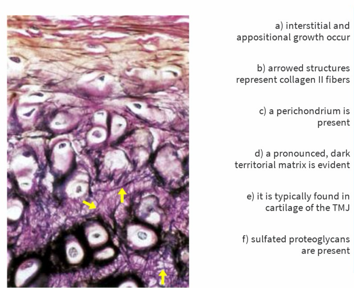
B, E
elastic cartilage
Inappropriately matched pair?

B
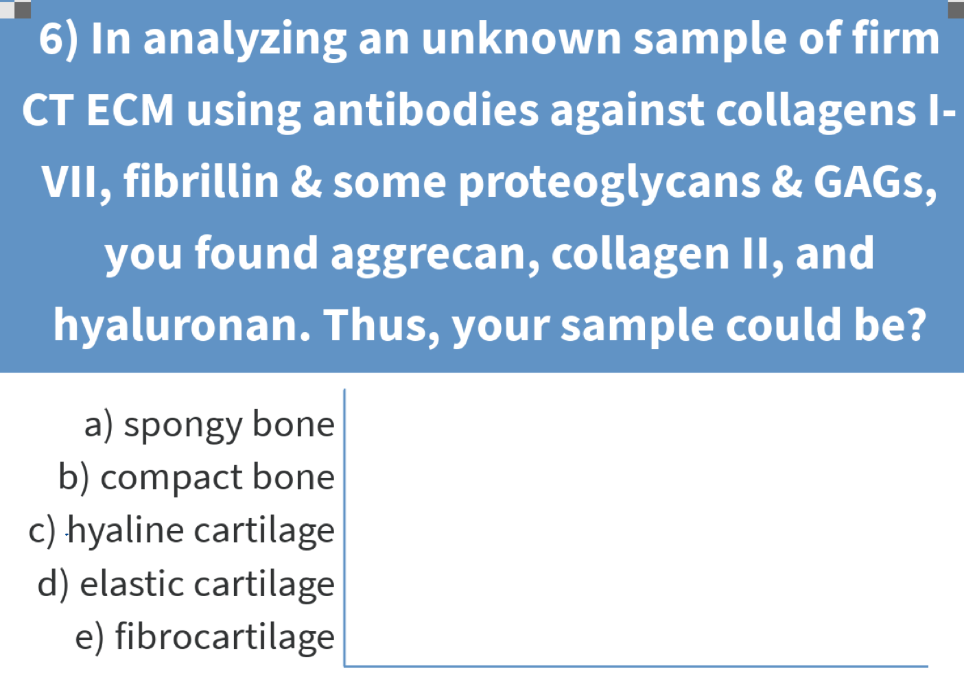
Antibodies find aggrecan, collagen II, hyaluronan. What is the sample?
C
Transverse canals are found at A, B, or C?

B
What are the arrows pointing at (ground section)?

A
not live osteocytes (dead in ground section)
Which INCORRECTLY describes this tissue?
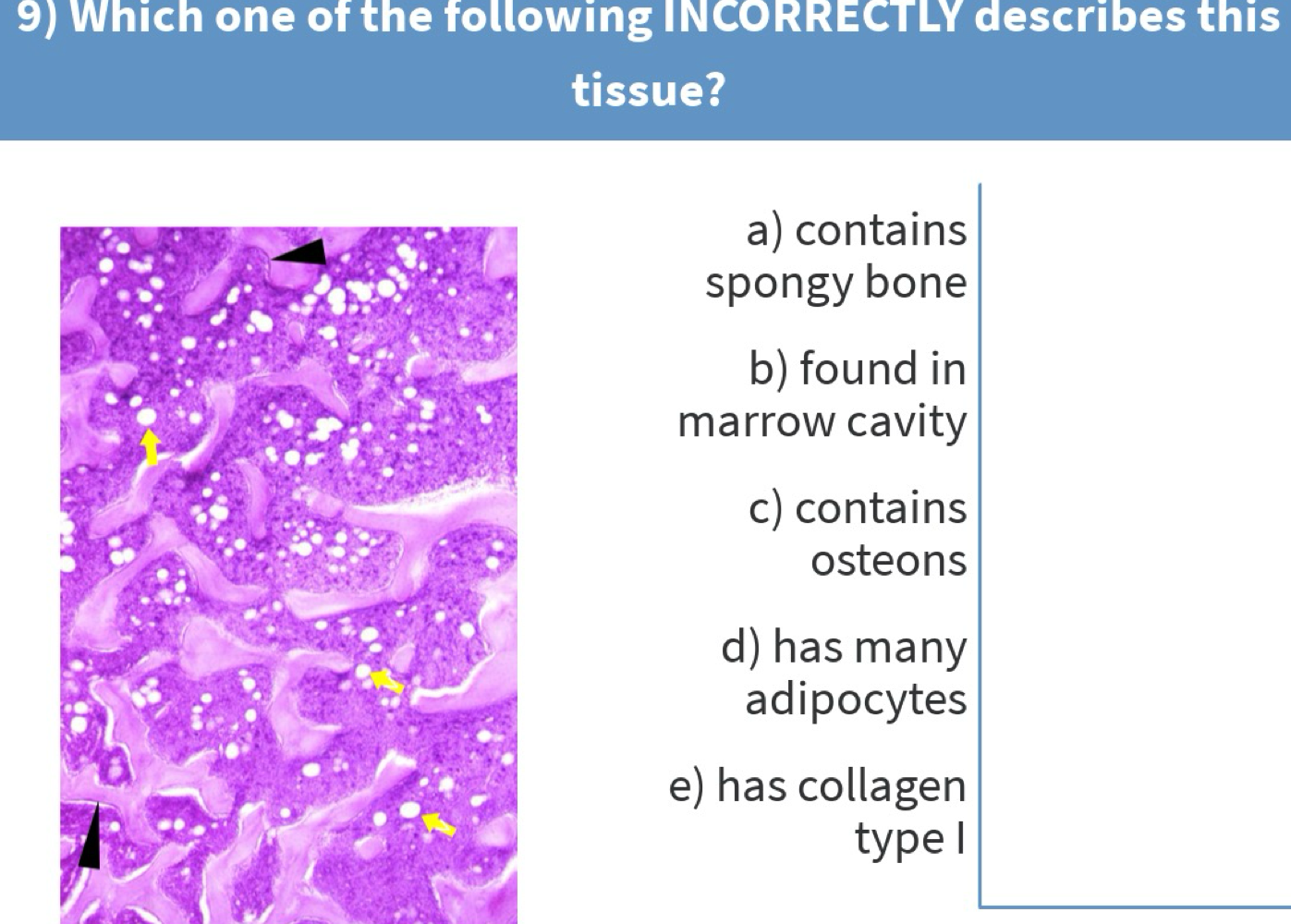
c) Contains osteons
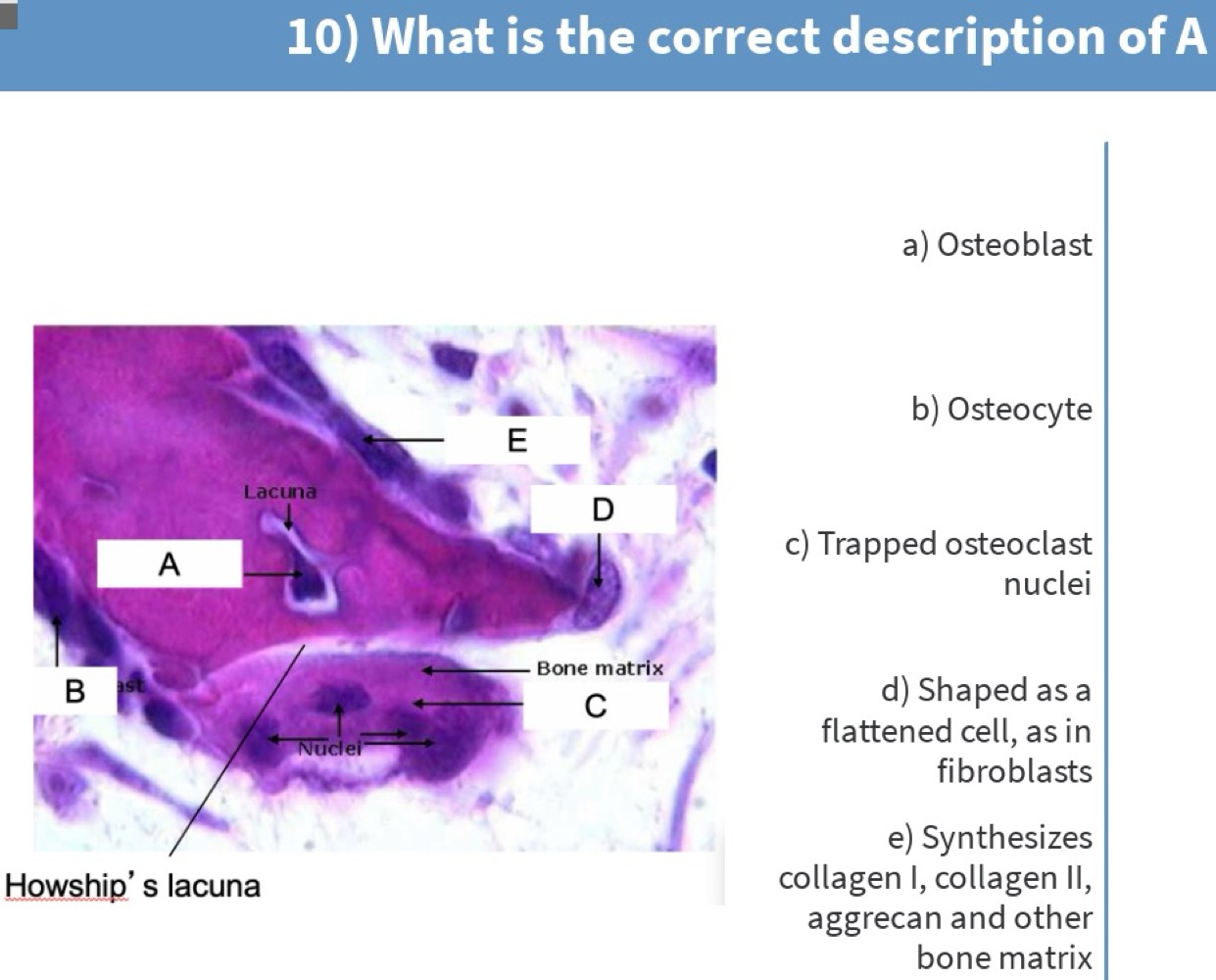
b) Osteocyte

e) Communicates with osteoblasts to maintain bone homeostasis
What is known about osteoporosis?

B, C
The 3 types of cartilage with locations?
Hyaline: fetal, articular, respiratory, costal.
Elastic: ear, larynx (epiglottis).
Fibrocartilage: IVD, meniscus, TMJ