Bones of the pelvic girdle and lower extremity
 Call Kai
 Learn
Learn Practice Test
 Spaced Repetition
 Match
Match Flashcards
 Knowt Play
1/63

There's no tags or description
Looks like no tags are added yet.
Name | Mastery | Learn | Test | Matching | Spaced | Call with Kai | Chat |
|---|
No analytics yet
Send a link to your students to track their progress
64 Terms

Ilium
forms the superior region of the hip bone and part of the acetabular surface


iliac crest, iliac fossa, arcuate line, sacroiliac joint, greater sciatic notch
Marking of the ilium


Iliac crest
superior border of the ilium, beginning at the sacrum and ending on the lateral aspect of the hip.


Iliac fossa
large fossa on the anteromedial surface, inferior to the iliac crest

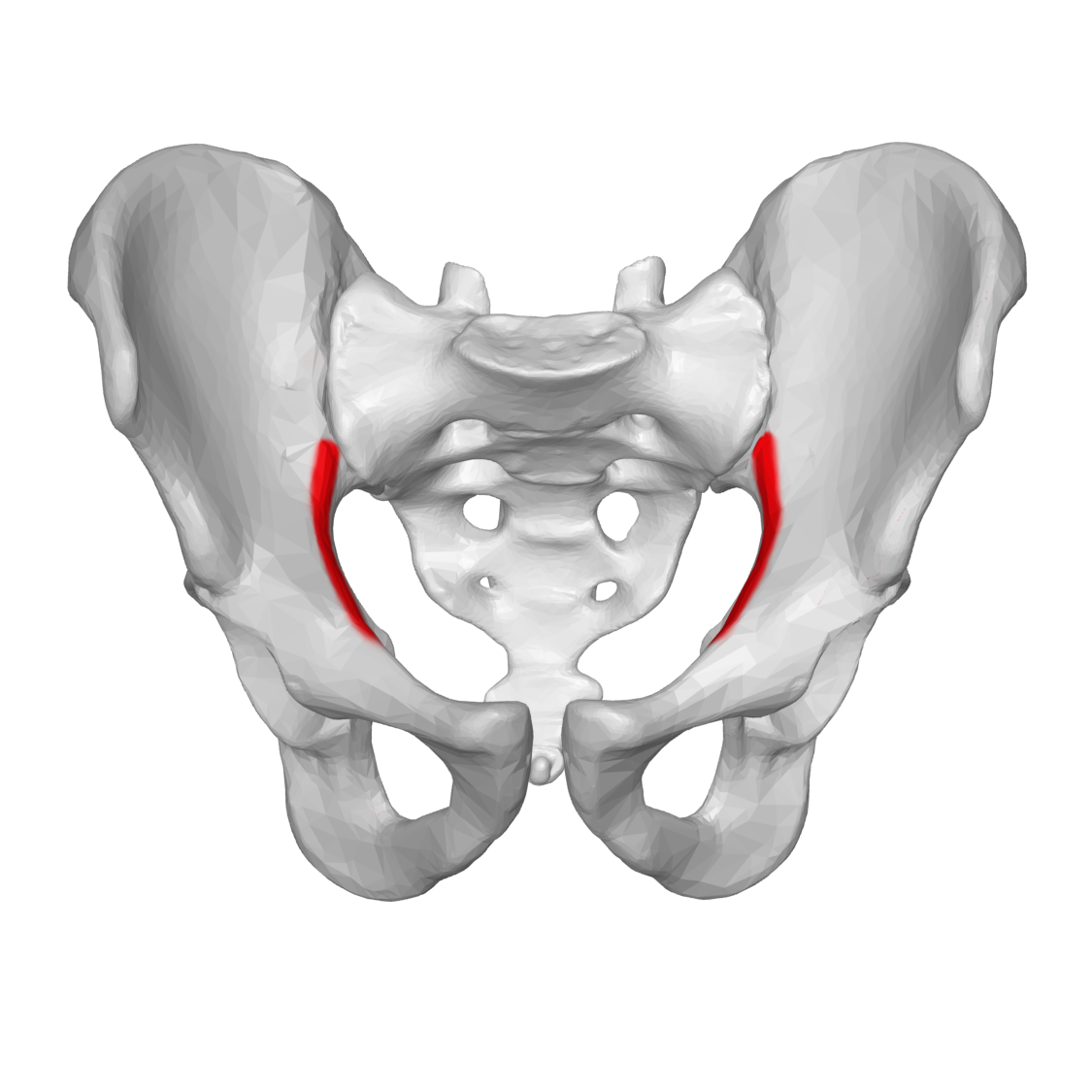
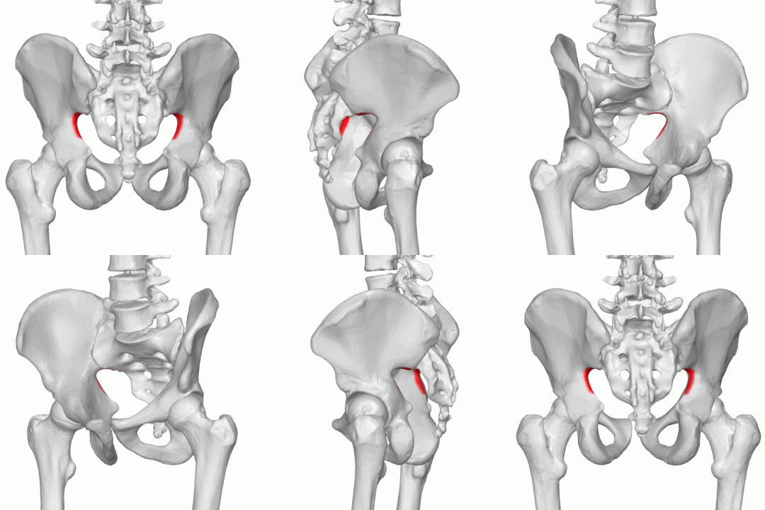
Arcuate line
oblique line b/w the ilium and ischium that composes the iliac part of the linea terminalis (pelvic brim) of the bony pelvis


Sacroiliac joint
where each hip bone articulates posteriorly w/ the sacrum


Greater sciatic notch
deep notch on the posterior surface of the ilium inferior to the posterior inferior iliac spine

Ischium
fuses w/ the ilium near the superior and posterior margins of the acetabulum

Markings of the ischium
ischial tuberosity, ischial spine, lesser sciatic notch

Ischial tuberosity
large, rough projection on the posterior, inferior surface of the ischium; attachment site for hamstring muscles


Ischial spine
small, sharp spine on the posterior aspect of the ischium b/w the greater and lesser sciatic notches


lesser sciatic notch
notch located immediately inferior to the ischial spine on the posterior surface of the ilium


Pubis
fuses w/ ilium and ischium at the acetabulum

Rami, pubic symphysis, pubic arch
Markings of the pubis

Rami
extends from the ischial tuberosity toward its anterior fusion w/ the pubis

Pubic symphysis
denotes site of articulation b/w the pubic bones


Pubic arch
angle formed when the left and right pubic bones are aligned at their symphysial surfaces. Females have a wider arch than males do

Acetabulum, obturator foramen
markings associated w/ os coxa when fused

Os coxa
hip bone. has left and right


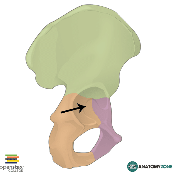
Acetabulum
deep bony socket; articulates w/ the head of the femur

Obturator foramen
large, oval hole in the inferior part of the hip bone


Femur
2 bones. longest, strongest, and heaviest bone in the body. thigh bone

head, neck, greater trochanter, lesser trochanter, intertrochanteric crest, linea aspera, lateral condyle, medial condyle, intercondylar fossa, patellar surface
Markings of the femur


Head of the femur
large, spherical structure on the proximal end of the femur; articulates w/ the acetabulum of hip bone

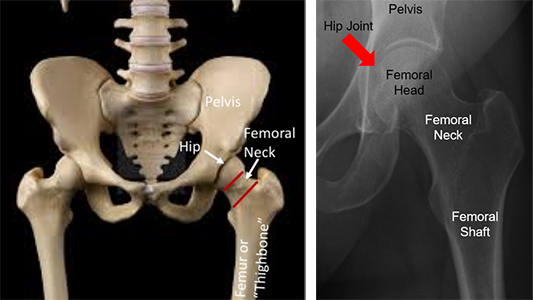
Neck of the femur
narrow portion where the head meets the shaft of the femur; part of the femur that’s fractured in a “broken hip”


Greater trochanter
very large projection on the proximal end of the femur; articulates w/ the acetabulum of hip bone


Lesser trochanter
large projection on the proximal, medial surface; attachment site of iliopsoas muscle


Intertrochanteric crest
large ridge that runs b/w the greater and lesser trochanters on the posterior surface; separates shaft and neck of femur


Linea aspera
rough line that runs vertically along the posterior surface of the shaft


Lateral condyle of the femur
large, rounded surface; articulates w/ the lateral condyle of the tibia


Medial condyle of the femur
rounded surface; articulates w/ the medial condyle of the tibia


Intercondylar fossa
depression on the distal end of the femur b/w the 2 condyles


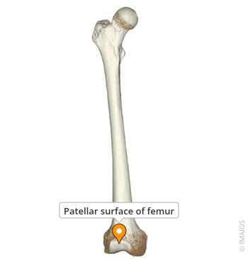
Patellar surface
smooth depression on the distal, anterior surface; articulates w/ the patella

Patella
2 bones. kneecap. large, roughly triangular bone. no markings to know


Tibia
2 bones. medially placed bone and the only weight bearing bone of the crural region


Medial condyle, lateral condyle, intercondylar eminence, tibial tuberosity, medial malleolus, anterior crest
Marking of the tibia

Medial condyle of the tibia
large, flat surface on the proximal, medial surface; articulates w/ the medial condyle of the femur


Lateral condyle of tibia
large, flat surface on the proximal, lateral surface; articulates w/ the lateral condyle of the femur


Intercondylar eminence
prominent projection b/w the 2 condyles on the proximal surface


Tibial tuberosity
projection on the proximal, anterior surface; attachment site for the patellar ligament (quadriceps femoris muscle attachment)


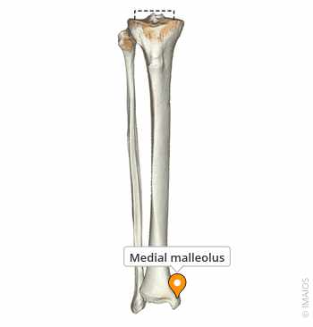
Medial malleolus
projection on the distal, medial surface; prominent bony landmark at the ankle

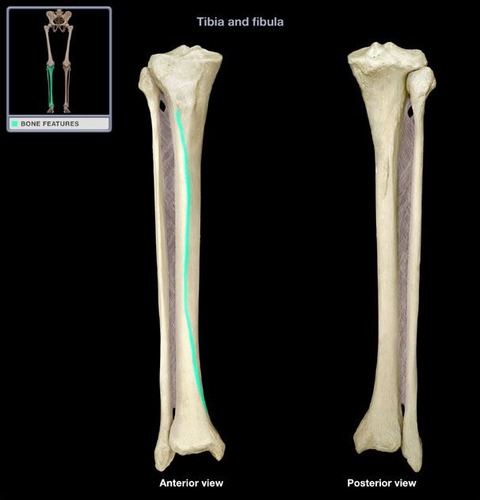
Anterior crest
aka the shin. ridge on the anterior surface extending distally from the tibial tuberosity

Fibula
2 bones. long, thin, lateral bone of the leg. doesn’t bear any weight

Head, lateral malleollus
Markings of the fibula

Head of the fibula
rounded proximal end of the fibula


Lateral malleolus
projection on the distal, lateral surface; prominent bony landmark at the ankle


Tarsals
makes up ankle and proximal foot
calcaneus, cuboid, intermediate cuneiform, lateral cuneiform, medial cuneiform, navicular, talus
Names of the tarsals

Calcaneus
bone that forms the heel of the foot; attachment site for calcaneal tendon


Cuboid
cube-shaped bone; located at the base of the 4th metatarsal

Intermediate cuneiform
wedge-shaped bone; located at the base of the 2nd metatarsal

Lateral cuneiform
wedge-shaped bone; located at the base of the 1st metatarsal

Navicular
bone shaped like a boat; located just anterior to the talus


Talus
major weight-bearing bone of the ankle; articulates w'/ the tibia and fibula


Metatarsals
5 bones in the arch of the foot; each bone consists of a head, body, and base


Phalanges
bones of the toes. all of the toes have 3 phalanges except the big toe

Proximal phalanges
phalanx closest to metatarsal bones


Intermediate phalanges
middle phalanx

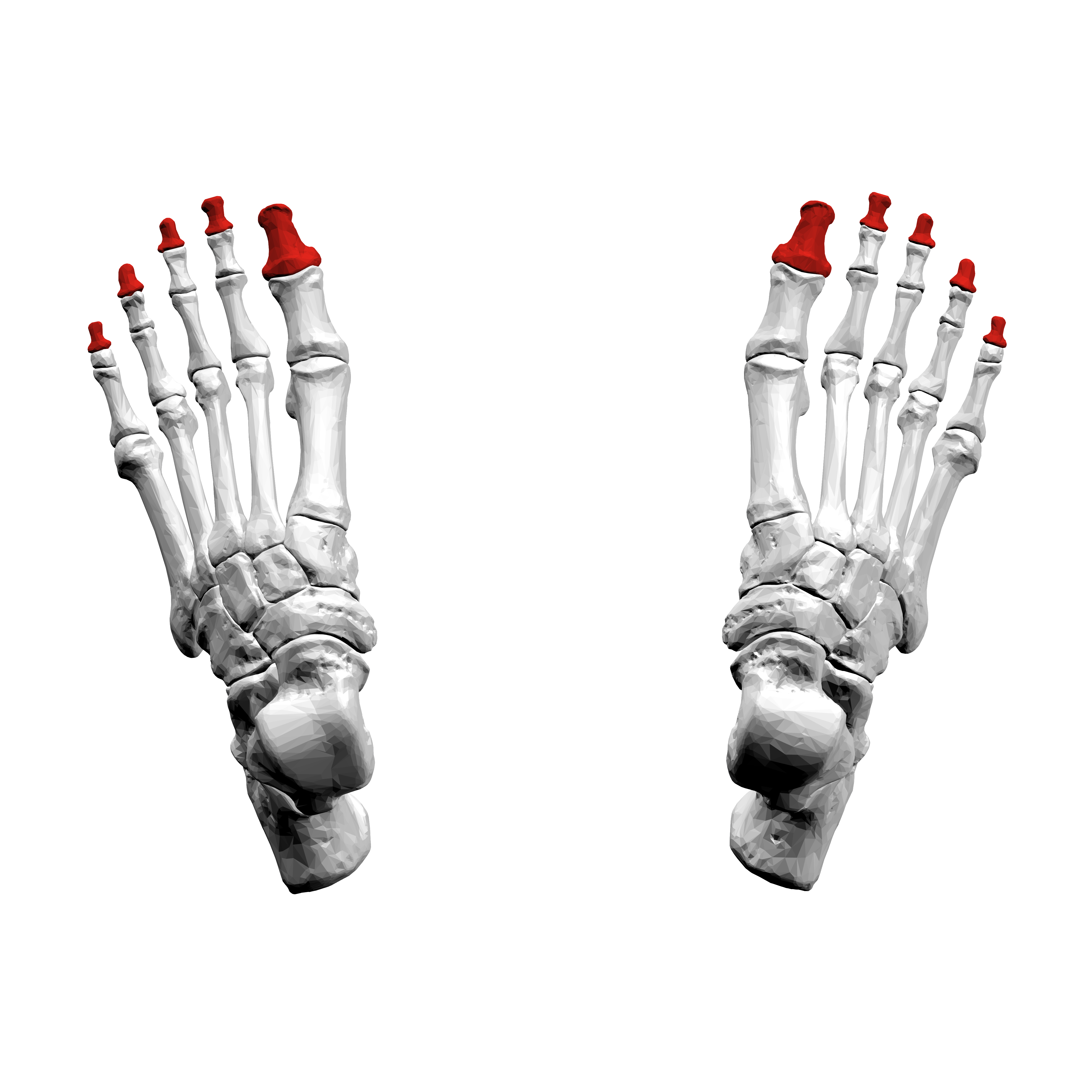
Distal phalanges
small, cone-shaped phalanx forming the end of the toes

Hallux
big toe
medial longitudinal, lateral longitudinal, distal phalanges
Markings of the arches of the foot

Medial longitudinal
highest of the 3 arches. extends from the heel to the big toe

Lateral longitudinal
not as high as medial arch so this part of the foot does contribute to the footprint. extends b/w the little toe and the heel
Transverse
runs perpendicular to the longitudinal arches. formed from the distal row of the tarsals and the bases of all 5 metatarsals