Chiolo Lecture 1
 Call Kai
 Learn
Learn Practice Test
 Spaced Repetition
 Match
Match Flashcards
 Knowt Play
1/27

Earn XP
Description and Tags
Name | Mastery | Learn | Test | Matching | Spaced | Call with Kai | Chat |
|---|
No analytics yet
Send a link to your students to track their progress
28 Terms
Chromosome
SINGLE DNA molecules & proteins

Bacterial chromosomes
Are usually circular
Occupy a nucleoli that LACKS a surrounding nuclear membrane
* Bacteria can also contain smaller circular plasmids
Doesn't have telomeres
Eukaryotic Nuclear Chromosomes
Are linear
Have telomeres at each end
Reside inside a membrane-bound nucleus
Each chromosome contains one long DNA double helix association with proteins
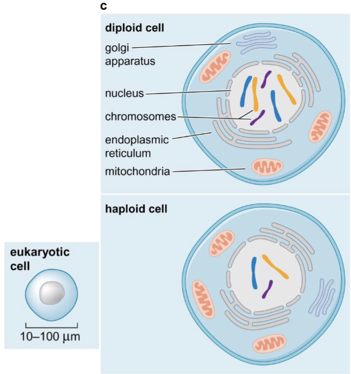
Diploid vs. Haploid Cells
Diploid cell = has 2 copies of each chromosome (one maternal & one paternal)
Haploid cell = only 1 copy of each chromosome…. example is a gamete
Different Types of DNA Sequences
Genes:
introns
exons
Inter-genes:
regulatory elements (ex: promoters)
genome-wide repeats (ex: transposons) **
micro-satellite repeats (ex: CACACACACA…) **
** = classes of repeated DNA
Gene Density
Genome size does NOT scale directly with the number of protein-coding genes… much of the larger human genome consists of regulatory DNA and repeated sequences, so human gene density is much lower (than E.coli)
more complex organisms = less gene density
E.coli =
E.coli genome is about 4.6 Mb and contains roughly 4,400 genes
High gene density, little inter-genic regions
Humans =
Human haploid genomes is about 3,200 Mb and has 20,000 protein-coding genes
Low gene density, longer inter-genic regions

Introns
Are removed during mRNA splicing… so not found in protein
Introns are NOT left“in”
A eukaryotic gene is transcribed into a primary transcript that contains both introns and exons… intron removal produced mature mRNA

DNA Sequence Percentages (in humans)
Genes (introns & exons) = 37.5%
Exons = <2%… protein-encoding exons account for less than 2%
Inter-genes = 62.5%
44% = genome-wide repeats (ex: transposons)… transposons make up a large fraction of genome
Repeated DNA (transposons, satellite repeats) are a large fraction of the human genome

“Beads on a string”
Looking at 10-nm fiber of chromatin structure… electron microscopy reveals repeating particles along DNA… these particles are nucleosomes… give extended chromatic a “beads-on-a-string” appearance
the bead corresponds mainly to DNA wrapped around a histone core
the connecting segment is linker DNA

Nucleosome
A nucleosome is composed of a histone core AND the DNA wrapped around it
the histone core is an octomer or “core”
is two layers, each divided into 4 parts
two copies each of: H2A… H2B… H3… H4
Core DNA = DNA wrapped around histone… about 146 base pairs
Nucleosome formation compacts DNA about 6-fold
Histones are very abundant
Histones are among the most evolutionary conserved eukaryotic proteins because their structural role is fundamental

Linker DNA vs. Core DNA
Core DNA
146 base pairs
wrapped around histone core
Linker DNA
20-60 base pairs
connects adjacent nucleosomes and varies in length

MNase Digestion
The experiment identifies the length of the DNA wrapped around a nucleosome and the linker DNA… the length the “core DNA” was determined to be 146 bp this way
(1) Microccocal nuclease (MNase) preferentially cuts exposed linker DNA… light digestion releases fragments containing 1, 2, 3, or more nucleosomes
(2) After proteins are removed, these fragments form a ladder separated by roughly 1 nucleosome repeat length
(3) More extensive digestion trims accessible DNA until the histone core protects about 146 bp
Histones (what are they?)
Small, positively charged (basic) proteins
Basic histones (positively charged) associate tightly and non-specifically with the DNA (negatively charged)
Contain many lysine (K) and Argenine (Ar) residues.. which carry positive charge at physiological pH
The opposite charge helps histones bind DNA strongly and with limited sequence specificity

Histones (structure)
H2A, H2B, H3, H4 histones contain a “histone fold domain” and an N-terminal tail
these core histones differ in size and sequence… but share a common general organization
Each core histone contains a histone-fold domain composed mainly of alpha helices… the domain mediates histone pairing and helps assemble the octomer (dimerization of histone pairs)
Also have distant N-terminal tails… act as regulatory regions

Histone-fold Domain vs. Histone Tail
Histone-fold domains stabilize the core
packed inside core, where create a stable scaffold for DNA wrapping
Tails have regularity functions
tails extend outward and remain accessible to modifying enzymes and binding proteins… protrude

DNA Wrapping
The DNA wraps around the histone octamer in a left-handed (left hand thumbs up) superhelical path
Histone tails (including those of H2B & H3) emerge between the DNA gyres
H2B & H3 histone tails direct DNA wrapping in a left-handed manner
What can post-translational modifications regulate?
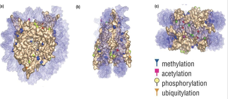
BOTH histone fold domains AND tails
Modifications can alter histone-DNA contacts, nucleosome-nucleosome interactions, or binding by regulatory proteins
Post-Translational Modifications
Cells covalently modify proteins after translation —> “P.A.M.U".”
Phosphorylation: often change protein activity or interactions
Acetylation: regulate interaction with other proteins
Methylation: regulate interaction with other proteins
Ubiquitination: attachment of a 76-AA (small) protein called ubiquitin to substrates in lysine (K) residues… some unqiuitin chains can target proteins for proteasomal degradation, while other ubiquitin signals regulate localization, function, and other features

Histone-fold Domain Modifications
Histone-fold domain modifications change the strength of interactions with DNA
Histone modifications can occur on residues in the structured core, including surfaces that contact DNA or other histones
A change in charge, size, or binding properties at these positions can strengthen or weaken histone-DNA association and affect nucleosome stability, positioning, or mobility
ex: phosphorylation causes addition of negative charge
ex: acetylation neutralizes positive charge of lysine
Histone Tail Modifications (DNA interactions)
Some histone tail modifications change the strength of interaction with DNA… alter chromatin interactions
Can weaken interactions with negatively charged DNA
Ex: acetylation neutralizes the positive charge of K (lysine)
Ex: phosphorylation adds negative charge
Enzymes add and remove these ‘marks’… chromatin can change dynamically during transcription, DNA replication, and DNA repair… called epigenetic regulation of genome

Epigenetic Regulation
Changes in gene function that do not require a change in DNA sequence
Ex: enzyme addition and removal of post-translational modification ‘marks’ which cause dynamic chromatin changes key ro the regulation of replication, transcription, and DNA repair
Histone Tail Modifications (protein interactions)
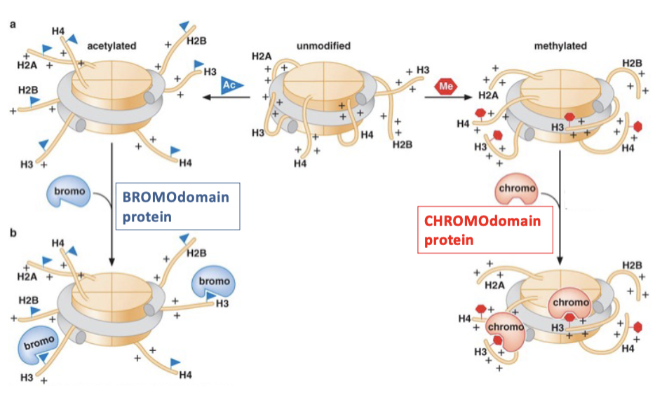
Histone tail modifications create platforms for recruiting specific proteins… can create substrates for the binding of regulatory proteins
Histone marks recruit reader proteins
Histone modifications can act as “docking sites”…
Bromodomains = recognize acetylated lysines (AB)
Chromodomains = recognize methylated lysines (CM)
Recruitment of proteins with these domains can then further alter the state of the chromatin… making it more or less accessible
Ex: to transcription, repair
What can histone tail modifications impact?
the strength of interaction with DNA… “dynamic chromatin changes”
protein recruitment… “docking sites”
Histone Variants
Histone variants = alternative versions of canonical histones… their incorporation can specify unique functions in the genome… can be incorporated into nucleosomes at special chromatin locations
CENP-A: an H3 variant found at centromeres… helps specify kinetochore assembly
H2AX: an H2A variant involved in the DNA-damage response… phosphorylated at sites of DNA damage

Chromatin Remodelers (site accessibility & more)
ATP - dependent chromatin remodelers control access/site accessibility
Chromatin-remodeling complexes use ATP hydrolysis to change histone-DNA contacts
slide nucleosome
eject nucleosome
exchange histone variants (dimer exchange)
Changing nucleosome position (slide/eject) exposes or occludes DNA-bindimg sites… therefore remodelers regulate access without changing underlying DNA sequence
OTHER: histone exchange, such as the replacement of H2A with the H2AX variant… not related to site accessibility

Chromatin Remodelers & Histone Modifiers (working together)
Chromatin remodelers cooperate with histone modifiers to change the chromatin status, altering chromatin accessibility
Mechanistic transition from relatively closed chromatin to more open chromatin during processes such as transcription or DNA repair (could also work opposite in other cases)
Ex:
1) Sequence-specific DNA binding protein can recruit a histone acetyletransferase… and then this acetylation of histone creates a binding site for bromodomain-containing proteins (recall A.B.)
2) Bromodomain protein then recruits a chromatin remodeler that slides or removes (ejects) nucleosomes… exposing additional binding sites for other DNA-binding proteins to occupy
3) so closed —> open… but sequence of events can vary by locus

Cancer Cells
Cancer cells lose the ability to regulate histone modifications… misregulation of these histone modifiers is frequently found in cancers
Cancer cells frequently carry mutations in chromatin regulators (ex: HDACs, HSMs, HATs, HMTs)… these changes can misrelate gene expression… resulting in dysregulated proliferation, differentiation, genome stability, and cell death

Cancer Therapies
Many cancer therapies target chromatin components… chromatin modifiers act as therapeutic targets… many compounds effective against cancer cells are inhibitors of chromatin modifiers
Understanding how chromatin regulators work = essential for interpreting their effectiveness in cancer therapies & roles in inducing secondary effects and cancer recurrences… altering chromatin state means altering access and thus transcription
