A&P CH. 16 - Special Mechanoreception & Photoreception
 Call Kai
 Learn
 Practice Test
 Spaced Repetition
 Match
 Flashcards
 Knowt Play
1/150

There's no tags or description
Looks like no tags are added yet.
151 Terms
What is mechanoreception?
Detection of mechanical stimuli, such as vibrations and movements

Which inner ear structure houses the receptive organs for hearing?
The cochlea

Which inner ear structures house the receptive organs for equilibrium?
The vestibule detects linear acceleration
the semicircular canals detect angular acceleration

What are the three main divisions of the ear?
Outer ear, middle ear, and inner ear

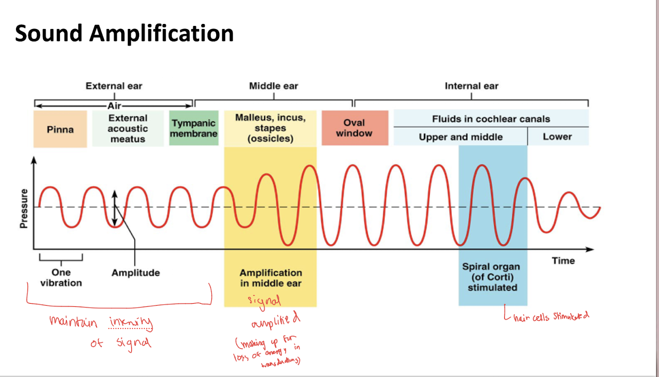
What is the function of the outer ear in hearing?
It collects sound waves and directs them toward the tympanic membrane


What is the function of the middle ear?
The tympanic membrane and auditory ossicles transmit and amplify vibrations toward the oval window.


Approximately how much do the auditory ossicles amplify sound, according to the lecture?
Approximately 20 times

What is the function of the inner ear in hearing?
Its fluid-filled labyrinths contain the sensory receptors that transduce mechanical stimuli
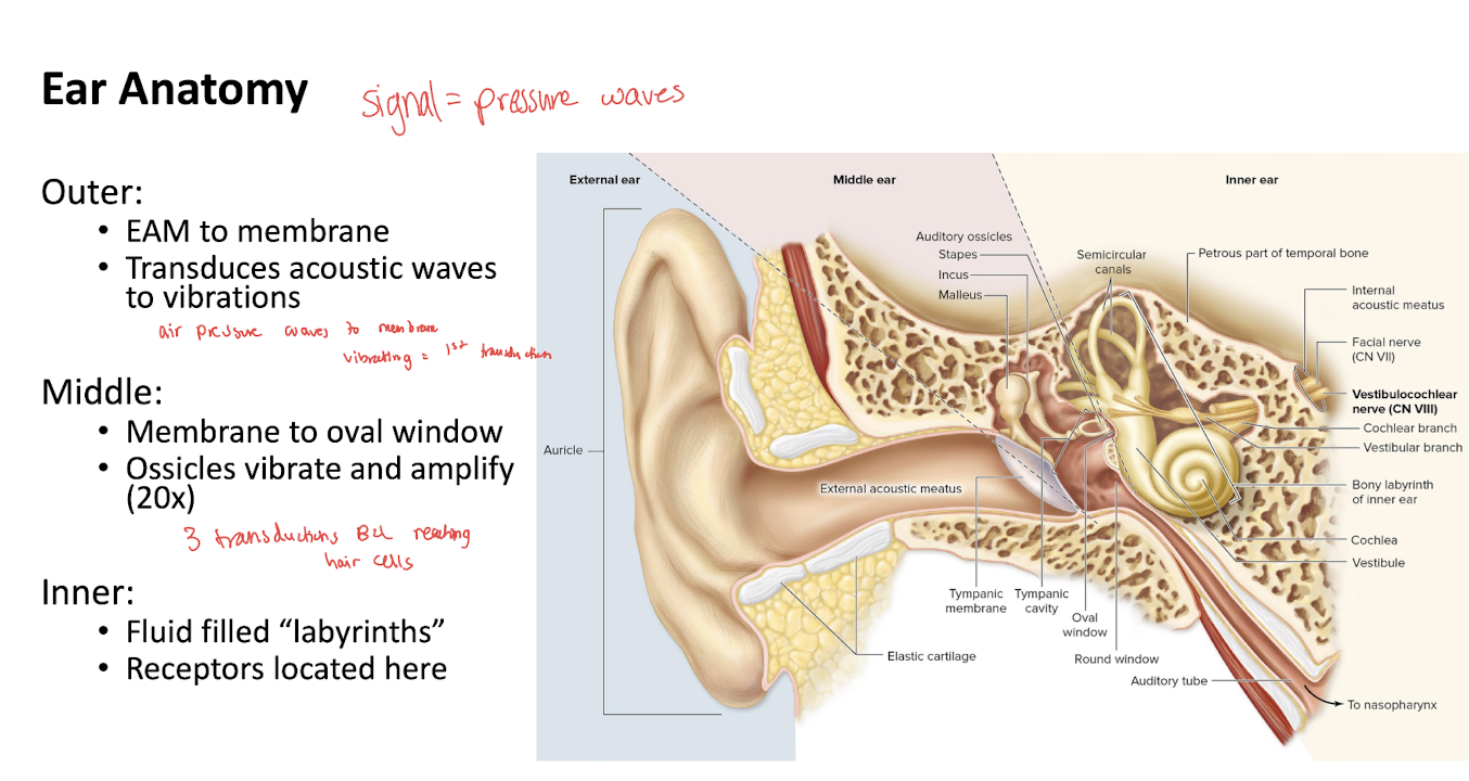
What is the difference between the bony labyrinth and membranous labyrinth?
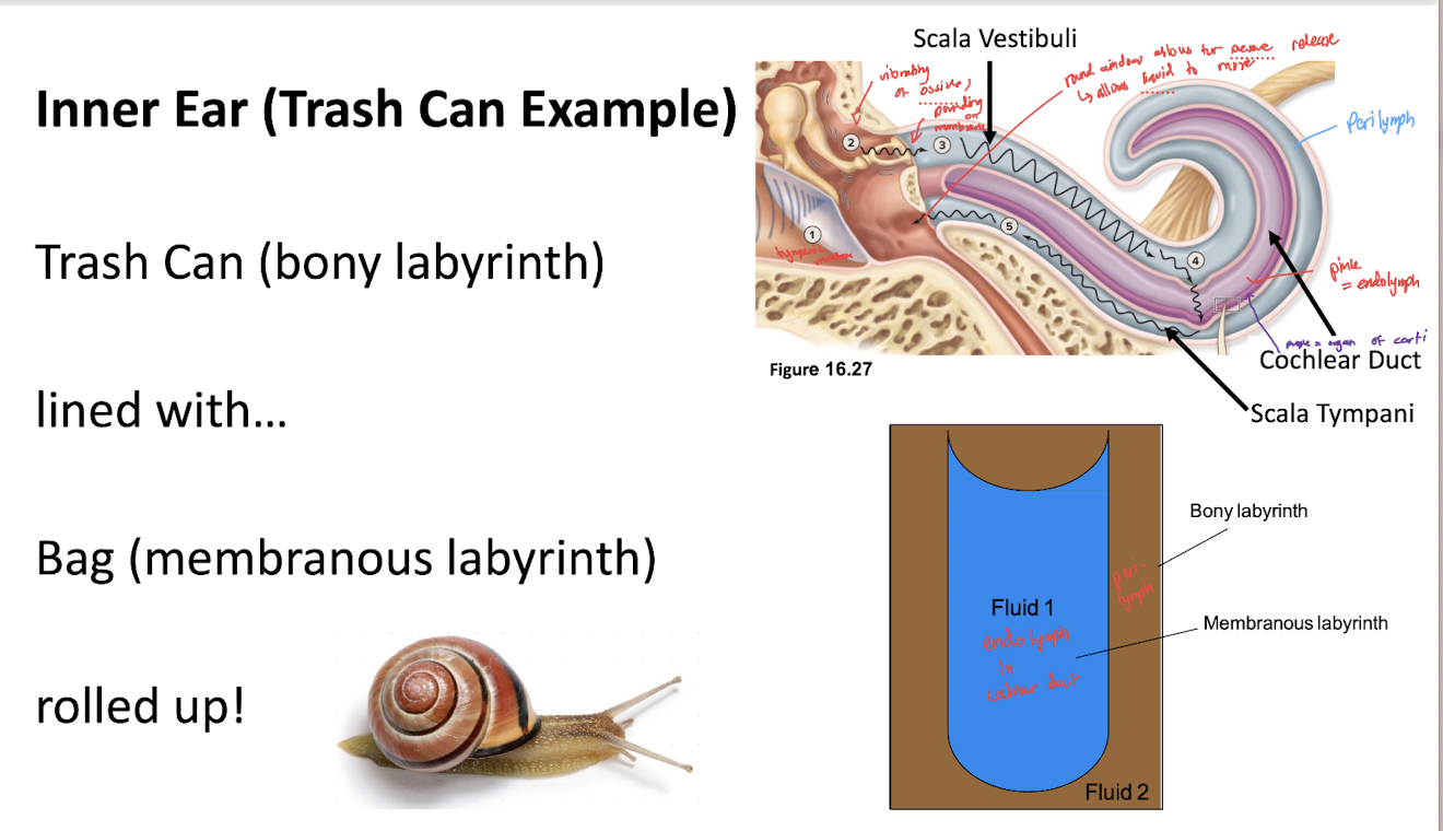
bony labyrinth is the surrounding system of spaces
membranous labyrinth consists of enclosed ducts and sacs within it
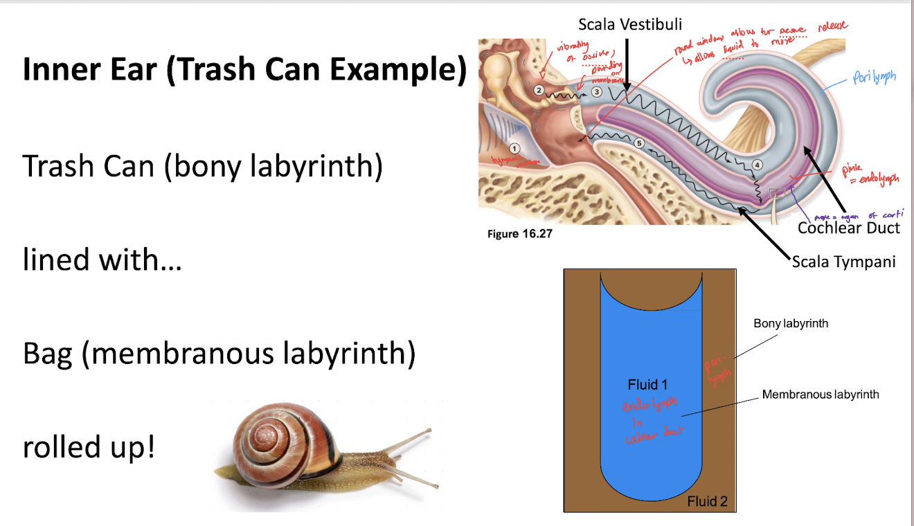
What fluid fills the bony labyrinth?
Perilymph, which is high in Na⁺ and low in K⁺.

What fluid fills the membranous labyrinth?
Endolymph, which is low in Na⁺ and high in K⁺.

What structures make up the cochlear portion of the bony labyrinth?
scala vestibuli: (upper chamber of the cochlea that receives sound vibrations from the middle ear bones via the oval window and channels them into the inner ear fluid)
scala tympani: (lower chamber of the cochlea that relays fluid vibrations back down to the round window, where the pressure is safely dissipated into the middle ear)
Both chambers are filled with perilymph
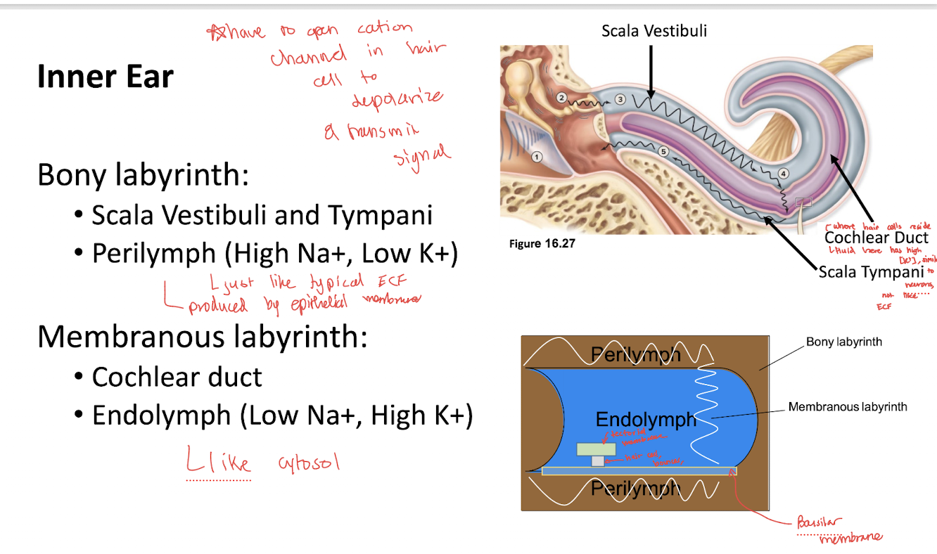
Where is the cochlear duct located, and what fluid does it contain?
Within the cochlea; it is part of the membranous labyrinth and contains endolymph

Why is the difference between perilymph and endolymph important for sensory transduction
Their different ion concentrations establish gradients that allow ion movement through mechanically gated channels in hair cells
What is the receptive organ for hearing?
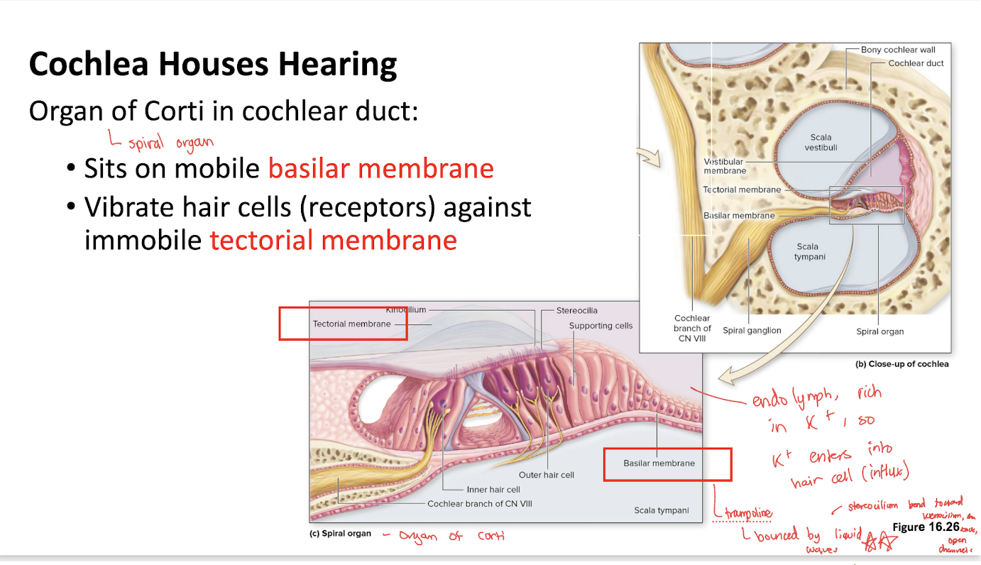
The organ of Corti, located in the cochlear duct

What sensory receptors are found in the organ of Corti?
Mechanoreceptive hair cells with stereocilia
What is the basilar membrane?
A mobile membrane supporting the organ of Corti
trampoline! bounced by liquid waves

What is the tectorial membrane?
A membrane positioned above the hair cells that provides a relatively stationary surface against which their stereocilia bend
vibrate hair cells (receptors) agaiinst immobile tectorial membrane

Which membrane moves during acoustic stimulation: the basilar or tectorial membrane?
The basilar membrane vibrates, while the tectorial membrane is treated as relatively stationary in the lecture model

How does movement of the basilar membrane stimulate hair cells?
It causes the hair cells and their stereocilia to move relative to the tectorial membrane, bending the stereocilia

What are stereocilia?
Hair-like projections on sensory hair cells that bend in response to mechanical movement.
What are tip links, and what is their function?
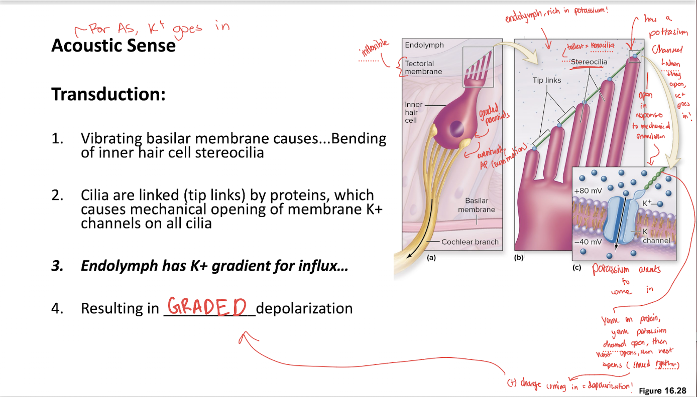
Protein links connecting adjacent stereocilia
bending the stereocilia tensions these links and mechanically opens ion channels

What happens when acoustic stimulation bends stereocilia in the excitatory direction?
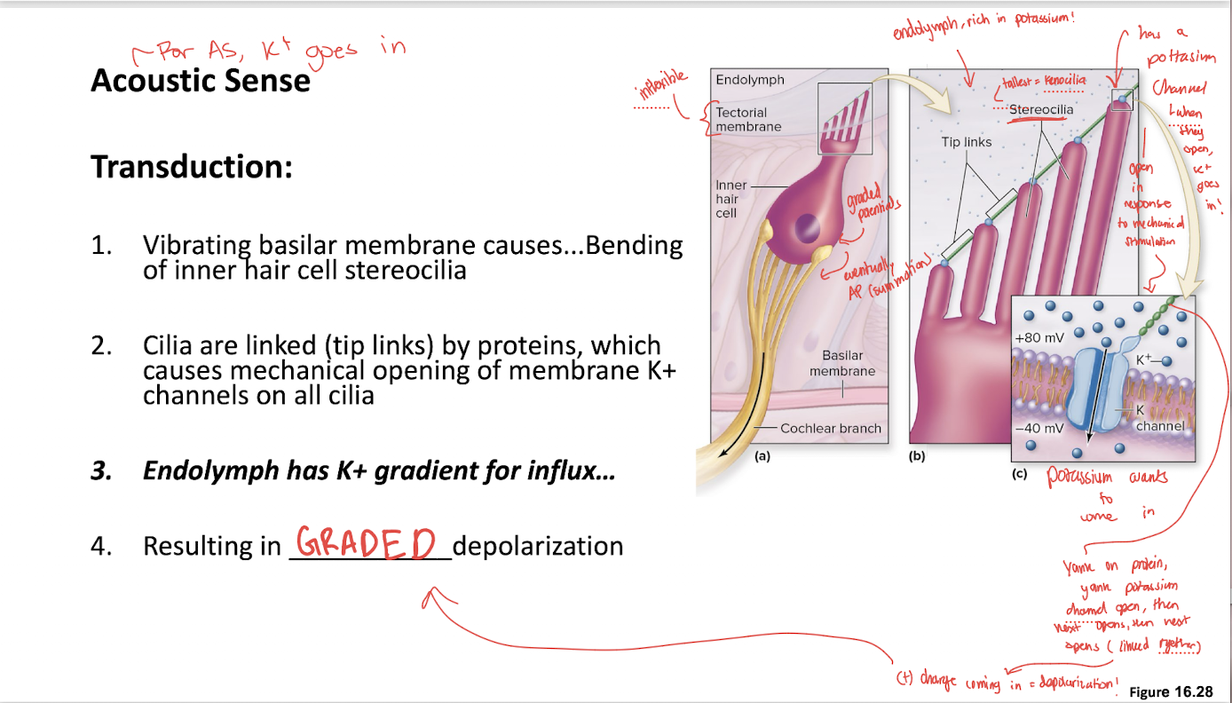
Tip links open mechanically gated K⁺ channels, allowing K⁺ to enter the hair cell from the K⁺-rich endolymph
bends toward Kinocilium
Why does K⁺ enter cochlear hair cells despite being a positively charged ion?
Endolymph has a high K⁺ concentration and an electrical gradient that favors K⁺ entry through open channels

What electrical change occurs when K⁺ enters an acoustic hair cell?
Depolarization, producing a graded receptor potential

What happens when the stereocilia bend in the opposite direction?
Mechanically gated channels close, reducing K⁺ influx and causing the hair cell to hyperpolarize relative to its resting state
Bending away from Kinocilium

Does a cochlear hair cell directly transmit an action potential along CN VIII?
No
The hair cell produces a graded potential that changes signaling by the associated afferent neuron
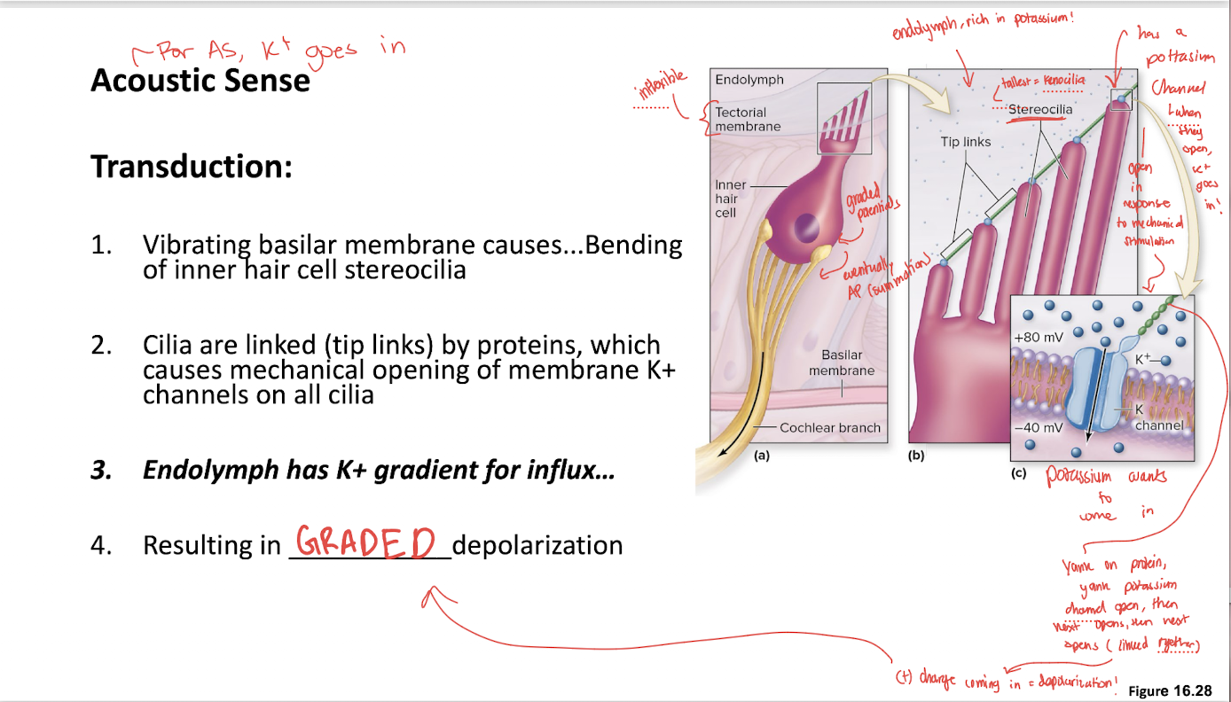
Trace the acoustic transduction mechanism from sound vibration to a receptor potential
Basilar membrane vibrates → stereocilia bend → tip links open K⁺ channels → K⁺ enters from endolymph → hair cell depolarizes (graded depolarization)

What is the difference between a graded potential in a hair cell and an action potential in an afferent neuron?
The hair cell's graded potential varies with stimulation
the afferent neuron's action potentials carry the signal toward the CNS
Which cranial nerve transmits acoustic information to the CNS?
CN VIII, the vestibulocochlear nerve, specifically its cochlear division
What is the general auditory pathway shown in the lecture?
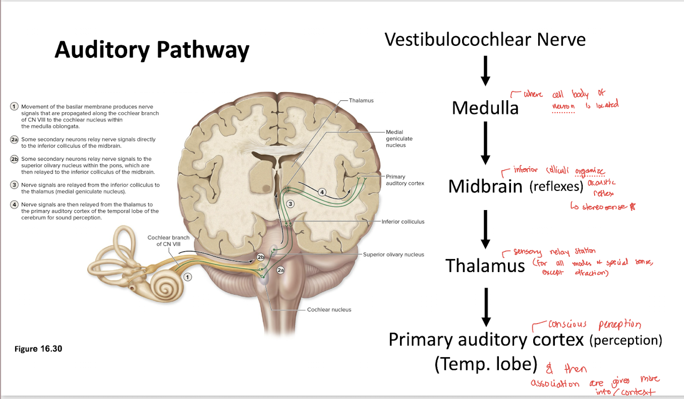
CN VIII → medulla/brainstem auditory pathways → inferior colliculi of the midbrain → thalamus → primary auditory cortex
What role does the inferior colliculus play in hearing?
It participates in auditory processing and acoustic reflexes

What is the role of the thalamus in the auditory pathway?
It relays auditory information toward the cerebral cortex

Where is the primary auditory cortex located?
In the temporal lobe

What is the function of the primary auditory cortex?
It supports conscious auditory perception, including processing sound characteristics such as pitch

What do auditory association areas contribute?
They provide additional interpretation and context for sounds beyond basic sensory perception

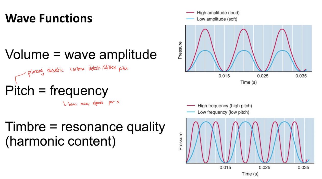
What determines the volume or loudness of a sound wave?
Its amplitude

What determines the pitch of a sound wave?
Its frequency (how many signals per second)
primary acoustic cortex detects/differs pitch

What determines the timbre of a sound?
Its resonance quality or harmonic content
What is tonotopy?
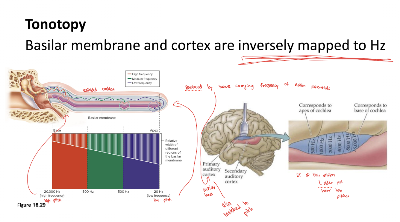
The spatial organization of sound-frequency information, with different locations representing different frequencies
Basilar membrane & cortex are inversely mapped to Hz

What physical property of the basilar membrane supports tonotopy?
Its mechanical properties vary along its length, causing different regions to respond best to different sound frequencies

How is tonotopy represented in the primary auditory cortex?
Different cortical locations respond preferentially to different sound frequencies, preserving an organized frequency map

How does tonotopy relate to frequency along the basilar membrane and in the auditory cortex?
Both structures have an organized spatial representation of frequency
the lecture describes the mapping as inversely related to Hz along the basilar membrane and cortex
What type of information does the vestibular system detect?
Acceleration and movement of the head, contributing to equilibrium and balance

What type of equilibrium is associated with the vestibule?
Static equilibrium, including linear acceleration and head tilt

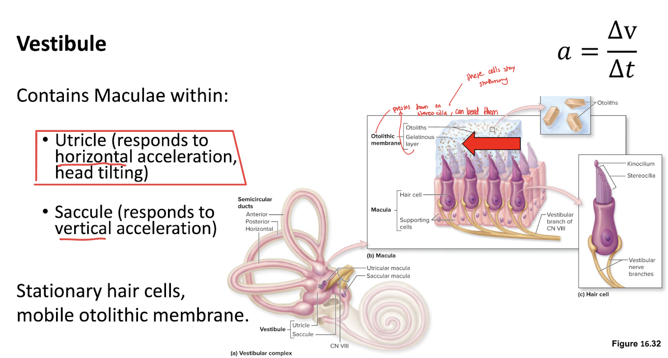
Which two structures within the vestibule contain the receptive organs for linear equilibrium?
The utricle and saccule

What is the receptive organ within the utricle and saccule?
The macula

What type of acceleration does the utricle primarily detect?
Horizontal linear acceleration and head tilt

What type of acceleration does the saccule primarily detect?
Vertical linear acceleration
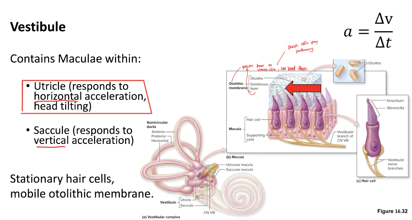
What is the otolithic membrane?
A gelatinous membrane overlying the hair cells of a macula that moves relative to the hair cells during linear acceleration or head tilt

What are otoconia, or otoliths?
Small calcium carbonate crystals associated with the otolithic membrane

What is the function of otoconia during linear equilibrium?
Their mass adds inertia and weight to the otolithic membrane, helping gravity and linear acceleration displace it relative to the hair cells
Why are otoconia important for detecting linear acceleration?
They add mass to the otolithic membrane so that head movement or gravity can shift the membrane and bend the stereocilia
Why does the cochlea not require otoconia for acoustic transduction?
Hearing uses sound-driven vibration of the basilar membrane and relative movement at the tectorial membrane, rather than gravity or linear acceleration acting on a weighted membrane

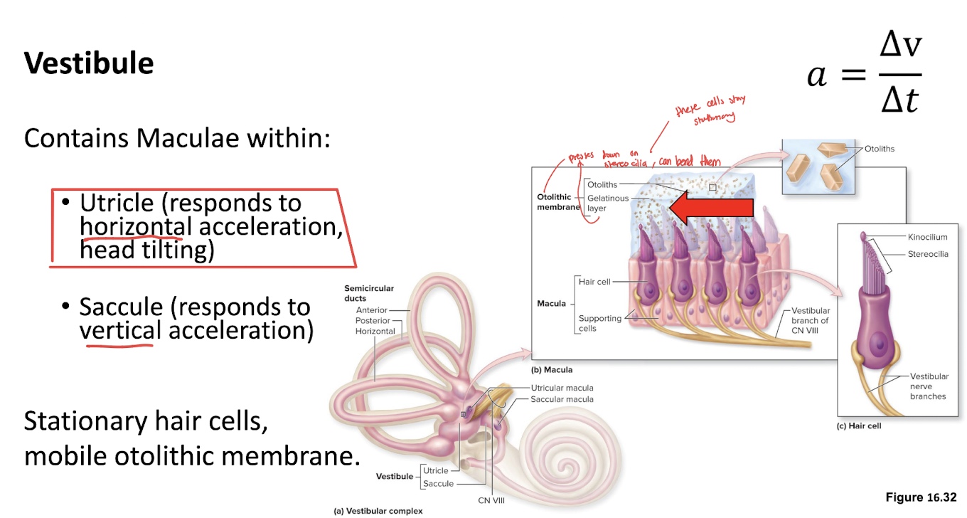
What happens to vestibular hair cells when the head tilts or accelerates linearly?
The otolithic membrane shifts relative to the hair cells, bending their stereocilia

What is the kinocilium?
The tallest hair-like projection in a vestibular hair bundle; the direction of stereocilia bending relative to it determines the response

What happens when vestibular stereocilia bend toward the kinocilium?
The hair cell becomes more excited, increasing its depolarization and the firing rate of the associated vestibular afferent neuron
Ca++ influx
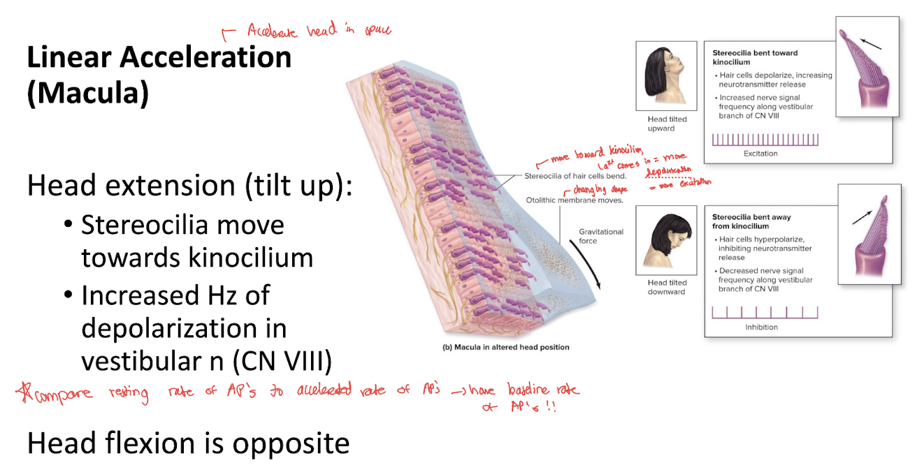
What happens when vestibular stereocilia bend away from the kinocilium?
The hair cell becomes less excited, reducing its depolarization and the firing rate of the associated vestibular afferent neuron

How does the otolithic membrane differ from the tectorial membrane?
The otolithic membrane moves over vestibular hair cells; the tectorial membrane is relatively stationary while the basilar membrane and cochlear hair cells move during hearing

What type of equilibrium is associated with the semicircular canals?
Dynamic equilibrium, particularly angular or rotational acceleration

What is the receptive organ for angular equilibrium in the semicircular canals?
The crista ampullaris

What is the crista ampullaris?
A sensory ridge in the ampulla of a semicircular canal containing vestibular hair cells

What is the ampullary cupula?
A gelatinous structure overlying the hair cells of the crista ampullaris
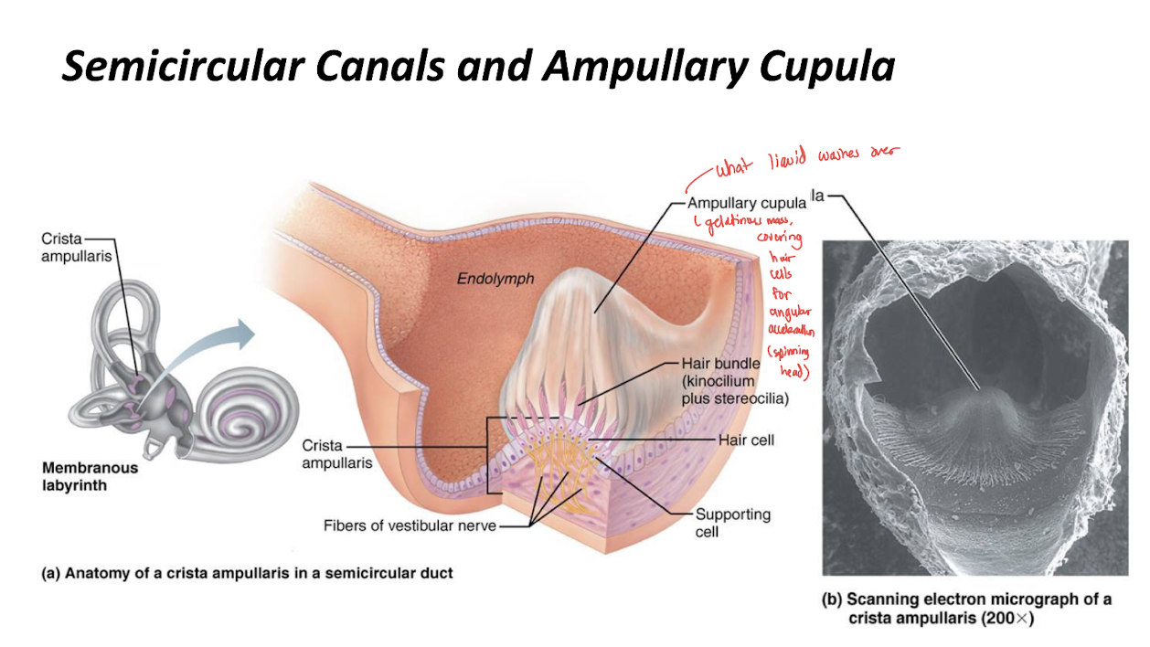
What happens to the ampullary cupula when the head rotates?
Inertia causes the endolymph to move relative to the canal, deflecting the cupula and bending the hair cells' stereocilia

How does the cupula differ from the otolithic membrane?
The cupula detects angular acceleration through endolymph movement; the otolithic membrane detects linear acceleration and head tilt with help from otoconia

Why can semicircular canals detect rotational acceleration?
Endolymph lags behind head rotation because of inertia, creating relative fluid movement that deflects the cupula

What happens when the cupula bends vestibular stereocilia toward the kinocilium?
The hair cell becomes more depolarized and vestibular afferent firing increases

What happens when the cupula bends stereocilia away from the kinocilium?
The hair cell becomes less depolarized and vestibular afferent firing decreases

What cranial nerve carries equilibrium information?
CN VIII, the vestibulocochlear nerve, specifically its vestibular division
How does CN VIII differ from a sensory nerve that is inactive until a stimulus occurs?
Vestibular afferents normally fire at a baseline rate even at rest
movement can increase or decrease this firing rate

What happens to vestibular nerve firing when a stimulus excites the hair cell?
Action potential frequency increases above baseline
What happens to vestibular nerve firing when a stimulus inhibits the hair cell?
Action potential frequency decreases below baseline
How does the lecture distinguish acoustic from equilibrium hair-cell ion movement?
It emphasizes K⁺ influx for acoustic hair cells and Ca²⁺ influx for equilibrium hair cells
Compare the hair-cell response to hearing a sound versus accelerating forward in a car.
Sound vibrates the basilar membrane, bending cochlear stereocilia and increasing K⁺ influx during excitation
Forward acceleration shifts the otolithic membrane, bending vestibular stereocilia and changing vestibular hair-cell excitation and afferent firing
Compare the moving structures in acoustic, linear-equilibrium, and angular-equilibrium transduction.
Acoustic: basilar membrane vibrates relative to the tectorial membrane
Linear equilibrium: otolithic membrane shifts relative to macular hair cells
Angular equilibrium: endolymph movement deflects the cupula over hair cells in the crista ampullaris
What does BPPV stand for?
Benign paroxysmal positional vertigo

Which sense is specifically affected by BPPV?
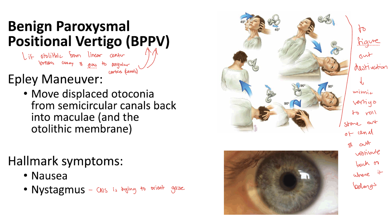
Angular equilibrium, producing abnormal vestibular signals associated with head position and movement

What are the hallmark symptoms of BPPV listed in the lecture?
Nausea and nystagmus, often with positional vertigo

Is BPPV a peripheral or central problem?
A peripheral vestibular problem involving the inner ear, not primarily a central brain lesion

What causes BPPV according to the lecture?
Otoconia detach from the otolithic membrane and enter a semicircular canal, where they abnormally affect endolymph and cupula movement

Why can displaced otoconia cause vertigo?
They create abnormal mechanical stimulation of the semicircular canal's angular-equilibrium receptors, sending misleading movement signals to the CNS

What is the Epley maneuver used for?
It is a treatment for BPPV that repositions displaced otoconia
guide displaced otoconia out of the semicircular canal and back into the vestibule, toward the otolithic membrane where they belong
Therapeutic. Positional tests are used to help diagnose BPPV; the Epley maneuver repositions the otoconia
What are the two broad components of vision?
Optical components focus light; neural components transduce light and transmit visual information to the brain

What is refraction?
The bending of light as it passes between media with different optical properties

Optical component =
focuses light onto receptor cells

Neural…
transduces light into graded potentials (then APs)
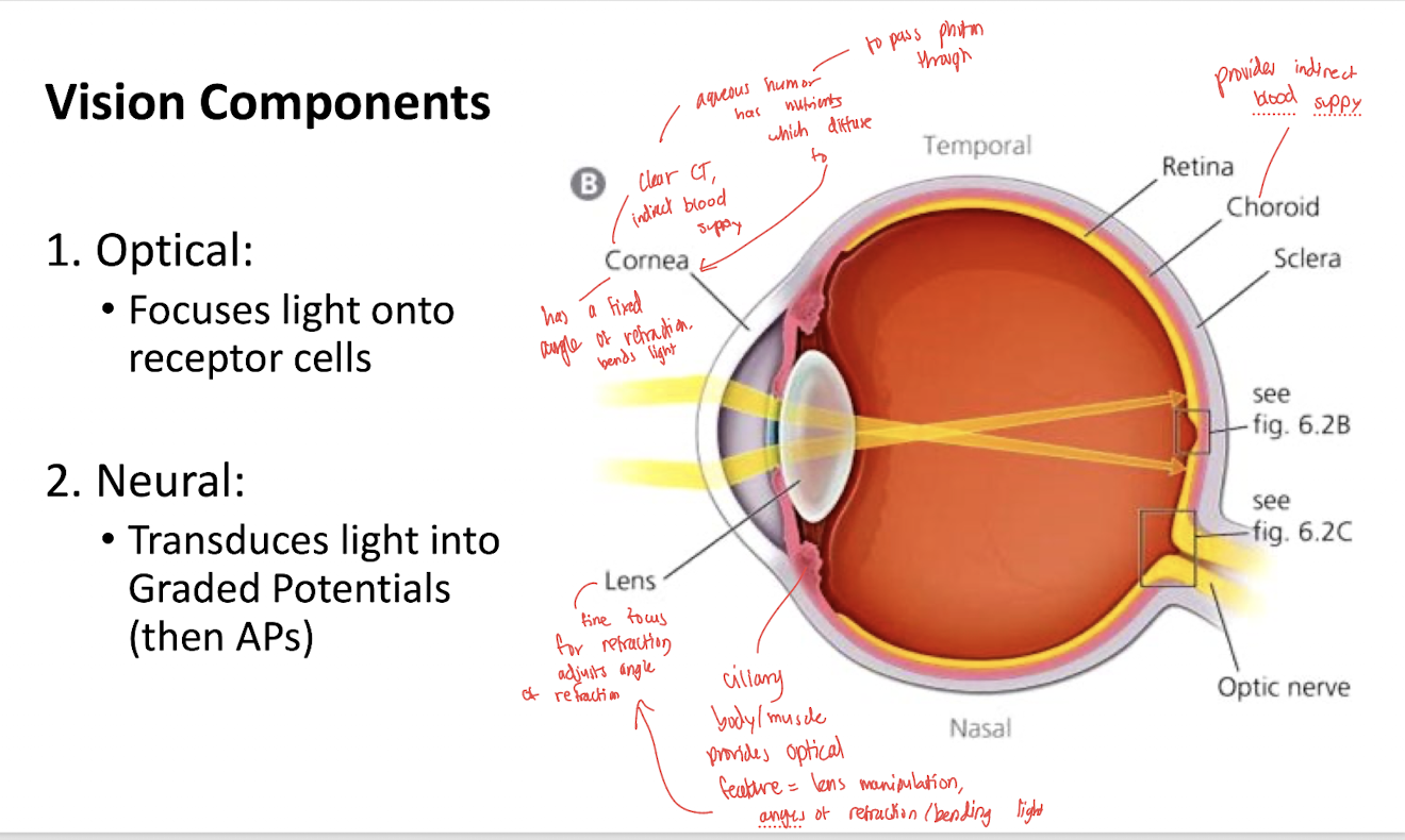
How do the cornea and lens contribute to refraction?
Both are convex and refract light

What is the function of the cornea?
It provides much of the eye's fixed refraction, bending incoming light toward the retina

What is the function of the lens?
adjusts its shape to fine-tune refraction and focus images onto the retina

The iris…
controls light volume

What is visual accommodation?
Changing lens shape to focus images from objects at different distances onto the retina
What happens to the lens when shifting focus from a far object to a near object?
The lens becomes more convex or “fat,” increasing its refractive power

What happens to the ciliary muscle and suspensory ligaments when focusing on a near object?
The ciliary muscle contracts, suspensory ligaments slacken, and the lens becomes more convex

What happens to the ciliary muscle and suspensory ligaments when focusing on a far object?
The ciliary muscle relaxes, suspensory ligaments become taut, and the lens becomes flatter

What is the function of the iris?
It controls pupil diameter and regulates how much light enters the eye

What is the pupil?
The opening in the iris through which light enters the eye

What are the two autonomically controlled muscles of the iris?
The sphincter pupillae and dilator pupillae

Which autonomic division controls the sphincter pupillae, and what does it do?
The parasympathetic division; contraction constricts the pupil, reducing its diameter

Which autonomic division controls the dilator pupillae, and what does it do?
The sympathetic division; contraction dilates the pupil, increasing its diameter

How does pupil diameter typically change in bright light versus darkness?
Bright light promotes parasympathetic constriction; darkness promotes sympathetic dilation
