Biology of the skin - structure & function
 Call Kai
 Learn
 Practice Test
 Spaced Repetition
 Match
 Flashcards
 Knowt Play
1/68

There's no tags or description
Looks like no tags are added yet.
69 Terms
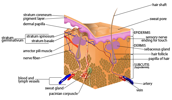
What is the very basic structure of the skin?
epidermis
dermis
subcutis/hypodermis
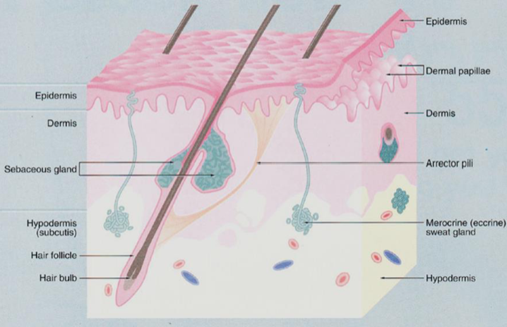
What are the main functions of the skin?
physical barrier
physiological functions
appendages
site of pathology
What does the skin act as a physical barrier against?
Mechanical damage
water
chemicals
particles
infectious agents
what physiological functions is the skin responsible for?
temp regulation
sensation & nerve signalling
vit D synthesis
immune defence
protection against UV radiation
Describe the histology of the epidermis?
Composed of keratinized stratified squamous epithelium, cornified (dead) = KETARINOCYTES
MELANOCYTES - produce melanin
Langerhans Cells
Merkel Cells
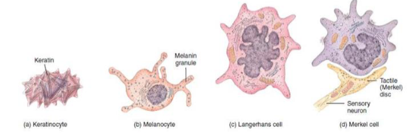
which pigment/cell in the skin absorbs UV light?
Melanin/ melanocytes
How do melanin granules protect DNA in keratinocytes from damage?
Cluster around the nucleus, forming a protective veil. this protects the nuclear DNA from damage
Describe the histology of the dermis
dense irregular connective tissue supported by fibroblasts
Dense in the papillary region (contains small blood vessels)
contains glands ect, sensory nerves, some immune cells
Describe the histology of the subcutis
loose connective tissue with adipose cells
Larger blood vessels, nerves
Thick vs thin skin - which is more abundant?
Thin skin - lines most of the body
What are the 5 layers of the epidermis?
Cornified layer - stratum corneum
Stratum Lucidum - in thick skin only
Granular layer - stratum granulosum
Spinous layer - striatum spinosum
Basal layer - stratum basale

What are the two fates of stratum basale stem cells?
Remain as stem cells - capable of mitosis
OR
differentiate for form epidermal layer above
Are cells in the stratum spinosum capable of cell division?
NO - post mitotic cells only
What holds together cells in the stratum spinosum?
Prominent intracellular keratin filaments and desmosomes

Describe the differentiation of cells in the stratum granulosum
Protein Keratohyalin is produced and lamellar bodies form
Function of keratohyalin?
in stratum granulosum - binds to and aggregates keratin filaments
Function of lamellar bodies
Contain lipid bodies important for the hydrophobic barrier to water
in what layer of the epidermis does apoptosis occur?
Stratum granulosum and stratum corneum
During apoptosis what happens to cells?
They shed their nuclei and organelles - forming the tough layer of the skin (eventually dead cells fall apart)
Describe the shape of melanocytes in the epidermis
Dendritic in shape
What is the embryological origin of melanocytes?
Ectoderm - neural crest more specifically
What kind of cells are Langerhans cells?
APCs - dendritic in shape
where do Langerhans cells arise from?
Bone marrow, and migrate to the epidermis via blood.
if presented with a pathogen they migrate to LNs
What are Merkel cells?
Specialised cells found in the basal layer
Role of Merkel cells?
Associated with nerve fibres = responsible for fine touch sensation
which layer of the epidermis is only present in thick skin?
Stratum lucidum - between the stratum corneum and the stratum granulosum
Describe stratum lucidum
4-6 layers of flattened clear, dead keratinocytes = large amounts of keratin and thickened plasma membranes
List the pathological changes that can occur in the epidermis
Imbalance between cell renewal and cell loss
e.g. scalingBreakage of cell-cell junctions
e.g. blistersTrauma and wound repair
Basal layer needed for regenerationFailure of barrier functions
Usually consequence of aboveInfection and inflammation
Various external and systemic causesDevelopment of tumours
Keratinocytes or melanocytes
What are the two regions of the dermis?
Papillary dermis
Reticular region

Describe the structure of the papillary dermis
Superficial portion of dermis
relatively loose connective tissue (fine collagen & elastin fibres)
Extensive capillary network, lymphatics and nerve endings
Describe the structure of the reticular dermis
Deeper, thicker layer = majority of skin overall
dense irregular connective tissue (prominent bundles of collagen fibres)
fibroblasts and immune cells (mast cells, macrophages, lymphocytes)
blood vessels, nerves and nerve endings
site of various skin appendages (hair follicles, sweat glands…)
What are the main roles of the dermis? why?
Sensation, protection, thermoregulation
site of nerves, glands and skin vasculature
Pathological changes in the dermis
Trauma and wound repair
Collagen synthesis by fibroblasts, initial scar effectInflammatory and immune responses
Can be due to infection (e.g. of skin appendages or following trauma) or other immune processes (e.g. hypersensitivity, autoimmune)Vascular involvement
Important in both wound healing and inflammatory response-
Describe the composition of the subcutis /hypodermis
Mature adipose tissue, can be separated by fibrous septa
the thickness of subcutis varies between sites and individuals

What are the functions of the subcutis/hypodermis?
Insulator, protector and energy store
List skin appendages
`Hair and nails
Glands
sebaceous glands
eccrine glands
apocrine glands
What is the nail plate?
It is a hard keratinised plate at the end on each digit
What does the nail plate rest on?
On a stratified squamous epithelium structure formed from 4 layers of the epidermis
What forms the nail matrix?
Continual renewal of cells within the nail root proliferate to form the nail matrix

What layer of the epidermis does the hair follicle reside in?
From the stratum basale

Describe a hair follicle
Tubular structure formed from basal layer of epidermis
hair/dermal papilla is contained in the bulbous expansion at base of hair follicle - providing root with oxygen and nutrients
isthmus - narrowing of follicle above the bulb
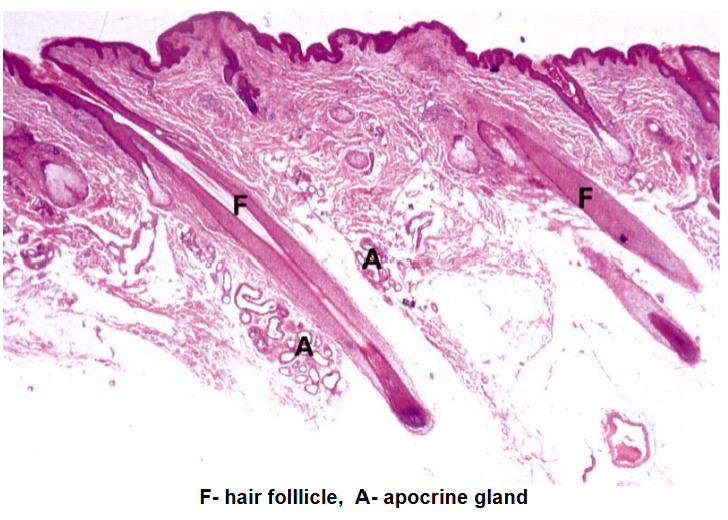
Anatomy of a hair
The hair shaft itself is composed of three concentric layers of keratinised cells
From innermost to outermost:
medulla
cortex
cuticle

What makes up hairs?
Dead epidermal cells that have replaced their internal structures with the protein keratin
Structure of hair medulla
Central core = loosely arranged cells, can be missing in fine hair
Structure of hair cortex
Thickest, middle layer = rich in keratin filaments and pigment granules
structure of hair cuticle
Thin, outermost layer = overlapping keratin scales that interlock with inner root sheath
What do sebaceous glands secrete?
Sebum - lipid containing substance coating hair to keep it soft, supple and waterproof
Sebaceous gland structure
Sebaceous glands have a branched acinar structure
The acini filled with lipid filled vacuoles converge into a small duct which empty into a hair follicle.
Acinar cells become distended with increasing lipid contents and die releasing their contents in a process termed holocrine secretion

What is the role of eccrine glands?
Secrete sweat directly onto skin surface by merocrine secretion - evaporation of sweat is essential for thermoregulation
Contents of sweat
Water
sodium & chloride ions
urea
some metabolites
Where are eccrine glands located in the body?
Most abundant in palms, soles, forehead & axillae ( but found in all tissue)
Which layer of the skin are eccrine sweat glands located?
Dermis - deep reticular dermis
What is the role of apocrine glands?
Secretion of viscous cloudy substance that contains pheromones and causes body odour upon reaction with skin bacteria
How does secretion form apocrine glands take place?
In an apocrine manner - discharged into upper part of hair follicle
When do these coiled tubular apocrine glands become functional?
during puberty
Where in the skin are apocrine glands located?
Deep reticular dermis and subcutis/hypodermis
What are the 5 main physiological functions of skin?
Thermoregulation
sensation
Vit D synthesis
Immunological role
Protection against UV
What kind of control is thermoregulation using the skin?
autonomic control
How does the skin control thermoregulation?
Skin surface temperature is controlled via alterations in blood flow through vessels in papillary dermis (vasoconstriction/dilation)
Sweat released from eccrine glands aids heat loss via evaporation
Which cells carry out vit D synthesis?
Keratinocytes in the presence of UV light - further conversion to active hormone needed in liver and kidney
Which cells carry out immune responses in the skin?
Langerhans cells (APCs) and lymphocytes in the epidermis
Macrophages, mast cells and lymphocytes in dermis
what layer of the skin do ‘superficial’ wounds extend to?
Basal layer of the epidermis
How do superficial wounds heal?
Basal cells in the epidermis detach from the basement membrane
cells migrate as sheet until opposite sides meet
cells stop migrating dur to cellular response = contact inhibition
Epidermal growth factor = stimulates cell division to build new strata - resulting in thickened epidermis
What layer of the skin do ‘deep’ wounds extend to?
Into the dermis
What are the phases of deep wound healing?
Inflammatory phase
Migratory phase
Proliferative phase
Maturation phase
Inflammatory phase
formation of fibrin clot (stop bleeding & filling wound)
Removal of bacteria/ damaged tissue through vascular (vasodilation & increased permeability) and cellular response (phagocytic neutrophils: monocytes)
Mesenchymal cells develop into fibroblasts
Migratory phase
Clot becomes scab
fibroblasts migrate along fibrin threads and lay down new collagen accompanied by angiogenesis
Epithelial cells migrate to bridge gap between scab and wound
Tissue filling wound = granulation tissue
proliferative phase
Extensive growth of epithelial cells beneath scab
Deposition by fibroblasts of collagen fibres
Continued angiogenesis
Maturation phase
Scab sloughs off
collagen fibres become more organised
fibroblast decrease in number