Biology A-Level: Cell Membrane & Transport
 Call Kai
 Learn
 Practice Test
 Spaced Repetition
 Match
 Flashcards
 Knowt Play
1/17

There's no tags or description
Looks like no tags are added yet.
18 Terms
What is the the fluid mosaic model of membrane structure?
The fluid mosaic model describes the cell surface membrane as a phospholipid bilayer with proteins embedded in it. Hydrophilic phosphate heads face the watery environments inside and outside the cell, while hydrophobic fatty acid tails face inwards, away from water, and interact with each other. Intrinsic proteins are embedded in the bilayer, with their hydrophobic regions interacting with the fatty acid tails and hydrophilic regions facing the watery environments. The membrane is fluid because phospholipids and some proteins move laterally, and mosaic because proteins are scattered throughout the bilayer, in a pattern resembling a mosaic.
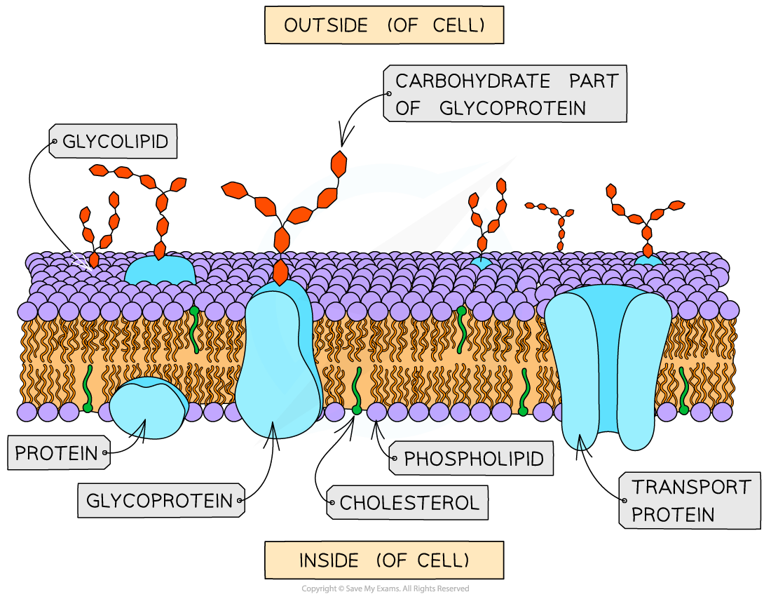
How are cholesterol, glycolipids and glycoproteins in cell surface membranes arranged?
Cholesterol is found between the phospholipid molecules, with its hydrophobic region interacting with the fatty acid tails and its hydrophilic region near the phosphate heads. Glycolipids consist of carbohydrate chains attached to phospholipids, while glycoproteins consist of carbohydrate chains attached to proteins. Both are found on the outer surface of the membrane, with their carbohydrate chains projecting outwards and acting as cell recognition sites.
What are the roles of phospholipids, cholesterol, glycolipids, proteins and glycoproteins in cell surface membranes?
1. Phospholipids
Stability: Form the basic structure of the cell surface membrane as a phospholipid bilayer.
Permeability: The hydrophobic interior prevents most water-soluble substances and ions from passing directly through the membrane.
Fluidity: Phospholipids can move laterally within the bilayer, allowing the membrane to remain fluid and flexible.
2. Cholesterol
Stability: Fits between phospholipid molecules and helps stabilize the membrane.
Fluidity: Restricts phospholipid movement at high temperatures, reducing fluidity, and prevents phospholipids from packing too closely at low temperatures, helping maintain fluidity.
Permeability: Reduces the permeability of the membrane to water-soluble molecules and ions by filling spaces between phospholipids.
3. Proteins
Channel proteins
- Transport: Form hydrophilic channels through the membrane, allowing specific ions or polar molecules to cross by facilitated diffusion, down their concentration or electrochemical gradients.
Carrier proteins
Transport: Bind to specific ions or molecules and change shape to transport them across the membrane.
Can carry out facilitated diffusion down a concentration gradient or active transport against a concentration gradient using energy, usually from ATP.
Receptor proteins
- Cell signaling: Bind to specific signaling molecules, such as hormones, causing a response inside the cell. Binding may trigger a series of reactions within the cell.
4. Glycoproteins
Cell recognition: Carbohydrate chains project from the outer surface of the membrane and act as recognition sites.
Cell surface antigens: Some glycoproteins act as antigens, allowing the immune system to distinguish between cells, such as recognizing foreign cells.
Cell signaling: Some glycoproteins act as receptor proteins that bind to specific signaling molecules and trigger a cellular response.
What is the process of cell signaling?
Cell signaling begins when a cell secretes specific chemical messengers called ligands, such as hormones. The ligands are transported to target cells, for example, through the bloodstream. The ligands bind to specific complementary receptors on the cell surface membrane of target cells, forming ligand-receptor complexes. This triggers a series of reactions inside the target cell, leading to a specific cellular response.
What is simple & facilitied diffusion?
Simple diffusion is the net movement of particles from a region of higher concentration to a region of lower concentration, down a concentration gradient, due to their random movement. Particles pass directly through the phospholipid bilayer without transport proteins or energy from ATP. Examples include oxygen and carbon dioxide moving across cell surface membranes.
Facilitated diffusion is the net movement of particles from a region of higher concentration to a region of lower concentration through specific channel or carrier proteins in the cell surface membrane. Channel proteins provide hydrophilic pathways for ions or polar molecules, while carrier proteins bind to specific substances and change shape to transport them across the membrane.
What is osmosis?
Osmosis is the net movement of water molecules from a region of higher water potential to a region of lower water potential through a partially permeable membrane. Water moves down its water potential gradient because the membrane allows water molecules to pass through but restricts some dissolved solutes. Osmosis is a passive process and does not require ATP.
What is active transport?
Active transport is the movement of substances across a membrane against their concentration gradient, from a lower concentration to a higher concentration, using energy from ATP. Specific carrier proteins bind to the substances and change shape to move them across the membrane. ATP provides the energy needed for this process. An example is the uptake of mineral ions by root hair cells.
What is endocytosis?
Endocytosis is the process by which a cell takes substances into the cell by engulfing them with its cell surface membrane. The membrane folds around the substance and pinches off to form a vesicle inside the cytoplasm. It requires energy from ATP and allows cells to take in large particles or bulk quantities of substances.
What is Exocytosis?
Exocytosis is the process by which a cell releases substances outside the cell using vesicles. Vesicles containing substances move toward and fuse with the cell surface membrane, releasing their contents outside the cell. It requires energy from ATP and is used to secrete substances such as hormones, enzymes, and neurotransmitters.

How do you investigation simple diffusion using agar?
Aim: To investigate how far substances diffuse into agar.
Method:
Prepare agar containing an indicator such as phenolphthalein and sodium hydroxide, producing pink agar.
Cut the agar into cubes of different sizes.
Place the cubes in dilute hydrochloric acid for the same length of time.
Remove the cubes, cut them in half and measure the depth of the colorless region.
Results and explanation: Hydrochloric acid diffuses into the agar and neutralizes the alkali, causing the pink color to disappear. Smaller cubes become colorless more quickly because they have a larger surface area-to-volume ratio and a shorter distance for diffusion.
How do you investigate diffusion using a Visking Tube?
Visking tubing (sometimes referred to as dialysis tubing) is a non-living, selectively permeable membrane made from cellulose.
Pores in this membrane are small enough to prevent the passage of large molecules (such as starch) but allow smaller molecules (such as glucose) to pass through by diffusion.
This can be demonstrated by:
Filling a section of Visking tubing with a mixture of starch and glucose solutions
Suspending the tubing in a boiling tube of water for a set period time
Testing the water outside of the visking tubing at regular intervals for the presence of starch and glucose to monitor whether diffusion of either substance out of the tubing has occurred
The results should indicate that glucose, but not starch, diffuses out of the tubing

How do you investigate osmosis using Visking Tube?
Aim: To investigate osmosis using a partially permeable membrane.
Method:
Tie one end of a piece of Visking tubing and soak it to make it flexible.
Fill it with concentrated sucrose solution and tie the other end.
Blot the tubing dry and measure its initial mass.
Place it in distilled water for a fixed time.
Remove it, blot it dry and measure its final mass.
Results and explanation: The tubing gains mass because water moves from the distilled water, which has a higher water potential, into the sucrose solution, which has a lower water potential, through the partially permeable membrane.
Visking tubing is a model of a partially permeable membrane. Unlike a living cell membrane, it does not contain phospholipids or transport proteins.
How do you investigate osmosis using plant tissue?
Aim: To investigate the effect of solution concentration on the movement of water into or out of plant tissue.
Method:
Cut equal-sized potato cylinders using a cork borer.
Blot them dry and measure their initial mass.
Place them in different concentrations of sucrose solution for the same amount of time.
Remove them, blot them dry and measure their final mass.
Results and explanation: In a dilute solution, water enters potato cells by osmosis, so their mass increases. In a concentrated solution, water leaves the cells, so their mass decreases. At the isotonic concentration, there is no net movement of water and the percentage change in mass is approximately zero.
How do you investigate the effect of changing surface area to volume ratio on diffusion using agar blocks of different sizes?
Method
Prepare agar containing sodium hydroxide and phenolphthalein indicator, making the agar pink.
Cut the agar into cubes of different sizes, for example, 1 cm, 2 cm and 3 cm.
Calculate the surface area, volume and surface area to volume ratio of each cube.
Place the cubes in the same volume and concentration of dilute hydrochloric acid for the same length of time.
Remove the cubes, cut them in half and measure the depth of the colorless region, where the acid has diffused into the agar.
Calculate the percentage of each cube that has become colorless and compare the results.
Results and explanation
Hydrochloric acid diffuses into the agar and neutralizes the sodium hydroxide, causing the pink indicator to become colorless. Smaller agar cubes have a larger surface area to volume ratio, so a greater proportion of their volume is reached by the acid in the same time. Larger cubes have a smaller surface area to volume ratio, so a smaller proportion of their volume becomes colorless.
How do we investigate the effects of immersing plant tissues in solutions of different water potentials, using the results to estimate the water potential of the tissues?
1. Method
Use a cork borer to cut equal-sized cylinders of potato and trim them to the same length.
Blot the potato cylinders dry and measure their initial masses.
Prepare a range of sucrose solutions with different concentrations and place equal volumes into labeled test tubes.
Place one potato cylinder into each solution for the same length of time, keeping the temperature constant.
Remove the cylinders, gently blot them dry using the same method, and measure their final masses.
Calculate the percentage change in mass for each cylinder.
Results and explanation
Dilute sucrose solution: Water enters the potato cells by osmosis because the solution has a higher water potential than the cell sap. The mass increases.
Concentrated sucrose solution: Water leaves the potato cells by osmosis because the solution has a lower water potential than the cell sap. The mass decreases.
Isotonic solution: There is no net movement of water because the solution and the tissue have approximately equal water potentials. The percentage change in mass is zero.
Estimating the water potential of the tissue
Plot a graph of percentage change in mass against sucrose concentration.
Find the sucrose concentration where the graph crosses 0% change in mass. At this point, the solution and the potato tissue have approximately equal water potentials.
Use the known water potential of that sucrose solution, obtained from a suitable reference table at the experimental temperature, to estimate the water potential of the potato tissue.
What is the movement of water between cells and solutions?
Water moves by osmosis from a region of higher water potential to a region of lower water potential through a partially permeable membrane. Water moves down its water potential gradient until equilibrium is reached, when there is no net movement of water.
Higher water potential outside the cell: Water enters the cell by osmosis.
Lower water potential outside the cell: Water leaves the cell by osmosis.
Equal water potential inside and outside: There is no net movement of water.
How does the movement of water affect plant cells?
If a plant cell is placed in pure water or a dilute solution, water will enter the plant cell through its selectively permeable cell surface membrane by osmosis. As water enters the vacuole of the plant cell, the volume of the plant cell increases. The expanding protoplast (living part of the cell inside the cell wall) pushes against the cell wall and pressure builds up inside the cell. The inelastic cell wall prevents the cell from bursting. The pressure created by the cell wall also stops too much water entering and this also helps to prevent the cell from bursting. It thus becomes turgid, providing strength and support for the plant.
If a plant cell is placed in a solution with a lower water potential than the plant cell (such as a concentrated sucrose solution), water will leave the plant cell through its selectively permeable cell surface membrane by osmosis. As water leaves the vacuole of the plant cell, the volume of the plant cell decreases. The protoplast gradually shrinks and no longer exerts pressure on the cell wall, beginning to pull away from the cell wall, thus the plant cell is plasmolysed or flaccid.
How does the movement of water affect animal cells?
When an animal cell is placed in a solution with a lower water potential than the cell (such as a concentrated sucrose solution), water will leave the cell through its selectively permeable cell surface membrane by osmosis. This will cause the cell to shrink and shrivel up.
When an animal cell is placed in pure water or a dilute solution, water will enter the cell through its selectively permeable cell surface membrane by osmosis, as the pure water or dilute solution has a higher water potential. The cell will continue to gain water by osmosis until the cell membrane is stretched too far and the cell bursts (cytolysis), as it has no cell wall to withstand the increased pressure created.