28 SupPostMediastinum Lymph
 Call Kai
 Learn
Learn Practice Test
 Spaced Repetition
 Match
Match Flashcards
 Knowt Play
1/28

There's no tags or description
Looks like no tags are added yet.
Name | Mastery | Learn | Test | Matching | Spaced | Call with Kai | Chat |
|---|
No analytics yet
Send a link to your students to track their progress
29 Terms
Superior + inferior borders of superior mediastinum
superior border: TV1 - Rib 1
inferior border: sternal angle
Contents summary of superior mediastinum
retrosternal
thymus + great veins
Intermediate structures
aortic arch + vagus n. + phrenic n. + sympathetic branches
Prevertebral
trachea + esophagus
left recurrent laryngeal n.
thoracic duct + lymph nodes
Thymus
lymphoid organ that gradually becomes replaced by fat
differentiation of lymphocytes
Left brachiocephalic v.
great vein in retrosternal mediastinum formed by union of internal jugular + subclavian veins
Right brachiocephalic v.
great vertical vein in retrosternal mediastinum that joins w SVC + azygous v.
Ligamentum arteriosum
embryonic remnant of ductus arteriosus
connected pulmonary trunk/arteries + aortic arch when lungs were still in development
Anastomoses
superior epigastric a. ←→ inferior epigastric a.
anterior intercostal a. ←→ posterior intercostal a.
Trachea characteristics
btwn CV6 - sternal angle
can be compressed by aortic arch + brachiocephalic trunk
Contents of retrosternal mediastinum
great veins
left brachiocephalic v.
right brachiocephalic v.
lymphatic ducts (L + R jugular angles)
Contents of intermediate mediastinum
aortic arch
ligamentum arteriosum
Nerves
phrenic n.
more superficial n.
vagus n.
more lateral n.
sympathetic branches
left recurrent laryngeal n.
runs behind ligamentum arteriosum
Contents of prevertebral mediastinum
trachea
left recurrent laryngeal n.
esophagus
behind heart + trachea
thoracic duct + lymph nodes
Sup/ant/post borders of posterior mediastinum
Superior border: TV4-5 disc / below sternal angle
Anterior border: pericardium
Posterior border: vertebrae
Summary contents of posterior mediastinum
thoracic aorta
thoracic duct
primary bronchi
tracheobronchial nodes
azygous system
esophagus
ANS/ Splanchnic n.
vagus n.

posterior mediastinum from right side
can see azygous system
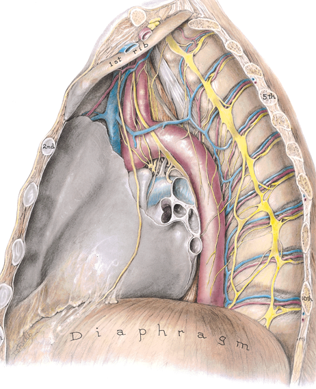
posterior mediastinum from left side
can see aortic arch + descending aorta
Branches form thoracic aorta
bronchial a.
pericardial a.
posterior intercostal a.
subcostal a. (last intercostal a.)
superior phrenic a.
goes to diaphragm
esophageal a.
mediastinal a.
Thoracic aorta vertebrae levels
TV4 - TV12
Esophagus innervation
vagus n.
Esophageal anatomose
upper portion: caval system
lower portion: portal system
Azygous system of veins
drains body wall + provides alternative if IVC is blocked
intercostal veins
hemizygous veins
accessory hemizygous veins
Vulnerable to metastasis

thoracic duct
major lymphatic channel
Cisterna chyli
lymphatic sac at TV12 in abdomen that ascends towards left brachiocephalic v.
Somatic lymph nodes (for body wall)
axillary
parasternal
intercostal
diaphragmatc
Visceral lymph nodes (for viscera)
anterior mediastinal
posterior mediastinal
tracheobronchial
Lymphatic drainage of breasts
inferior breast → abdominal/inferior phrenic lymph nodes
medial breast → parasternal nodes
most of breast → axillary nodes
Lymphatic drainage of thoracic wall
superior half → axillary nodes (+ parasternal nodes)
inferior half → superior inguinal nodes
Lymphatic drainage of heart
right → brachiocephalic nodes
left → tracheobronchial nodes
Lymphatic drainage of lungs
right lung → tracheobronchial nodes + R paratracheal nodes
left superior → superior tracheobronchial → L paratracheal
left inferior → inferior tracheobronchial/ carinal nodes → R paratracheal » L paratracheal
Tracheobronchial node branches
pulmonary
within lungs
bronchopulmonary (hilar)
around hilum of lungs
inferior tracheobronchial (carinal)
below carina
superior tracheobronchial
above tracheal bifurcation
tracheal (paratracheal)
along trachea
