Axial and Appendicular Skeleton
 Call Kai
 Learn
Learn Practice Test
 Spaced Repetition
 Match
Match Flashcards
 Knowt Play
1/177

There's no tags or description
Looks like no tags are added yet.
Name | Mastery | Learn | Test | Matching | Spaced | Call with Kai | Chat |
|---|
No analytics yet
Send a link to your students to track their progress
178 Terms
Frontal Bone (skull)
forehead bone

Parietal Bone (skull)
Bones that form the sides and top of the cranium.

occipital bone (skull)
back of skull

Temporal Bone (skull)
Side of skull

coronal suture (skull)
the suture between the parietal and frontal bones of the skull

sagittal suture (skull)
the suture in middle vertical, separates parietal bones

squamous suture
the suture between parietal and temporal bones

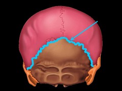
lambdoid suture
the suture between parietal bones and occipital bone
Sphenoid bone (lateral view)
forms part of the base of the skull and parts of the floor and sides of the orbit

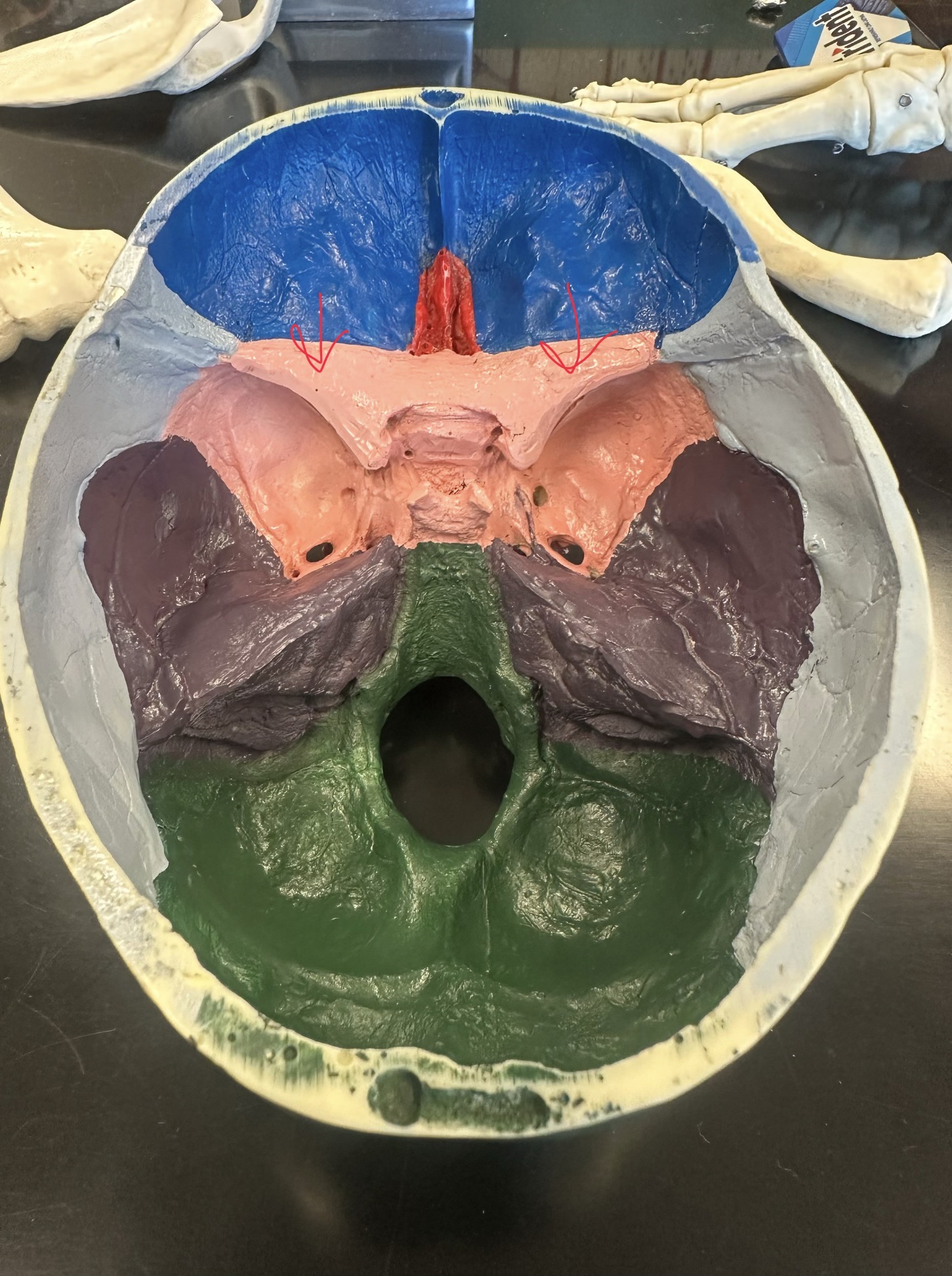
Sphenoid bone (superior view)
Butterfly shaped
-Greater wing
-Lesser wing

Sphenoid greater wing

Sphenoid lesser wing
Ethmoid Bone (Medial View)
the inside portion of the eye socket, goes through behind the nose, and pops out at other inside portion of eye socket

Ethmoid bone

frontal sinus
cavity within the frontal bone (swimming pool in the frontal bone)

sphenoid sinus
cavity located in body of sphenoid bone

styloid process (temporal bone)
pole-like process extending downward from the temporal bone on each side of the skull

mastoid process (temporal bone)
round projection on the temporal bone behind the ear
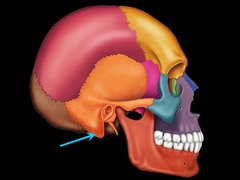
external auditory meatus (temporal bone)
tube-like opening for ear canal in temporal bone

Petrous portion of temporal

Squamous portion of temporal

Mastoid portion of temporarl

Zygomatic process

Mandibular fossa

internal auditory meatus of temporal bone
tube-like hole inside of the skull connecting to the ear canal

external occipital protuberance (occipital bone)
bump on back of head

foramen magnum (occipital bone)
opening of the occipital bone through which the spinal cord passes

occipital condyles (occipital bone)
rounded processes that articulate with the atlas

sella turcica (sphenoid bone)
houses the pituitary gland (swimming pool)

superior nasal concha (ethmoid bone)
Most cranial and posterior; harder to see.

middle nasal concha (ethmoid bone)
Bone between the superior and inferior conchae

inferior nasal concha (ethmoid bone)
Below middle nasal concha

crista galli (ethmoid bone)
superior projection in the middle of the cribriform plate of the ethmoid bone; the horn

cribriform plate (ethmoid bone)
horizontal plate to crista galli that is the textured bit perpendicular to the horn

perpendicular plate of ethmoid
top portion of nasal septum

optic canal
allows the optic nerve to pass to the eye

Maxilla Bone
upper jaw, where upper teeth are in

zygomatic bone
cheek bone

zygomatic process of temporal bone
extension from the temporal bone that forms the posterior portion of the zygomatic arch

nasal bone
forms the bridge of the nose

lacrimal bone
small fragile bone making up part of the front inner walls of each eye socket (the tear drop area)
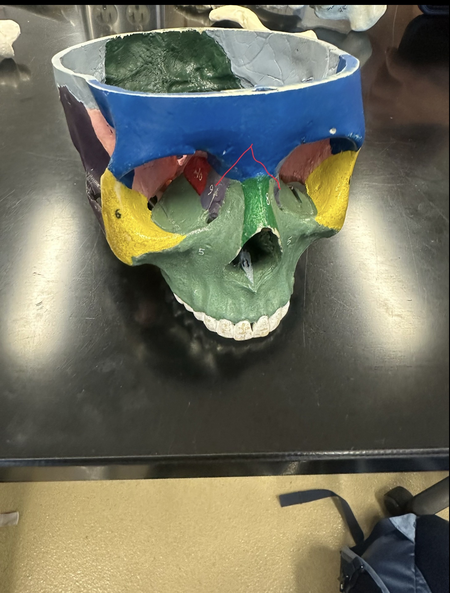
palatine bone
either of two irregularly shaped bones that form the back of the hard palate and helps to form the nasal cavity and the floor of the orbits

vomer bone
forms the base for the nasal septum

Mandible Bone
lower jaw bone

mandible condylar process
articulates with temporal bone

coronoid process
the shark tooth part of the mandible

Mandibular angle

Mandibular body

Maxilla alveolar process

Maxilla alveolus socket

Zygomatic arch

Sphenodial fotanelle

Mastoid fonatalle

Cervical Vertebrae (C1-C7)
first set of seven bones, forming the neck

Thoracic Vertebrae (T1-T12)
second set of 12 vertebrae; they articulate with the 12 pairs of ribs to form the outward curve of the spine

thoracic curve
A primary curve, accommodates the thoracic organs

lumbar vertebrae
L1-L5 lower back

lumbar curve
a secondary curve; balances the weight of the trunk over the lower limbs; it develops with the ability to stand

sacral curve
primary curve, accommodates the abdominopelvic organs

Sacrum
5 fused vertebrae at base of spine

Medial sarcal crest

Sarcal hiatus

Coccyx
tailbone

Atlas (C1)
supports the head, no body

Axis (C2)
second cervical vertebrae. Allows the head to shake "no"

vertebral body
main portion of the vertebra, separate from the arches of the vertebra

Hyoid bone greater horn

vertebral foramen
canal through which spinal cord passes

spinous process
sharp, slender projection

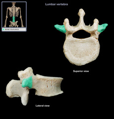
transverse process
two lateral projections from the vertebral arch
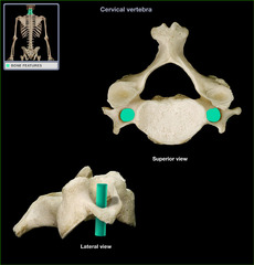
transverse foramen
only found in the cervical vertebrae and allow passage of the vertabral artery, vein, and nerve
pedicles of thoracic vertebrae
walls of the vertebral arch

Laminae of cervical vertebrae

intervertebral disk
fibrocartilage pads that separate and cushion the vertebrae

dens of axis
acts as pivot for rotation of atlas and the skull

bifurcated spinous process
cervical vertebrae process

Sternum
breastbone

manubrium
upper portion of the sternum

sternum body
main long part of sternum

xiphoid process
lower portion of the sternum

rib cage
12 pairs of ribs

True ribs (1-7) vertebrosternal
have a direct attachment to the sternum via cartilage

false ribs (8-12) veterbrochondral
ribs that do not have a direct attachment to the sternum

Vertebral ribs (floating)

costal cartilage
connects ribs to sternum

head of rib
Articulates with the costal facet of a thoracic vertebral body.

neck of rib
between head and tubercle

tubercle of rib
articulates with transverse process of thoracic vertebra
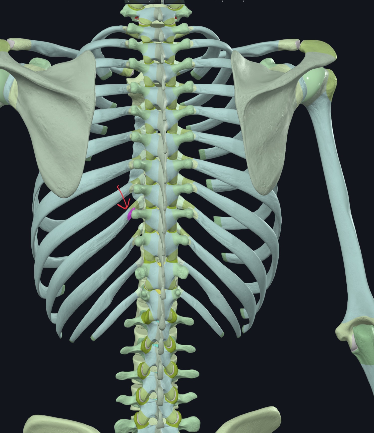
shaft of rib
main part of rib

hyoid bone
a U-shaped bone in the neck that supports the tongue.

Laminae of Thoracic vertebrae

Lumbar vertebrae

Laminae Cervical vertebrae

Costal facet of thoracic vertebrae

Scapula
shoulder blade

scapular spine
Divides posterior aspect of scapula into supraspinatus fossa (above) and infraspinatus fossa (below)

glenoid cavity
socket in scapular that receives head of humerus

acromion process
the highest portion of the shoulder

coracoid process
process above the glenoid cavity that permits muscle attachment

supraspinous fossa of scapula
smooth groove fossa above scapula spine
