3 radio- physics physical characteristics of equipment and ray beams
 Call Kai
 Learn
Learn Practice Test
 Spaced Repetition
 Match
Match Flashcards
 Knowt Play
1/184

There's no tags or description
Looks like no tags are added yet.
Name | Mastery | Learn | Test | Matching | Spaced | Call with Kai | Chat |
|---|
No analytics yet
Send a link to your students to track their progress
185 Terms

LOOK
done

In the production of X-rays, what type of tube is required?
A vacuum-sealed tube that accelerates electrons.

In an X-ray tube, what is the function of the cathode filament?
It emits electrons by thermionic emission.

In an X-ray tube, what is thermionic emission?
The release of electrons from an electrode due to its temperature.
the production of X-rays required a vacuum-sealed tube that accelerated electrons (emitted by thermionic heating of the cathode filament) towards what
high-atomic number metal anode
In an X-ray tube, what does the generator establish between the cathode and anode?
A potential difference (V)
the maximum kinetic energy that electrons can acquire is given by the …..
potential difference between the cathode and anode
In what unit is the potential difference applied between the cathode and anode expressed?
Kilovolt peak (kVp).
what does this equation mean
Emax [ keV] = kVp
the maximum energy of the emitted proton

in intraoral dentistry units usually operate at what voltage
60-70 kVp
in orthopantomography what is the voltage value
60-90 kVp
what is the voltage value for CBCT
70–120 kVp
What kVp range is typically used for panoramic dental X-rays?
60–90 kVp.
What controls the total amount of radiation generated in dental X-ray equipment?
mA and exposure time.
What does the product mA × time represent in dental X-ray equipment?
mAs.
What does mAs control in dental X-ray equipment?
The total amount of radiation generated.
cathode
What is the function of the cathode filament in an X-ray tube?
When heated, it emits electrons by thermionic emission.

cathode
What does the focusing cup do in an X-ray tube?
It helps direct the electron cloud toward a small area of the anode.
cathode
At what potential is the focusing cup in an X-ray tube?
The filament potential.
anode
What is the anode known as compared to the cathode in an X-ray tube?
the target
anode
What is the anode target typically made of?
Tungsten (W, Z = 74).
anode
Why is tungsten typically used for the anode target?
Because of its high melting point, good thermal conductivity, and low vapor pressure.
anode
Which materiasl can be used for the anode target in some special systems?
Molybdenum (Mo, Z = 42).
Gold (Au, Z = 79).
tube window and housing
What does the thin window of an X-ray tube allow?
The useful beam to exit.
tube window and housing
What is the function of the housing and shielding of an X-ray tube?
They reduce leakage radiation.
vacuum
Why is a vacuum essential inside an X-ray tube?
To avoid energy losses through collisions with gas.
What happens to most of the electron-beam energy when it strikes the target?
It is converted into heat.
What happens to a small fraction of the electron-beam energy at the target?
It is transformed into X-radiation (characteristic and bremsstrahlung)
read
Focal Spot Size, Sharpness, and Heel EffectSimple explanation
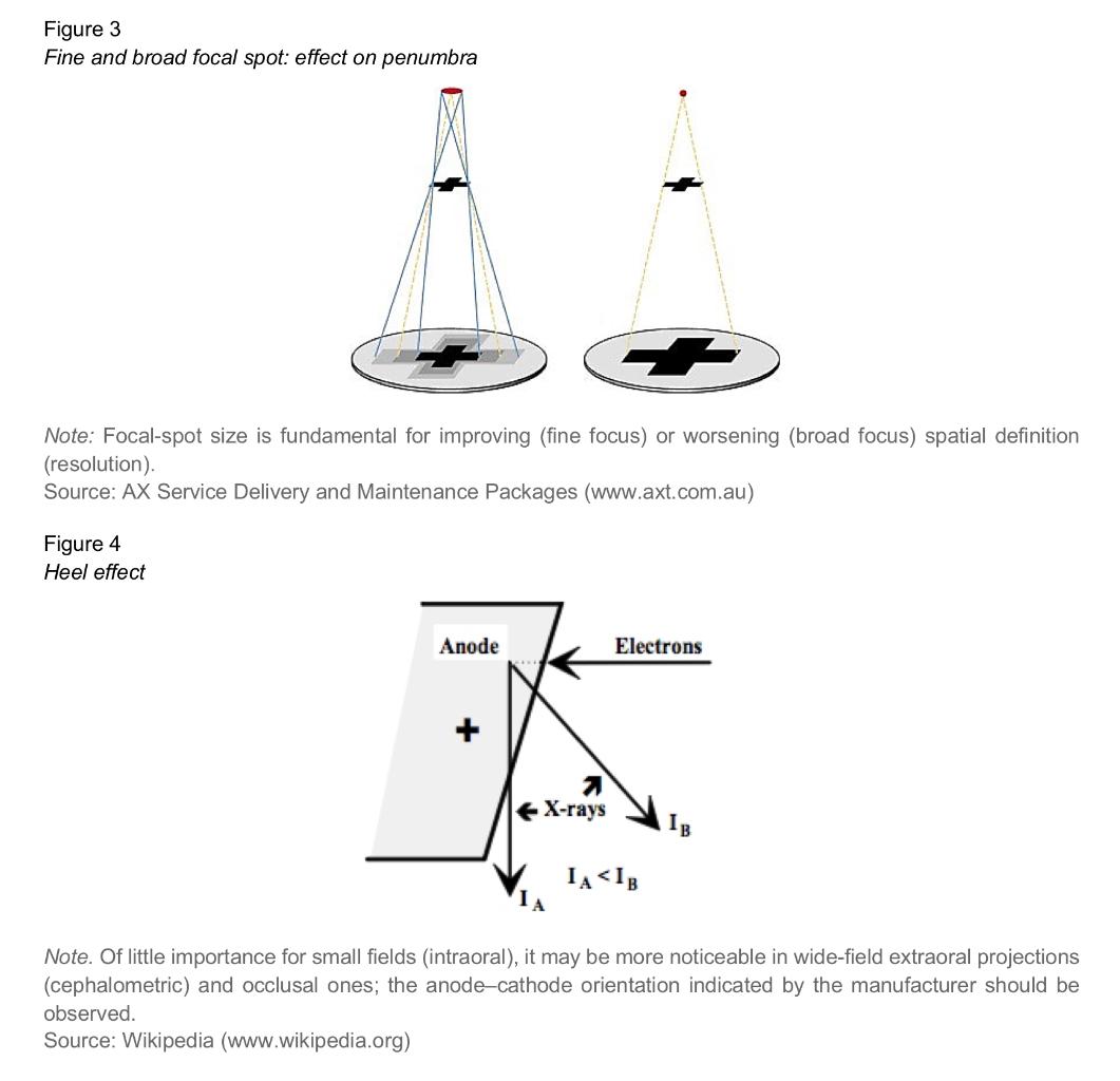
The focal spot is the area of the anode where the electron beam converges. A smaller focal spot improves spatial definition (resolution), but it also concentrates more thermal load.
The size of the focal spot also affects penumbra: a larger focal spot produces more penumbra and therefore reduces sharpness.
The heel effect means that X-ray intensity is greater on the cathode side than on the anode side. This happens because of self-absorption within the target. It matters more when using wide fields
done
What is the X-ray focal spot?
The area of the anode where the electron beam converges.
What effect does a small focal spot have on spatial definition?
It improves spatial definition (resolution).
What effect does a small focal spot have on thermal load?
It concentrates thermal load.
What type of focal spots does modern dental equipment use?
Fine focal spots (Approximately 0.4 mm)
What is the fine focal spot size in high-resolution applications?
Approximately 0.4 mm.
What determines the penumbra in X-ray imaging?
The focus–object–detector geometry.
What effect does a larger focal spot have on penumbra?
It increases penumbra.
What effect does increased penumbra have on image sharpness?
It reduces sharpness.
ANS
decrease focal spot = sharpness/resolution
decrease focal spot = heat load
decrease focal spot = penumbra
increase resolution
increase heat load
decrease penumbra
what is the heel effect
effect that causes higher emitted intensity on the cathode side
What causes the heel effect in an X-ray tube?
Self-absorption within the target itself.
How does the heel effect affect X-ray intensity?
Intensity is greater on the cathode side than on the anode side.
When should the heel-effect intensity gradient be considered
In wide-field projections.
look
done
beam filtration
the beam emerging from the tube is what
polychromatic
beam filtration
what does filtration do
removed low-energy photons
beam filtration
why is it important to remove low energy photons
otherwise they would be absorbed into the skin without contributing diagnostic information
beam filtration
filtration removed low-energy photos - and so doing what to the beam itself
hardening the beam (increasing its mean energy) while reducing its intensity
beam filtration
total filtration
What is total filtration in an X-ray tube?
The sum of inherent filtration and added filtration.
beam filtration
total filtration
What makes up the inherent filtration of an X-ray tube?
The tube window and fixed components.
beam filtration
total filtration
What is the typical equivalent thickness of inherent filtration?
Approximately 0.5 mmAl.
beam filtration
total filtration
What can cause inherent filtration to increase over time?
Tungsten deposits.
beam filtration
total filtration
What is added filtration in an X-ray tube?
Aluminium foils placed between the tube exit and the collimator.
beam filtration
total filtration
What thickness of aluminium foils is used for added filtration?
1–2 mm aluminium.
beam filtration
total filtration
Where are the added filtration aluminium foils positioned?
Between the tube exit and the collimator.
beam filtration
total filtration - legal requirements exam
What is the minimum total filtration for dental units operating at ≥70 kVp?
2.5 mmAl.
beam filtration
total filtration - legal requirements exam
What is the minimum total filtration for dental units operating at lower voltages?
1.5 mmAl.
beam filtration
total filtration - legal requirements exam
read
TF ≥ 2,5 mmAl for ≥ 70 kVp and ≥ 1,5 mmAl for < 70 kVp
done
What happens to maximum photon energy when filtration is added?
It does not change.
What happens to the position of characteristic lines when filtration is added?
It does not change.
What happens to the relative intensity of characteristic lines when filtration is added?
Their relative intensity decreases.
collimation
what is the function of a collimator
It delimits the irradiated area, reducing dose and scattering radiation
collimation
What type of collimator is recommended for intraoral radiography?
A rectangular collimator.
collimation
What is the approximate size of a rectangular intraoral collimator?
35 × 45 mm².
collimation
What type of cone is used with the rectangular intraoral collimator?
A long cone, strictly speaking a PID (Position Indicating Device).
collimation
What is the recommended length of the PID for intraoral radiography?
20–30 cm.
collimation
What type of collimation is used in orthopantomography?
Double collimation.
collimator
for orthopantomography we use a double collimator where are they paced
one in the front of the tube and another in the front of the detector/image receptor
automatic exposure control (AEC)
What does automatic exposure control (AEC) do?
It interrupts the X-ray exposure when a preset optical density or signal level is reached at the receptor.
automatic exposure control (AEC)
What does AEC effectively adjust when the optical density/present signal level is reached
The mAs (milliampere-seconds, and it represents the total amount of X-ray radiation produced during an exposure)
automatic exposure control (AEC)
In which type of dental X-ray units are AEC systems common?
Extraoral units.
automatic exposure control (AEC)
Why do intraoral units typically regulate only exposure time?
Because intraoral units usually have fixed mA.
What do intraoral units with fixed mA usually regulate?
only the exposure of time
image receptors - film
What do image receptors do in dental radiography?
They convert differences in transmitted-beam intensity into diagnostic information.
image receptors - film
What is the base of radiographic film made from?
Polyester.
image receptors - film
What is the polyester base of radiographic film coated with?
A gelatine emulsion containing silver halides and a protective overcoat.
image receptors - film
What silver halides are contained in radiographic film?
Silver bromide (AgBr) and silver chloride (AgCl).
image receptors - film
How is intraoral radiographic film enclosed?
In a light-tight packet (opaque packet)
image receptors - film
What does the intraoral film packet contain?
Black paper and a posterior lead foil.
image receptors - film
What is the function of the posterior lead foil in intraoral film?
It reduces backscatter.
image receptors - film
Where is extraoral radiographic film placed?
In a light-tight cassette with intensifying screens.
image receptors - film
What happens to some Ag⁺ ions when radiographic film is exposed?
They are reduced to metallic silver.
image receptors - film
What is formed when Ag⁺ ions are reduced to metallic silver during exposure?
A latent image.
image receptors - film
What is the latent image proportional to?
The exposure.
image receptors - film
What happens to the latent silver during development?
It catalyses the selective reduction of more Ag⁺ to Ag⁰.
image receptors - film
What does the reduction of more Ag⁺ → Ag⁰ during development make visible?
The dark areas of the image.
image receptors - film
What is the function of the stop bath during film processing?
t halts the action of development.
image receptors - film
What is the function of the fixer during film processing?
It dissolves unexposed halides and stabilizes the image.
image receptors - film
What determines the different grey shades on processed radiographic film
Greater or lesser amounts of retained metallic silver.
digital systems
What are the two main approaches used in digital dental image receptors?
Photostimulable phosphor plates (PSP) and electronic CCD/CMOS sensors.
digital systems
What do photostimulable phosphor plates (PSP) do with X-ray energy?
They store the energy deposited by the X-ray beam.
digital systems
How is a PSP plate read?
With a laser.
digital systems
What does the laser release when reading a PSP plate?
Light proportional to the latent signal.
digital systems
What is the light released from a PSP plate proportional to?
The latent signal.
digital systems
What happens to the signal from a PSP after laser reading?
It is converted into a digital image.
digital systems
What do electronic CCD/CMOS sensors convert X-rays into?
Electrical charges.
digital systems
How can CCD/CMOS sensors receive X-rays?
Directly or through a scintillator.
digital systems
What happens to the electrical charges produced by CCD/CMOS sensors?
They are read out and displayed immediately on the monitor.
digital systems
What is the main difference in processing between PSP and CCD/CMOS systems?
PSP is read with a laser; CCD/CMOS charges are read out immediately.
X-ray emission spectrum
What does an X-ray emission spectrum represent?
number of photons versus their energy.
X-ray emission spectrum
What are the two components of the X-ray emission spectrum?
A discrete component and a continuous component.