fetal monitoring CH14
 Call Kai
 Learn
 Practice Test
 Spaced Repetition
 Match
 Flashcards
 Knowt Play
1/43

There's no tags or description
Looks like no tags are added yet.
44 Terms
what are the goals of fetal surveillance?
maximize uterine & umbilical blood flow
assess & maximize fetal oxygenation
detect abnormal changes in FHR/fetal oxygenation
guide decision making & evaluate effectiveness of interventions
maintain appropriate uterine activity
support coping & labor progress
what is intermittent auscultation? what do we use? when is it used? who are the best candiates for this?
using a fetoscope or doppler to listen to FHR
IA is used for low risk pregnancies & normal FHR (110-160 bpm w/ regular rhythm)
auscultate FHR before, after, during a UC
assess UC pattern by palpation/maternal report
best candidate:
low risk pregnant individuals, full term, spontaneous labor on its own w/o medical inductions, absence of maternal or fetal comorbidities (HTN, diabetes
what are the 2 types of continuous electronic fetal monitoring? where is everything placed (describe steps)? how do they differ from one another?
external
uses EFM & uterine monitors use ultrasound to detect FHR & uterine pressure
FHR & UC are recorded & displayed on monitor
steps:
place toco transducer (uterine contractions) near fundal area b/c strongest contractions are there → button surface looking & placed higher up
apply gel to ultrasound (FHR) transducer & place it where the fetus’ back is (locate w/ leopold maneuver) → smoother surface looking → placed on lower abd
secure with a belt or faha
wireless EFM & toco are available → allows pt freedom of movement while in labor
internal
invasive devices
fetal scalp electrode (FSE) on fetal head or presenting part of the fetus to detect FHR
ex. applied to buttocks if pt is breech
IUPC (intrauterine pressure catheter) placed in uterine cavity
only used when someone is not progressing & unsure if contractions are inadequate
IUPC measures intensity of uterine contraction
what are the limitations to external monitoring?
unable to measure intensity & must be palpated to compare to nose, chin, forehead
correlated with higher operative deliveries (using tools)
sores occur if worn for a long time
what conditions need to be met for internal monitoring? what are the risks of internal monitoring? what is it contraindicated in?
conditions that need to be met:
spontaneous or artificial ROM
internal equipment can’t bypass or puncture an intact sac
cervix must be open enough to all clinician’s fingers & instrument to pass through
minimum of 1-2 cm dilation
fetal presenting part must be low enough in the pelvis
it must attach to a bony surface; avoid face, fontanelles, or genitals
risks:
contraindicated in: chorioamnionitis (infection of fetal membranes & amniotic fluid), active maternal genital herpes, HIV, +GROUPB STREP, placenta previa, undiagnosed vaginal bleeding
intrauterina infections (increases risk of chorionamnionitis)
injury where it can cause trauma, lacerations, uterine perforation
what can we do if we do not have amniotic fluid?
infuse NS through intrauterine pressure catheter → amnioinfusion
can tx thick meconium → fluid just leaks out of uterus
what is frequency of FHR assessment based on?
risk status, stage of labor, ongoing clinical assessment
for intermittent auscultation, when is it used? how often is it used during the latent, active, and pushing phase?
used for no risk factors
Q1 hr latent phase
Q30 min active phase
Q15 min pushing phase
switch to continuous EFM if abnormal FHR
for continuous EFM when is it used? how often is it used during normal tracing, decelerations, 2nd stage of labor?
used for when risk factors are present
ex. chronic HTN, GDM, pain meds, etc
Q1 hr normal tracing
Q30 min for decelerations
Q15 min 2nd stage of labor
what are the components to identify in EFM tracing?
FHR baseline & variability
identification of accelerations & decelerations
interpret uterine contractions
what is the fetal heart rate baseline? what is normal, bradycardia, tachycardia?
baseline = mean FHR
rounded to increments of 5
excludes accelerations, decelerations, or marked variability
at least 2 mins of identifiable baseline segments (not necessarily continuous) or it is indeterminate
normal baseline FHR 110-160 bpm
bradycardia <110 bpm for 10 mins or >
tachycardia >160 for for 10 mins or >
what is tachycardia? what are the causes? what nursing actions to take?
baseline above 160 for at least 10 min
causes:
infection, dehydration, certain drugs
nursing actions:
VS, abx, antipyretics, ice packs, IV hydration
what is bradycardia? what are the causes & some example? what nursing actions to take?
baseline below 110 for at least 10 min
causes:
fetal hypoxia caused by abrupt interruption of oxygenation (ex. maternal hypotension, abruption, cord prolapse, tachysystole)
nursing actions:
fetal resus, move to the OR
what can influence fetal heart rate?
uteroplacental unit
disruption to the intervillious space → decreases amount of O2 to fetus
ex. contractions, cord compression problems w/ placenta
a decrease in oxygen to the fetus results in deceleration of FHR
autonomic nervous system
PNS (vagus nerve) → decrease HR
SNS → increase HR
baroreceptors
stretch receptors that detect pressure changes → deceases HR, CO, BP
incr. BP → baroreceptors trigger vagus nerve → decrease HR
CNS
regulates variability
meds (ex. mag sulfate & fentanyl) can cross placenta → decrease HR
chemoreceptors
stimulates vagus nerve → decrease HR
hormonal regulation
decrease in oxygenation → hormones are released
epinephrine, norepinephrine, vasopressin, catecholamines are released to facilitate hemodynamic changes to maximize blood flow to vital organs
what does baseline variability mean? what is it an important indicator of? what does it reflect?
variability: small fluctuations in fetal heart rate
fluctuation in baseline FHR that are irregular in amplitude & frequency
shows peak & trough of HR within baseline
most important predictor of adequate fetal oxygenation & fetal reserve during labor
reflects intact CNS
describe the fetal heart rate variabilities. how does it look?
absent (worrisome)
little to no fluctuation in FHR
variability less than 5 bpm or basically 0
look: straight line
minimal (kind of concerning)
FHR detectable but less fluctuation than 5 bpm
look: not moving a lot
moderate (happy baby)
fluctuation in FHR ranges from 6 to 25 bpm
marked (may be concerning)
fluctuation in FHR >25 bpm
look: going crazy
what are some influences on variability?
fetal wake/sleep cycle
fetus less active during sleep
maternal & fetal oxygenation
decreased O2 → reduces variability
placental function
poor blood flow
ex. pre-e or pregnancy beyond due date
umbilical function
cord compression disrupts blood flow
narcotic effects
meds can depress CNS
fetal causes: anomalies, neuro function, cardiac function, prematurity, etc
what are the 2 characteristics of fetal heart rate?
acceleration
deceleration
early, late, variable, prolonged
what is acceleration? what could absence of it mean? how long are prolonged accelerations?
predictive of adequate oxygenation
absence of accels in intrapartum in fetal acidemia (baby’s blood loss has low pH & high acidity)
> 32 GA increase in FHR at least 15 beats above baseline for at least 15 secs
accels may be 10 beats above baseline & lasts 10 secs < 34 wks
prolonged accels are >2 mins < 10 mins
what is early deceleration? what are the shape & timing characteristics? what are the causes? what is the clinical significance (do they require intervention, what does it indicate)?
gradual decrease in FHR that start before the peak of a contraction & returns to baseline by end of contraction
characteristics
shape: visually apparent gradual decrease of >30 secs to the nadir (peak), smooth & symmetrical
timing: occurs at the same time as contraction, beginning before & returning to baseline with contraction (mirrors contraction)
causes:
head compression, which stimulates vagal nerve & results in decrease in HR
clinical significane:
usually benign & a normal physiological response during labor, do not indicate fetal distress, do not require intervention

what is late deceleration? what are the shape & timing characteristics? what are the causes? what is the clinical significance (do they require intervention, what does it indicate)?
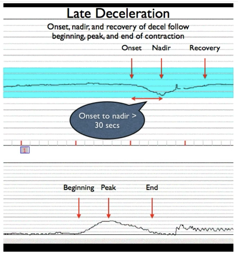
gradual decreases in FHR that begin after the peak of contraction & return to baseline after the contraction has ended
characteristics:
shape: gradual & smooth, visually apparent symmetrical
onset is gradual w/ onset of nadir >30 secs
onset, nadir, recovery of decel occurs after onset of UC
timing: begin after peak of contraction & return to baseline after contraction ends
cause:
uteroplacental (placental) insufficiency, which reduces blood flow & oxygen to fetus
clinical significance:
indicative of fetal distress & requires prompt evaluation & intervention
persistent late decels → need to change maternal positions, increased IV fluids, or even expedited delivery
what are the nursing actions for late deceleration?
fetal resuscitation
consider tocolytics (relax uterus from making excessive UC)
consider delivery
consider fetal scalp stimulation or VAS
what is variable deceleration? what are the shape & timing characteristics? what are the causes? what is the clinical significance (do they require intervention, what does it indicate)? what are the nursing actions?
abrupt decrease (periodic or episodic) in FHR that varies in shape, timing, duration, depth
often irregular & may or may not coincide w/ contractions
recurrent variables that become deeper = fetal acidemia
characteristics:
shape: V, U, or W shaped
visually apparent of ABRUPT decrease in FHR of <30 secs from baseline to nadir with drop of >=15 bpm below baseline & lasts for >15 seconds but < 2 mins
timing: occur at any time during contraction, often beginning & ending suddenly
cause:
umbilical cord compression, which reduces blood flow & oxygen to fetus
clinical significance:
can be benign but can indicate issues aka cord compression OR severe cases → fetal hypoxia
continuous monitoring & assessment are needed
nursing actions
fetal resuscitation
consider amnioinfusion
oliohydramnios → less cushioning → cord compression → variable decels

what are the characteristics of concerning variable deceleration?
prolonged
> 60 bpm lasting for more than 60 secs
absent variability
what is prolonged deceleration? what is it due to/caused by? what are the nursing actions?
decrease in FHR below baseline >15 bpm lasting >2 mins but <10 mins
due to profound change in fetal O2:
interruption of placental perfusion
tachysystole
too many contractions
excessive UC reduces time for placental blood flow to recover
maternal hypotension
abrupted placenta
placenta separates → reducing exchange
interruption of umbilical blood flow
cord compression
cord prolapse (cord is between cervix & vagina aka ahead of the baby)
nursing action:
fetal resuscitation
consider amnioinfusion
consider tocolytics
consider delivery
turn to the side → reposition
anything to promote circulation

what does veal chop stand for?
variable → cord compression
early → head compression
accels → okay (oxygenated)
late → placental insufficiency
what are the 4 components to interpret uterine contractions?
frequency: time elapsed from the beginning of 1 contraction to the beginning of the next
assessed on fetal monitor or by palpation to ensure contractions occur at appropriate intervals for the stage of labor
duration: time elapsed from the beginning of a contraction & its end, measured in seconds
important to monitor b/c excessive long contractions → uterine hyperstimulation & potential complications
intensity: strength of contraction at its peak palpation (mild, moderate, strong → nose, chin, forehead)
determines effectiveness of contractions in dilating the cervix
measured by palpation or IUPC (mmHg)
resting tone: baseline pressure of the uterus before contractions begin (soft or firm)
how relaxed or tense the uterus is when not contracting
measured using IUPC or palpation (described as soft if relaxed)
IUPC normal resting tone is <20 mmHg
normal resting ensures proper fetal oxygenation & uterine recovery between contractions
if fetal HR goes down = worry about deceleration

what is the normal uterine contractions? what is tachysystole & what is the worst outcome it can result in?
normal: <= 5 UCs in 10 mins averaged over 30 minutes
tachysystole (excessive uterine activity → progressively effects fetus)
> 5 UCs in 10 mins over 30 minutes
excessive UC → decreased blood flow in the intervillous (where O2 exchange occurs) → decreased O2 to fetus → fetal hypoxemia → abnormal FHR
tachysystole may result in abruption or uterine rupture
what are the nursing actions for tachysystole?
turn off pictocin
IV fluids → corrects hypotension
reposition pt to side lying
administer a tocolytic (terbutaline) → relaxes uterine muscle
how are episodic & periodic changes related to UCs?
episodic → not associated w/ contractions
ex. baby moves causing FHR to increase even though mom does not have contraction
periodic → ARE associated w/ contractions
how to classify/interpret fetal HR?
by categories 1, 2, 3of the NICHD
what is characteristics of category 1 (baseline HR, variability, accelerations, decelerations)? what is the clinical significance?
category 1: reassuring/normal
baseline HR: 110-160 bpm
baseline variability: moderate (6-25 bpm)
accelerations:
present or absent
accelerations are generally reassuring if they occur w/ fetal movement or contractions
decelerations:
no late or variable
early decels may be present & considered normal
clinical significance
indicates well oxygenated fetus (normal acid basis status), normal CNS function
no immediate concerns
continue monitoring & care are generally appropriate
what is characteristics of category 2 (baseline HR, variability, accelerations, decelerations)? what is the clinical significance?
category 2: indeterminate
baseline HR:
within or outside normal range (ex. less than 110 or more than 160)
baseline variability:minimal or absent variability
accelerations:
may or may not be present
decelerations:
early: gradual decreases that coincide w contractions
late: gradual decreases that begin after the peak of contraction
variable: irregular in shape & timing
clinical significance
fetal status is uncertain & requires further eval
may require additional monitoring, interventions, or assessment to determine the cause & address potential issues
what is characteristics of category 3 (baseline HR, variability, accelerations, decelerations)? what is the clinical significance?
category 3: abnormal
baseline HR:
either less than 110 (brady) or more than 160 (tachy) w/ persistent abnormalities
baseline variability: absent variability accompanied with either recurrent late decels, recurrent variability, or bradycardia
accelerations:
absent, esp in the presence of concerning decels
decelerations:
recurrent late: gradual decreases that begin after the peak of contraction
recurrent variable: abrupt decreases that vary in shape & timing, often associated w cord compression
prolonged decel: decrease in FHR lasting more than 2 mins but less than 10 min
clinical significance
indicate possible fetal distress or hypoxia
immediate intervention is necessary, which includes repositioning mom, increase maternal O2, or preparing for an expedited delivery (c-section), increase fluids
what heart rate pattern is part of category 3? what is it associated with?
sinusoidal (smooth, repeating waves)
associated w severe fetal anemia, ominous often premorbid finding

what are the nursing managements and interventions for category 3?
reposition
change mother’s position can alleviate cord compression & improve blood flow
increase fluids
administer fluids can help improve uterine blood flow & oxygen delivery to the fetus
oxygen admin
providing supplemental oxygen to the mother may help improve O2 to the fetus
assessing uterine activity
ensuring that uterine contractions are not too frequent or too strong can help reduce the risk of decelerations
discontinue or decrease the oxytocin (pitocin) then notify MD
immediate delivery
in cases of persistent or severe late decels or other indications of fetal distress, expedite delivery via c-section may be needed
when should we intervene?
recurrent late decels
recurrent variability decels
minimal variability > 30 min
fetal tachycardia
fetal bradycardia
prolonged decels
where does O2 exchange occur in the umbilical cord? what blood travels to the fetus?
occurs in intervillous space
oxygenated blood travels to fetus
what is the umbilical cord acid base balance & what do we do? what does it evaluate? how is it measured?
objective analysis of the pH blood levels
after birth, take blood from the umbilical artery & the vein to examine acid base
evaluates whether a poor neonatal outcome is due to hypoxia as evidenced by an acidic pH
normal levels are compared to the cord blood sample:
acidic pH → fetus moves from aerobic metabolism to anaerobic
normally, can the fetus maintain aerobic metabolism if there is slight decrease in blood flow & how? when does anaerobic respiration occur & what can it cause?
normally, it can maintain aerobic metabolism even though there is decrease in blood flow to uterus d/t fetal reserve (more O2 in placenta’s intervillious space than the fetus actually needs)
anaerobic respiration occurs when O2 in intervillious space falls below 50% of normal levels
can cause…
redistributes blood to vital organs (heart, brain, adrenal glands)
chronically altered O2, fetal growth will decelerate & lead it intrauterine growth restriction
fetal myocardium changes → changes in HR
what is fetal distress? how do we address this & what does it prevent? what are the nursing interventions?
an emergency pregnancy, labor, delivery complication where the baby experiences birth asphyxia (O2 deprivation) → changedsin baby’s HR, movement, meconium in amniotic fluid
address by having c-section to prevent hypoxic ischemic encephalopathy (brain injury d/t lack of O2 & blood)
interventions:
reposition
increase fluids
o2 admin
assess uterine activity
d/c or decrease oxytocin & notify MD
immediate delivery
what is uterine irritability?
a condition where the uterus experiences frequent or irregular contractions that are not yet strong enough to be considered true labor contractions

what is coupling?
the occurrence of 2 consecutive contractions w/ minimal or brief resting period in between followed by a slightly longer period of relaxation

what is reactive twin strip?
pattern on FHR that indicates a reassuring or normal FHR for both twins → the term is used to describe fetal heart rate patterns & indicative of well being during labor or before (antepartum) monitoring