Anatomy & Physiology (McGraw Hill) - Chapter 4
 Call Kai
 Learn
 Practice Test
 Spaced Repetition
 Match
 Flashcards
 Knowt Play
1/39

Earn XP
Description and Tags
Cells
40 Terms
light microscope
passes light through specimen to produce 2D image

transmission electron microscope (TEM)
beam of electrons passes though specimen to produce magnified/high resolution 2D image

scanning electron microscope (SEM)
beam of electrons passes through and across surface of specimen to produce 3D image

integral proteins
proteins embedded within/extend across phospholipid bilayer of cell membrane

peripheral protein
proteins loosely attached to external or internal surface of membrane

diffusion
passive transport of solutes/ions across membrane down the concentration gradient
simple - small, nonpolar solutes move unassisted
facilitated - small, polar solutes require channels (leak or gated) and carrier proteins

osmosis
passive movement of water across semipermeable membrane based on concentration gradient, either directly through phospholipid bilayer or via aquaporins (protein water channels)

osmotic pressure
pressure exerted by movement of water across semipermeable membrane (caused by different solution concentrations)

hydrostatic pressure
pressure exerted by a fluid on the inside wall of its container

tonicity
ability of a solution to change the volume or pressure of a cell by osmosis
isotonic
hypertonic
hypotonic

active membrane transport
substances move across membrane against concentration gradient (requires energy)
primary - uses energy directly from ATP via phosphorylation
secondary - uses energy from movement of additional substance down its gradient

ion pump
cellular protein pumps that move ions across membrane
phosphorylation of ATP changes protein structure
Ex: Ca²+ pumps prevent calcium accumulation, cell remains flexible

symport
(secondary active transport) two substances moved in the same direction

antiport
(secondary active transport) two substances moved in the opposite direction

vesicular transport
transporting large substances across plasma membrane by a vesicle (requires energy)
exocytosis
endocytosis

phagocytosis
“cellular eating”
cell engulfs a large particle (ex: white blood cell engulfs microbe)

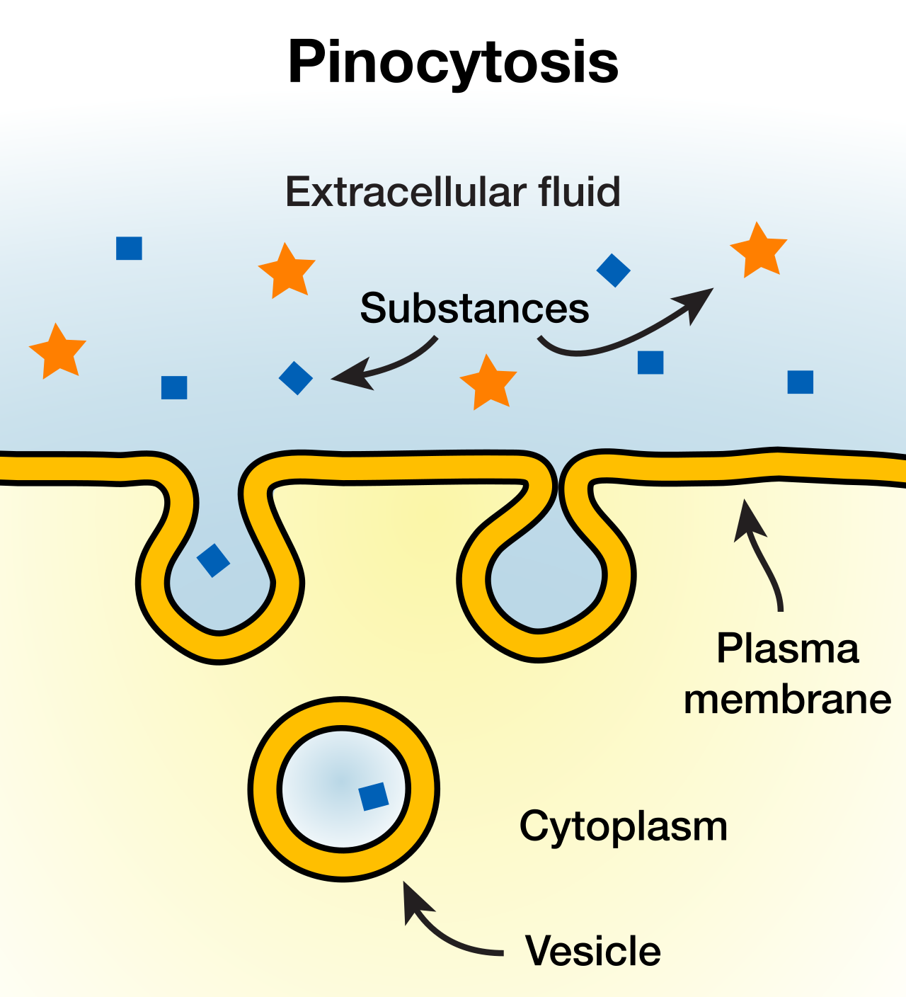
pinocytosis
“cellular drinking”
cell internalizes droplets of interstitial fluid containing dissolved solutes
receptor-mediated endocytosis
cell uses receptors on plasma membrane to bind external molecules and trigger endocytosis

resting membrane potential
electrochemical gradient when a cell is at rest
unequal distribution of ions across plasma membrane (more K+ in cytoplasm, more Na+ outside cell)
unequal charge (more positive+ inside cell)

channel-linked receptors
permits ion passage in or out of cell in response to ligand binding to gated channel

enzymatic receptors
ligand binds to receptor to activate protein kinase enzyme, which then phosphorylates other enzymes within the cell

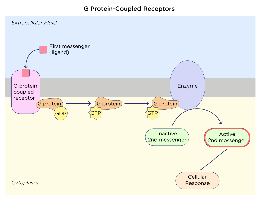
G protein-coupled receptors
indirectly activates protein kinase enzymes with use of g-protein and GTP
Ligand binds to receptor
G protein binds to activated receptor
GTP binds to G protein
GTP/G protein leave receptor, binds to/activates effector protein (i.e. enzyme, ion channel)
Activated effector protein generates secondary messenger
Secondary messenger activates protein kinase
Activated protein kinase phosphorylates other enzymes
rough endoplasmic reticulum
Synthesis: synthesizes proteins for secretion, plasma membrane, and lysosome enzymes
Processing molecules: modifies (i.e. add carbohydrates) and stores proteins
Organelle formation: helps form peroxisomes
Vesicle formation: forms transport vesicles for shipping proteins to Golgi apparatus

smooth endoplasmic reticulum
Synthesis: site of lipid synthesis (i.e. steroids)
Processing molecules: carbohydrate metabolism (i.e. glycogen synthesis)
Detoxification: detoxifies drugs, alcohols, poisons
Vesicle formation: forms transport vesicles for shipping to Golgi apparatus

Golgi apparatus
Synthesis: forms proteoglycans
Processing molecules: modifies and stores proteins from rough ER
Organelle formation: synthesizes digestive enzymes for lysosomes
Vesicle formation: forms secretory vesicles for delivering components to cell membrane and releasing contents from cell via exocytosis

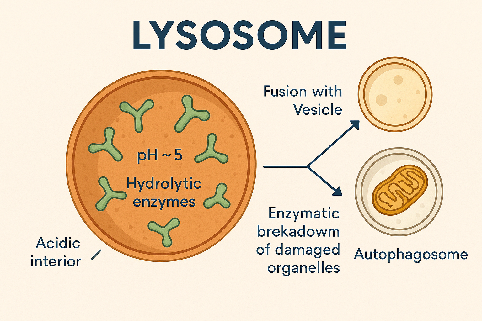
lysosomes
small, membranous sac that contains digestive enzymes (formed by Golgi)
break down molecules of endocytosed vesicles
remove damaged organelles and cellular components (autophagy)
break down cellular components following cellular death (autolysis)
peroxisomes
membrane-enclosed sacs that form from rough ER vesicles
Digestion: break down molecules (fatty acids, amino acids, uric acid) via beta oxidation
Synthesis: form specific lipids (i.e. bile salts)

mitochondria
“powerhouse” of the cell
digest organic molecules to produce ATP via aerobic respiration

ribosomes
Bound (attached to ER): synthesize proteins destined to be incorporated into plasma membrane, exported from cell, or housed within lysosomes
Free (suspended in cytosol): synthesize proteins for use inside cell
BOTH:
contain protein and RNA
arranged into small and large subunit (A, P, E sites)
made within nucleolus and assembled in cytoplasm

centrosome
pair of perpendicularly oriented cylindrical centrioles, surrounded by amorphous protein
Synthesis: organize microtubules (cytoskeleton proteins) and support their growth in nondividing cells
Cell division: direct formation of spindle fibers in dividing cells

proteasomes
protein-digesting organelles located in cytosol and cell nucleus
Protein digestion: degrade ubiquitin-tagged proteins that are damaged, incorrectly folded, or no longer needed
Quality assurance: control quality of exported cell proteins

cytoskeleton
Structural support and organization: maintains cell shape and organize organelles, provides internal support to plasma membrane and microvilli (microfilaments), stabilize desmosome cell junctions (intermediate filaments)
Cell division: separate chromosomes during cell division (microtubules), splits cell into two daughter cells via cytokinesis (microfilaments)
Movement: facilitate cytoplasmic streaming (microfilaments), serve as track for organelle/vesicle movement (microtubules), muscle contraction (microfilaments), control proteins of cilia and flagella (microtubules)

microfilaments
smallest component of cytoskeleton composed of two intertwined actin protein monomers
Functions - maintain cell shape, internal support, cell division

intermediate filaments
intermediate-sized, rigid filaments that provide structural support (i.e. cell junctions)

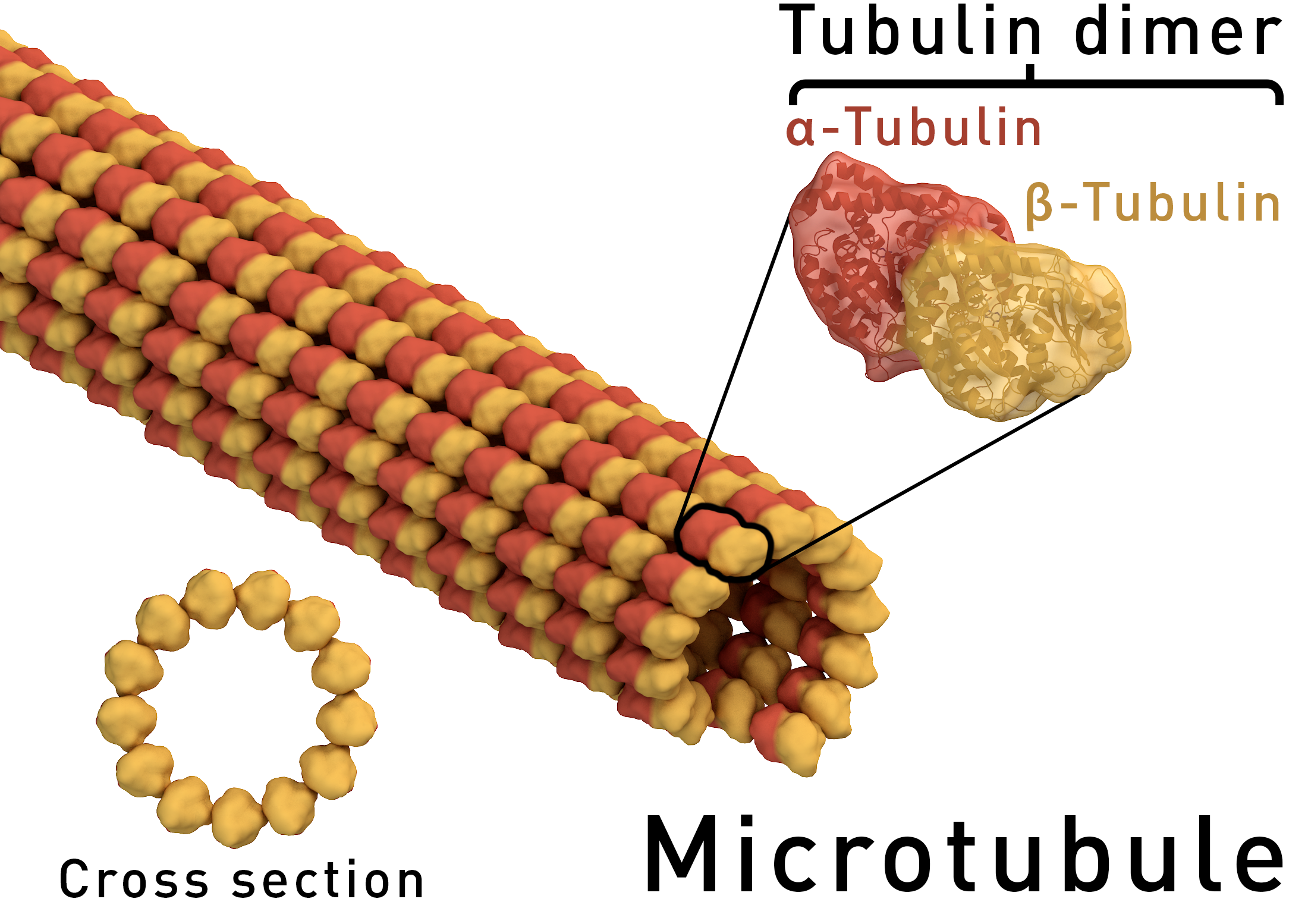
microtubules
largest component of cytoskeleton, composed of tubulin, may be elongated or shorted as needed
Function. - maintain shape, cell transport, cell division
cilia
hair-like projections that move substances along cell surfacef

flagella
long, whip-like cellular appendage that propels entire cell

microvilli
extensions of plasma membrane that increase surface area

tight junctions
strands or rows of transmembrane proteins linking cells, which prevent substances from passing between cells
linked to actin microfilaments
maintain polarity of epithelia
desmosomes
proteins that bind neighboring cells