Unit 2: LEV Pathophysiology
 Call Kai
 Learn
 Practice Test
 Spaced Repetition
 Match
 Flashcards
 Knowt Play
1/60

There's no tags or description
Looks like no tags are added yet.
Name | Mastery | Learn | Test | Matching | Spaced | Call with Kai | Chat |
|---|
No analytics yet
Send a link to your students to track their progress
61 Terms
deep vein thrombosis (DVT)
coagulates blood clot within a deep vein
most common serious sequelae (consequence of prev. disease/injury) is a pulmonary embolism (PE) that originated from a lower extrem. DVT
PE has significant morbidity and mortality if not recognized and/or treated early
risk factors:
age
women >60yr
remains stable for males across age groups
previous VTE (DVT and/or PE)
hereditary hypercoagulable conditions
acquired hypercoagulable conditions
blood stasis conditions

venous thromboembolism (VTE)
consists of DVT in conjunction w/ obstruction to flow to a pulm. artery or branch (PE) from thrombotic emboli
—
90% of pts. w DVT involve lower extremities
79% of pts. w/ a PE have evidence of DVT in their legs
up to 50% of pts. w/ DVT may develop PE
LEV DVT
usually originates from calf veins and propagates up into thigh
affects only lone leg at a time
bilateral LEV exams are rare, if both legs are swollen u/s is ordered for whichever leg is worse
rarely clinically important if isolated to calf veins
less likely to dislodge and travel up the smaller vessels
very clinically important if left untreated and propagates up into larger deep veins
blood
blood composition:
55% plasma
liquid component
mix of water, sugar, fat, proteins, enzyme nutrients, hormones, gases, and salts
maintains body’s fluid balance
45% hematocrit
3 types of blood cells:
RBC, WBC, platelets
plasma composition:
large contributor to blood viscosity
contributes to osmotic pressure
albumin
dec. lvls lead to fluid accumulation in body (hypoalbuminemia)
causes edema
fibrinogen
essential to blood clotting
dec. lvls lead to inc. risk of bleeding
inc. lvls can cause clot formation
red blood cells:
aka erythrocytes
transports O2 to tissue, and CO2 to lungs
COMMON DISORDERS
anemia
dec # of RBCs to carry O2 to tissues » reduces blood viscosity
sickle cell anemia
malformed and elongated RBCs » causes clumping of RBCs which leads to clot formation
polycythemia
elevated RBCs » inc. blood viscosity
platelets:
aka thrombocytes
small cell fragments
assists in clotting (coagulation)
COMMON DISORDERS
thrombocytopenia
dec. # of thrombocytes » can cause excessive bleeding » lowers blood viscosity
thrombocytosis
inc. # of thrombocytes » causes unnecessary clotting » inc. blood viscosity

clot formation
normal setting:
triggered by injured blood vessel
vasospasm
platelets form a temporary plug at site of injury (primary hemostasis)
series of rxns known as coagulation cascade
activates clotting factors
thrombin converts fibrinogen into sticky fibrin strands
fibrins strands weave a mesh » trapping more platelets and RBCs
forms a stable fibrin clot that seals injury and stops bleeding
intravascular thrombus formation:
triggered by endothelial injury or inflammatory signals (ie. surgery, cancer, stasis)
leads to platelet adhesion proteins and cluster to form a primary plug
series of rxns known as coagulation cascade
results in a clot that obstructs blood flow
—
clot formation itself can cause irritation to the intima layer, triggering inflammation
phlebitis: inflammation of a the vein
thrombophlebitis: inflammation of the vein due to a presence of clot

virchow’s triad
DVT etiology
named after German physician Rudolf Virchow
describes 3 primary conditions that lead to thrombosis:
—
hypercoagulability = conditions that inc. blood viscosity and stickiness
venous stasis = conditions that promotes slow moving/stagnant blood flow
endothelial damage = conditions that activate the coagulation casacde
hereditary hypercoagulable conditions
protein C and S deficiencies (antithrombin deficiencies)
factor V leiden » most common inherited hypercoagulable disorder
elevated plasminogen activator inhibitor-1
PAI-1: inhibits fibrinolysis (process of breaking down clots)
non-O blood group » higher lvls of von Willebrand factor
inc. lvls of clotting factor proteins (II, VII, IX, X, XI)
primary hyperhomocysteinemia: impairs the balance of clotting factors
antiphospholipid antibody syndrome: immune system mistakenly produces antibodies that makes blood more likely to clot
myeleodysplasia: elevated lvls of pro-clotting cells, inflammation, and a genetic pre-desposition to clotting
hyperviscosity syndromes
acquired hypercoagulable conditions
malignant cancer: tumours can directly make blood more viscous/sticky
chemotherapy: can dec. body’s natural proteins that thin the blood; some cancer drugs can cause more viscous/sticky blood
heparin-induced thrombocytopenia: body produces antibodies against heparin (which is a natural coagulant)
inflammatory bowel disease: chronic inflammation activates clotting factors, inc. platelets, and impairs fibrinolysis
obesity: promotes continuous, low-grade inflammatory state
pregnancy, oral contraceptive use, or hormonal replacement therapy: elevated lvls of clotting proteins and dec. lvls of proteins that prevent clotting
polycythemia vera: overproduction of RBCs » making blood thicker
blood stasis conditions
immobilization
post surgery <3 months
highest risk <2wks post-op
most DVT are small and isolated to calf veins
~50% of cases spontaneously resolved within 72hrs
slow flow conditions
congestive HF
liver disease
extrinsic compression
pregnancy (compression on iliofemoral region)
trauma (hematoma)
May-Thurner syndrome » compression of lt. CIV by the rt. CIA
clinical findings with DVT
+ D-dimer test » indicated thrombus formation somewhere in body
D-dimer lvls only remain elevated for ~7days post-thrombus formation, so not as useful for chronic thrombosis
>500µg/mL indicates +D-dimer
usually highly sensitive
less sensitive w/ small clots isolated in calf veins » results in low lvls of D-dimer
low specificity, could be (+) from other conditions..
mild bruising/hematoma, hemorrhage, cancer, sepsis
a (-) D-dimer is highly predictive for rling out DVT
—
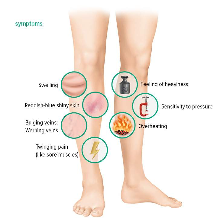
signs and symp:
swelling from edema
pain and tenderness (often focal)
redness & inc. warmth
symptoms inc. when walking & subside w/ rest
pain in buttocks and/or groin (w. iliofemoral DVT)
superficial thrombophelbitis
more common than deep vein thrombophlebitis, and the affected vein can feel like a tender palpable cord near skin’s surface; skin may be red or warm to touch
homan’s sign: discomfort in calf muscles on forced dorsiflexion of the foot w/ knee straight
non-specific, but presents in 1/3rd of DVT pts.

wells criteria
challenges w/ DVT signs and symp:
up to 50% of pts. are asymp.
up to 50% of pts. w/ “classic” DVT symptoms don’t have any DVT
—
to help w/ the symptoms being non-specific nor sensitive, we have the Wells scoring to help determine probability of the presence of DVT
=/>3 points = high
1-2 points = intermediate
=/<0 points = low
continuous flow seen in CFV findings for DVT
compressions:
main criterion for diagnosing presence of venous thrombosis is the inability to fully collapse a vein w/ compression
if DVT is evident by 2D, compressions should be done gently and w/ caution to avoid dislodging the clot
rouleaux flow:
can mimic thrombus
may indicate higher risk for thrombosis (venous stasis)
smoky appearance, shifts or moves w/ respiration or pressure
colour doppler:
partial (some blood flow) = non-occlusive DVT
complete (no blood flow) = occlusive
spectral findings:
ABOVE DVT SITE (above image)
continuous flow lacking phasicity (blocks relfections from diaphragm movement)
if continuous flow seen in CFV unilaterally » consider iliac vein obstruction
if continuous flow seen in CFV bilaterally » consider IVC obstruction
BELOW DVT SITE (below image)
calf compression/augmentation will not yield an increase in flow on the spectral tracing (in the absence of collaterals)

acute vs. chronic DVT
acute DVT:
expands vein (vein size > artery size)
anechoic - slightly hypo
collaterals can develop quickly
may be free floating or adherent to walls
chronic DVT:
more echogenic and retracts in size
more eccentric and focal
adherent to walls
can lead to “frozen” valves, synechia, calcification
partial recanalization of the lumen may be observed in 60-80% of DVT pts.
veins may collapse and fibrose (scar)
serpiginous collaterals
can lead to venous insufficiency from valvular damage
recurrent/new DVT
new areas of thrombus compared to prev. exam
>2mm inc. in vessel diameter during compression maneuvers
this is where careful mapping of baseline DVT is important
need to document length
distance to landmarks or deep/superficial system
non-occlusive vs. occlusive?
superficial thrombophlebitis
if thrombus is found in a superficial vein, it’s important to document its relationship to the deep venous sys.
thrombus within 3cm of the deep sys. is an indication for anticoagulation therapy
treatment for DVT
heparin:
intravenous anti-coagulant
gold standard for DVT treatment
permits fibrinolysis and prevents additional thrombus formation
contraindications..
active, uncontrollable bleeding
vitamin K antagonist (VKA):
type of anticoagulant medication that prevents blood clotting by interfering w/ the action of vitamin K (which is essential to produce certain clotting factors)
oral warfarin is the most widely prescribed VKA
taken in addition to heparin (usually over 5 days)
thrombolysis:
not commonly used as it comes w/ higher risk of bleeding
used for more serious extensive thrombus (ie. iliofemoral region) where there’s risk for post-thrombotic syndrome
compression therapy:
treatment for post-thrombotic syndrome NOT during active thrombus
wearing compression stocking (often 30-40mmHg) during the day can help prevent venous distension
sequelae of DVT
in most pts. w/ post DVT, the thrombosed vein shows at least partial recanalization over a period of months » allowing for adequate venous return
despite recanalization, venous valves can be permanently damaged in up to 60% cases
post-thrombotic syndrome (PTS)
long-term complication that can develop post-DVT
affects 20-50% of pts. post-DVT diagnosis
DVT may cause permanent damage to venous valves leading to..
venous reflux and blood pooling in veins
chronic swelling and leg pain
chronic leg swelling & reflux can result in ulceration of the gaiter zone (just above the ankle)
ulceration can develop spontaneously or post-trauma
underlying pathophysiology is complex and not entirely clear
poor circulation
skin changes (darkening, itchiness, dryness)
prolonged venous reflux leads to blood flow backup, ultimately to arterial side » resulting in diminished oxygenated blood to tissue
varicose veins
abnormally dilated & tortuous superficial veins
primary: develops in absence of coexisting DVT
secondary: associated w/ presence of DVT
critical forms of DVT
acute massive venous thrombosis that occludes major venous channels (iliofemoral)
can cause critical limb ischemia
may progress to limb loss
three stages
phlegmasia alba dolens:
stage 1
painful white inflammation (aka milk leg)
venous drainage is dec. but still present
patent small venous collateral channels
phlegmasia cerulea dolens:
stage 2
painful blue inflammation
venous obstruction is segmental and includes collateral channels
significant reduction in arterial inflow (cyanosis)
venous gangrene:
stage 3
ischemia leads to infarction & necrotic tissue
primarily affects toes and foot

Which of the following best describes the findings in this ultrasound images?
Acute DVT in the popliteal vein
Which of the following can contribute to edema accumulation?
Hypoalbuminemia

venous insufficiency
aka..
chronic venous insufficiency (CVI)
chronic venous valvular insufficiency (CVVI)
venous valvular insufficiency (VVI)
postphlebotic syndrome
—
the failure of the venous sys to provide adequate circulation (venous return to the heart), esp. from the lower extremity
occurs from venous valvular incompetence in the superficial, deep, perforating and/or the non-saphenous veins of the lower limb
distal superficial valve incompetence is more common than prox. incompetence
can also be found in pelvic veins
produces inc. ambulatory venous press.

varicose veins
distended, lengthened and tortuous superficial veins secondary to valve incompetence
superficial veins will distend outside of fascia where soft tissue doesn’t offer any external support
varicosities don’t affect deep veins bcuz fascia contains and provides external support to the deep sys
lower limb varicose veins are most often found along trunk of GSV
most are large (>4mm in diameter), palpable, and tortuous
chronic venous insufficiency (CVI)
affects ~40-50% of pop.
remains poorly understood
80% of pts. w/ CVI have reflux alone
clinical findings often not easily distinguishable from venous thrombosis

pathophysiology of venous insufficiency in lower limbs
venous valves are present in the deep, superficial, and perforating veins
greatest # of valves are in the calf
resting venous pressure ~90mmHg when standing (height dependent)
ambulation (walking) propels venous blood forward in the lower limbs
hemodynamics in a normal setting (competent valves)
valves:
open valves promote flow return to heart
brief flow reversal promotes valve closure
normal vein valve closure depends on..
reversal of blood flow to force valve leaflets to coapt
blood flow reversal exceeding 30cm/s
velo. of retrograde flow is related to external pressure applied to vein
calf muscle contraction:
distal valves and perforating valves clpse
venous press. in foot is reduced to 20-30mmHg
calf muscle relaxation:
venous sys is slowly refilled from arterial flow
calf venous pressure still remains low
blood flows from superficial to deep system via perforating veins
hemodynamics w incompetent valves
valves:
reflux is often secondary to vein wall weakening » vein wall dilation » valves no longer able to connect/coapt when closed
reflux occurs when valves are incompetent and a significant transvalvular PG is present:
during calf contraction and relaxation
performing valsalva
manual compression above or below incompetent valves
compression above will mimic diaphragm descent
compression below will mimic calf contraction and relaxation
calf muscle contraction:
blood moves from deep to superficial veins (reflux)
calf muscle relaxation:
blood can still move in a retrograde direction (reflux)
—
summary:
incompetent valves results in an uninterrupted column of blood under the influence of gravitational and hydrostatic pressure
leads to persistent elevation of venous pressure at rest and during exercise = venous HTN
etiology of venous insufficiency
primary venous insuffiency:
unrelated to venous obstruction
ETIOLOGY..
hereditary
congenital absence of valves
venous HTN
trauma
inflammation
secondary venous insufficiency:
damage secondary to obstruction/thrombosis
found in..
perforating, superficial, deep veins
sequelae of DVT
w/ recanalization:
DVT » recanalization (partial or complete) » permanent vein valve damage (60% of cases) » immobile leaflets fixed to wall » venous reflux when upright » post-thrombotic syndrome (PTS)
no recanalization:
DVT » no recanalization » chronic obstruction » post-thrombotic syndrome (PTS)
anatomical location of venous insufficiency
superficial:
reflux limited to superficial venous sys. alone
competent perforators
ie. GSV, SSV, superficial varicosities
deep:
usually sequela of previous DVT (secondary insufficiency)
deep valves damages post DVT
competent superficial and perforator veins
superficial & deep:
usually post DVT
involves both deep and perforator or superficial veins
risk factors for venous insufficiency
hx of DVT
inc. age
female
family hx
pregnancy
obesity
smoking
occupations that require long periods of standing or sitting (no calf contractions)
clinical findings of early stage venous insufficiency
edema & pain
ankle edema and leg pain that’s relieved by ambulation and/or limb elevation
relief w/ walking = reflux, pain w/ walking = obstruction/thrombus
limb pain (heaviness and achiness, worse when upright for long periods)
superficial vein dilation
dilated superficial or cutaneous calf veins
most commonly seen medial lower calf and ankle
aka varicose or spider veins (telangiectasia, reticular veins)
telangiectasia
broken capillaries appearing as small linear blood vessels
reticular veins
small visible blue-green veins
clinical findings of chronic stages of venous insufficiency
edema & pain
dilated calf veins become more prominent and tortuous
pitting edema (residual indent after pressing) that extends to mid-calf lvl
persistent venous pressure elevation promotes leakage of protein-rich fluid and RBCs thru capillary walls into intercellular spaces
chronic interstitial leakage
can lead to..
statis dermatitis
lipodermatosclerosis (LDS)
atrophie blanche
corona phlebectatica
statis dermatitis
aka varicose eczema or venous eczema
inflammation, redness, scaling, itching, and in severe cases » ulcers
results in brown (brawny) skin discoloration (aka stasis pigmentation or hemosiderin staining)
lipodermatosclerosis (LDS)
chronic skin inflammation w/ subcutaneous fibrosis
assoc. w/ slow-healing wounds
acutely
painful, red, warm to touch
often mistaken for cellulitis
chronically
thickening and hardening (induration)
hyperpigmentation
“inverted champagne bottle” or “bowling pin” appearance
atrophic blanche
porcelain white, stellate (star-shaped), and thin distinct scars from prev. healed venous ulcers
corona phlebectatica
fan-shaped dilated red and blue cutaneous veins
ankle ulceration
aka stasis ulceration
reduced tissue O2 intake » malnutrition/hypoxia
20% of pts. w/ CVI develop venous ulcers
recurrence common even after treatment
pts. have 3-4 incompetent segments involving the deep and superficial venous systems (segmental venous valve incompetence)
venous vs. arterial ulcerations
venous:
near medial malleolus
mild discomfort
shallow and irregular
venous ooze and likely to bleed
may also see..
statis dermatitis
brawny discolouration
varicosities
arterial:
near tibial areas, toes, bony prominences
severe discomfort
deep and regular shape
less likely to bleed
may also see..
shiny skin
loss of hair
thickened toenails
basics of ultrasound in venous insufficiency
role:
defining anatomical lvl and source(s) of incompetence
determining if pathological process involves both valve incompetence and/or venous obstruction
initial assessment to rule out thrombus should be performed
primarily focused on superficial veins, major tributaries, and perforating veins
pt. position:
standing on a platform 18-24in in height surrounded in railing
avoid bearing weight on examined leg during exam
if pt. can’t remain standing, they should be supine in an exaggerated reverse Trendelenburg position
anatomy assessed:
CFV
femoral vein(s)
SFJ
GSV and its tributaries
SSV
pop v
varicosities in pelvis and thigh:
assess these if varicosities are found..
IVC
pelvic veins
common and external iliac veins
normal sonographic findings (no insufficiency)
gradual dec. in venous diameter distally
full opening of valves and complete coaptation
GSV:
lies within saphenous compartment b/w echogenic superficial and muscular fascia » “Egyptian eye” in TRV
“terminal valve” present near saphenofemoral junction (SFJ), and a preterminal valve is freq. seen ~2cm below SFJ
superficial circumflex iliac vein, superficial inferior epigastric vein, and superficial & deep pudendal veins join the GSV b/w these two valves
these tributaries are often source of reflux if the terminal GSV valve is incompetent
SSV:
normally ends above popliteal crease
usually joins Pop V laterally @ saphenopopliteal junction (SPJ)
may alternatively join a thigh extension vein (Giacomini vein) and join the GSV or deep veins
thigh extension (TE) vein:
found in ~95% of pop.
often considered the prox. extension of SSV into thigh
giacomini vein (GV) usually refers to part of TE that communicates the SSV w/ GSV
TE may be source of reflux from more prox. veins
may terminate into GSV, posterior thigh muscle veins, femoral vein, or branches of the iliac vein
perforator veins:
perforator diameter <1mm
may be duplicated or have a accompanying artery
four main groups:
posterior, anterior, medial, lateral
doppler findings:
spontaneous, antegrade flow that’s phasic w/ respiration (above calf)
flow that’s augmented in cephalad direction w/ distal manual compression or foot flexion and flow cessation post-compression
flow arrest w/ valsalva that returns to cephalad flow w/ release
abnormal sonographic findings (venous insufficiency)
2D:
veins >7mm diameter » inc. incidence of reflux
GSV..
dilation commonly seen at entry point of an incompetent tributary
dec. in size commonly seen distal to entry point of an incompetent tributary
perforator veins..
2mm = 50% chance of dysfunction
>3mm = confirms dysfunction
doppler:
prominent retrograde flow when distal compression is released
can be seen in both colour and spectral
retrograde flow w/ proximal manual compression (abdo or upper limb)
retrograde flow during deep inspiration
prominent retrograde flow during Valsalva
prolonged duration of reflux (documented along spectral timeline)
clinically significant superficial or deep reflux - >0.5-1s
significant perforator reflux =/>0.35s
longer the reflex = more severe
treatment options for venous insufficiency
compression stockings:
only treatment available for deep vein valve incompetence
conservative treatment to help promote appropriate valve function
transilluminated powered phlebectomy (TIPP):
uses illuminated endoscope to visualize varicose veins
removes varicose veins w/ powered suction device thru small incisions
phelbectomy:
aka surgical saphenectony or vein avulsion
via a small surgical incision, a wire is used to pull out and disconnect the incompetent vein
endovenous ablation:
non-surgical method for chronic valvular incompetence
vein segments are ablated by either..
heat (laser or radiofrequency)
chemical (aka sclerotherapy)
glue
causes veins to close off and scar over time
endovenous therapy advantages
performed using local anesthesia
minimally invasive
reduced risk of post-op scarring or infection
shorter procedure duration
less post-procedure discomfort compared w/ vein stripping or avulsion
majority of pts. can resume normal activity within days of procedure

ultrasound and treatment for venous insufficiency
pre-procedure:
determine incompetent valve sites, diameter, and depth of veins, detect tortuous sites, detect anatomical variations
can use skin markers to note location incompetent perforating veins and varicosities to help identify secondly by doppler & to reassess at time of treatment
procedure:
help guide wire and catheter placement
venous ablation occurs when RF or laser energy is applied to endothelial surface of vein wall and..
causes heating and vaporization of venous blood
results in carbonization and thermal destruction of the vein wall
seen on u/s as vasospasm followed by formation of thrombus
coagulation usually seen within 10-20s of applying RF or laser energy
post-op:
IMMEDIATELY
pt. scanned to confirm ablation and then compression bandages applied
DAYS LATER
confirm successful ablation of superficial and perforating veins
confirm continued patency of CFV and prox. FVs
rule out thrombus prox. to terminal GSV valve at SFJ
successful ablation appears as heterogeneous echoes throughout ablated vein lumen
may see arterialized spectral waveforms along vein walls, assoc. w/ intraluminal neovascularization
EXTENDED TIME LATER
veins will become retracted and non-compressible w/ thickened walls and an echogenic, cord-like appearance
should be no evidence of flow
Venous incompetence in the distal leg is more common than in the proximal leg.
true
Which of the following is NOT true with varicose veins?
Deep veins will distend outside the deep muscular fascia.
Which of the following symptoms is NOT consistent with lower extremity venous insufficiency?
Increase in pain with ambulation.
Match the following:
Telangiectasia = Superficial red linear vessels
—
Reticular veins = Superficial blue-green veins
—
Lipodermatosclerosis = Hyperpigmentation
Which of the following statements is NOT true for performing a duplex examination of the lower limbs to assess for venous insufficiency?
Reflux is primarily demonstrated by the Valsalva maneuver.
arterial stenosis/occlusion
may see significant plaque in lower extremity arteries
typical stenosis u/s findings
pre-stenosis » higher resistance flow
at stenosis » high velocities and turbulence
post-stenosis » turbulence
distal to stenosis » tradus parvus flow
—
occlusion = absence of flow

popliteal artery aneurysm
most common peripheral artery aneurysm
can be mistaken for Baker’s cyst by 2D alone
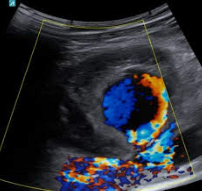
pseudoaneurysm
often seen as complication post-percuatenous arterial catheterization
seen in groin area w/ typical PSA u/s findings..
neck with to-and-fro flow
cystic mass w/ yin-yang colour flow & possible internal debris/thrombus
treated w/ ultrasound guided thrombin injection
most common treatment method
resolution is quick
soft tissue edema
common cause of limb swelling
bilateral leg swelling is more common w/ venous congestion (rt HF)
excessive inc. in venous transmural pressure = dec. PG b/w arterioles & venules = dec. capillary perfusion = inc. filtration of fluids into tissue (edema)
etiology:
DVT
venous HTN
venous insufficiency
HF » venous congestion
extrinsic venous compression/obstruction
fluid overload
renal failure
iatrogenic over-hydration
2D findings:
marbled, reticulated pattern when found in subcut. fat
small fluid collections may be seen within superficial tissues
can obstruct ultrasound’s signals and make visualization of deeper structures more challenged (inc. attenuation)
doppler findings:
extrinsic venous compression/obstruction:
continuous non-phasic flow below obstruction
HF » venous congestion
inc. transmission of cardiac phasic changes in peripheral veins
waveforms are even more pulsatile than usual in upper leg veins w/ more prominent bidirectional venous
—
pulsatile venous flow v. arterial flow:
venous patterns are less periodic than typical triphasic flow
venous flow direction is primarily still towards heart
venous flow pattern is affected by Valsalva
lymphadema
caused by obstruction in lymph channels » resultings in excess fluid in the interstitial tissue
may present as uni or bilateral leg swelling and pitting edema
swelling os often recurrent, less in the morning & more severe later in the day
etiology:
malignancy, radiation, infection, trauma, surgery
two types:
primary
less common
inherited; occurs independently
secondary
secondary to disease or other condition
sono findings:
relatively thick subcutaenous tissue w/ “ant farm” appearance
grainy appearance of subcutaneous tissue
degraded image quality » inc. attenuation
app. is typically indistinguishable from soft tissue edema
may or may not see enlarged lymph nodes
lymphatic system
lymph nodes are commonly seen in groin area, neck, and axillary regions
typically, <1cm in AP diameter & enlarge w/ inflammatory or malignant infiltration
inflamed lymph nodes are tender and can be the cause of lower limb pain
adenopathy = enlarged lymph nodes
can cause swelling if associated w/ lymphatic or venous obstruction
should be assessed for any abnormal characteristics
normal sono app:
hypoechoic mantle of peripheral tissue surrounding central hyperechoic hilum containing fat & feeding vessels
feeding vessels have low resistance arterial flow (doesn’t matter is benign or malignant)
benign appearance:
almond-shaped or kidney shaped
echogenic central hilum maintained
SAX diameter = <1/2 LAX diamater
maintains normal feeding vessels
extended from periphery to central hilum
well organized
malignant appearance:
abnormal architecture & more rounded than oblong
SAX diameter = >1/2 LAX diameter
central hilum may be replaced by tumour
dec. echogenicity
altered vascularity
irregular vessels follow disorganized pattern
cellulitis
diffuse soft tissue inflammation of the skin & subcutaneous tissues
usually caused by bacterial infection post-break in the skin from trauma, surgery, or bite
limb presents w/ redness, warmth, & shiny stretched skin app.
may also see skin thickening, edema, & swelling of subcutaneous tissues
often a clinical diagnosis that requires differential DVT diagnosis exclusion
sono findings:
mimics soft tissue edema
hyperemesis
lymphangitis
bacterial infection in the lymphatic vessels
presents as painful red streaks below skin surface
potentially serious
can rapidly spread to bloodstream (sepsis)
potentially fatal
sono findings:
mimics cellulitis & soft tissue edema
abscess
implies enclosed collection of pus
caused by bacterial infection
presents w/
focal swelling, skin erythema, pain, tenderness
sono findings:
may be well-circumscribed or ill-defined borders
fluid collection within soft tissues containing complex fluid & echogenic internal debris (that may or may not include shadowing from tiny gas bubbles)
may see surrounding hyperemic tissue
hematoma
blood accumulation within tissue post-injury
may present w/ focal or diffuse leg swelling
injury is accompanied initially by bleeding, then inflammation
can cause compression on surrounding veins
MRI is superior for viewing soft tissue contrast and extent of hematoma
assoc. with:
trauma
vigorous exercise or abrupt straining
prior injury/intervention
sono findings:
focal mass; can be cystic, complex, or solid
hyperechoic, and then either homo or hetero
7 days post-injury: intermediate to low lvl echogenicity & heterogeneous » persists for 3wks
area is often focal & fusiform
appears to “split” muscle strata
tends to spread cephalocaudal in the facial planes
absence of internal vascularity
may initially present w irregular borders
over time, mass will retract & becomes more well-defined and ovoid
comparison w/ contralateral anatomy can help highlight abnormal variations in muscular tissue
baker’s cyst
aka popliteal cyst or synovial cyst
results from fluid collections in the gastrocnemius-semimembranous bursa (an extension of the knee joint)
focal fluid collections along the pop fossa space, typically posteromedial to knee joint
seen posterior to the medial femoral condyle in b/w the tendons and gastrocnemius muscles
as these cysts enlarge, they may dissect thru the tissue planes and extend down into calf musculature
prone to spontaneous rupture
presents w/..
pain, tenderness, swelling, and similar symp. to DVT
typically in pts. >50-6oyrs
etiologies:
congenital
trauma
inflammation
local degenerative changes
usually related to inflammation or irritation
& assoc. w/ multiple conditions..
arthritis, trauma, infection, dialysis, hemophilia
differentials:
hematoma, abscess, tumour
soft tissue edema
popliteal artery aneurysm, popliteal vein aneurysm (2x inc. in size)
rare, associated w/ thromboembolic disease
sono findings:
CLASSIC
seen along posterior-medial aspect of joint, adj. to head of gastrocnemius muscle
simple cystic appearance w/ no internal echoes & well-defined walls
RUPTURED
irregular margins, heterogeneous contents and/or echogenic debris, & extension into calf
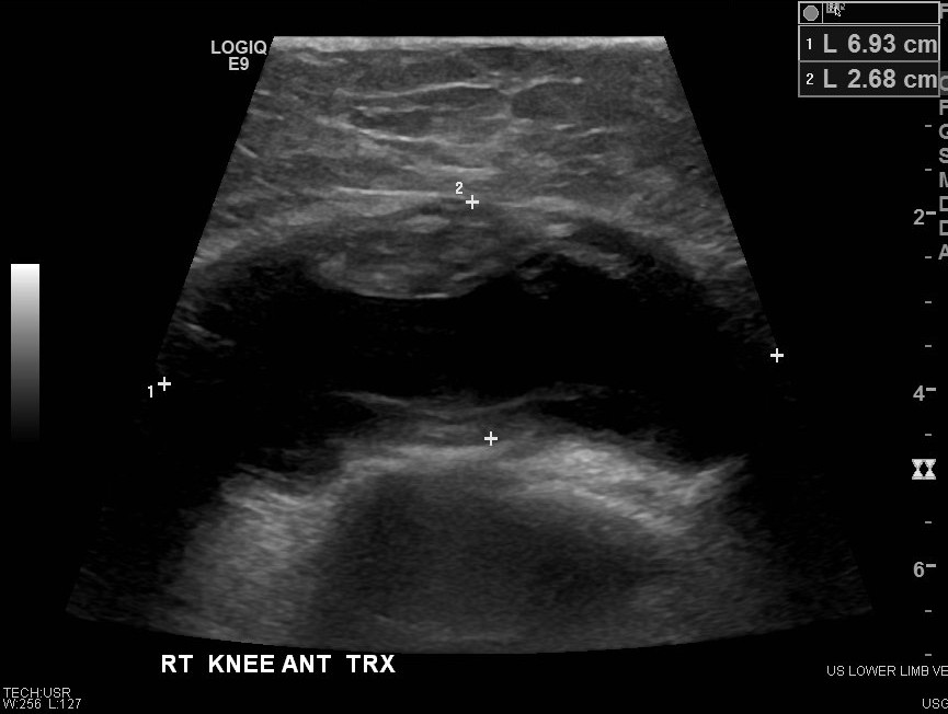
joint effusion
results from inflammation, infection, or trauma
may cause pain & swelling
distension of joint capsule w/ fluid
sono findings:
fluid collects laterally, medially, & superiorly to patella
fluid remains close to joint
fluid may be simple or complex
soft tissue tumours
unlikely cause of leg swelling and tenderness
solid soft tissue masses must be distinguished from superficial or deep vein thrombosis
large masses may be benign or malignant
better defined w/ CT or MRI
benign examples:
lipoma » most common benign lesion
fibroma
leiomyoma
desmoid tumour
neurofibromatosis
soft tissue hemangioma
malignant examples:
sarcoma
lymphoma
leukemia
sono findings:
variable appearance
may be solid or complex, depending on degree of necrosis
high velocity, low resistance arterial flow
Which of the following lower extremity venous Doppler findings is consistent with right heart failure?
Bilateral increased venous pulsatility and biphasic flow.

What is this ultrasound image showing?
Enlarged lymph nodes

The following patient presents to the ultrasound department with calf swelling and pain. Visual examination reveals obvious swelling, but no redness or warmth to the touch.
Large calf hematoma