BY 115 Spinal Cord, Cranial Nerves, Spinal Nerves, Autonomic Nervous System
 Call Kai
 Learn
 Practice Test
 Spaced Repetition
 Match
 Flashcards
 Knowt Play
1/264

There's no tags or description
Looks like no tags are added yet.
265 Terms
Spinal cord
Located within vertebral canal; runs length of body from foramen magnum to L1 vertebra; divided into gray matter and white matter
Five parts of spinal cord
Named for nerves that attach to them; superior to inferior: cervical, thoracic, lumbar, sacral, coccygeal
Cervical enlargement
Neurons that innervate upper limb
Lumbosacral enlargement
Neurons that innervate lower limb
Conus medullaris
Tapering inferior end of spinal cord; official “end” of spinal cord
Cauda equina
Inferiorly, groups of axons
Filum terminale
Within cauda equina; thin strand of pia mater
Meninges of spinal cord
Continuous with cranial meninges; three layers (superficial to deep): dura mater, arachnoid mater, pia mater
Spinal epidural space
Between vertebral canal and dura mater
Central canal of spinal cord
Lined with ependymal cells; contains CSF
Gray matter of spinal cord
Located centrally within spinal cord; contains neuron cell bodies, dendrites, and unmyelinated processes; organized into horns and the gray commissure
Anterior gray horns
Contains cell bodies of somatic motor neurons; innervates skeletal muscle
Lateral gray horns
Contains cell bodies of autonomic motor neurons; prominent in the T1-L2 regions of spinal cord; associated with sympathetic motor output
Posterior gray horns
Receive sensory information; contain sensory neuron axon terminals and interneurons
Gray commissure
Unmyelinated axons crossing from side-to-side; contains central canal
White matter of spinal cord
External to gray matter; contains bundles of primarily myelinated axons; organized into three regions called funiculi
Anterior funiculus
On anterior side of cord between anterior gray horns and anterior median fissure
Lateral funiculus
Lateral side of spinal cord
Posterior funiculus
Between posterior gray horns and posterior median sulcus
Spinal cord tracts
Bundles of axons that run in white matter of spinal cord; includes ascending tracts (sensory axons = traveling to brain) and descending tracts (motor neurons = traveling from brain)
Cranial nerves
12 pairs, part of the PNS, numbered I-XII, named primarily for structure or function
CN I
Olfactory nerve; sensory only, responsible for olfaction (smell); olfactory nerve fibers pass through the cribriform plate; synapse in the olfactory bulb
Anosmia
No sense of smell
CN II
Optic nerve; sensory only; responsible for vision; includes optic chiasm (axons from nasal halves of retinas cross to opposite side) and optic tracts (carry information toward brain)
CN III
Oculomotor nerve; somatic motor and parasympathetic motor functions
Oculomotor somatic motor functions
Supplies superior rectus (SR), inferior rectus (IR), medial rectus (MR), inferior oblique (IO), and levator palpebrae superioris
Oculomotor parasympathetic motor functions
Supplies sphincter pupillae (constricts pupil) and ciliary muscle (changes lens shape)
CN IV
Trochlear nerve; somatic motor only; innervates superior oblique; helps depress the eye, particularly when eye is adducted
Anopsia
Lack of vision
Diplopia
Double vision
CN V
Trigeminal nerve; mixed nerve: sensory + somatic motor; motor innervation to muscles of mastication
Trigeminal nerve sensory distribution
Sensory innervation from much of the face and anterior scalp, nasal and oral cavities, and teeth; distributed across ophthalmic branch (V1), maxillary branch (V2), and mandibular branch (V3)
CN VI
Abducens nerve; somatic motor; innervates lateral rectus; abducts eye (moves it laterally)

ID 1
Cervical plexus

ID 2
Cervical enlargement

ID 3
Brachial plexus

ID 4
Lumbosacral enlargement

ID 6
Conus medullaris

ID 7
Lumbar plexus

ID 8
Sacral plexus

ID 9
Filum terminale

ID 10
Atlas

ID 11
Cervical part

ID 13
Thoracic part

ID 14
Lumbar part

ID 15
Sacral part

ID 16
Cauda equina

ID 17
C1

ID 18
C2

ID 19
C3

ID 20
C4

ID 21
C5

ID 22
C6

ID 23
C7

ID 24
C8
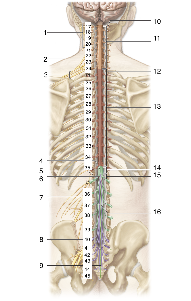
What nerve is between C8 and T2?
T1

ID 25
T2

ID 26
T3

ID 27
T4

ID 28
T5

ID 29
T6

ID 30
T7

ID 31
T8

ID 32
T9

ID 33
T10

ID 34
T11

ID 35
T12

What nerve is between T12 and L2?
L1

ID 36
L2

ID 37
L3
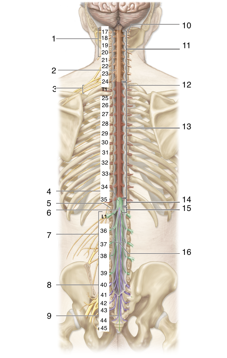
ID 38
L4

ID 39
L5

ID 40
S1

ID 41
S2

ID 42
S3

ID 43
S4

ID 44
S5
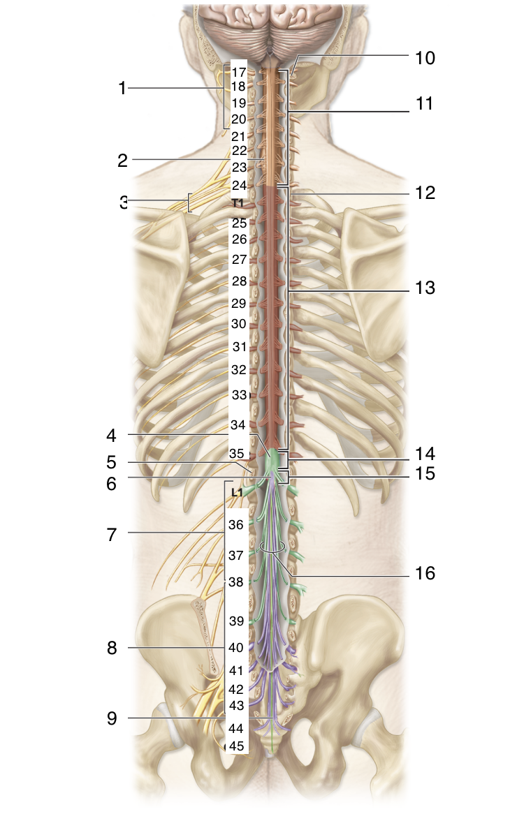
ID 45
Co1

ID 1
Spinal cord

ID 2
Posterior rootlets

ID 3
Conus medullaris

ID 4
Posterior root
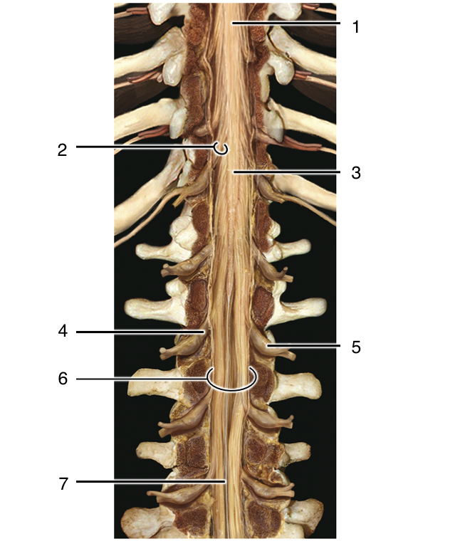
ID 5
Posterior root ganglion

ID 6
Cauda equina

ID 7
Filum terminale
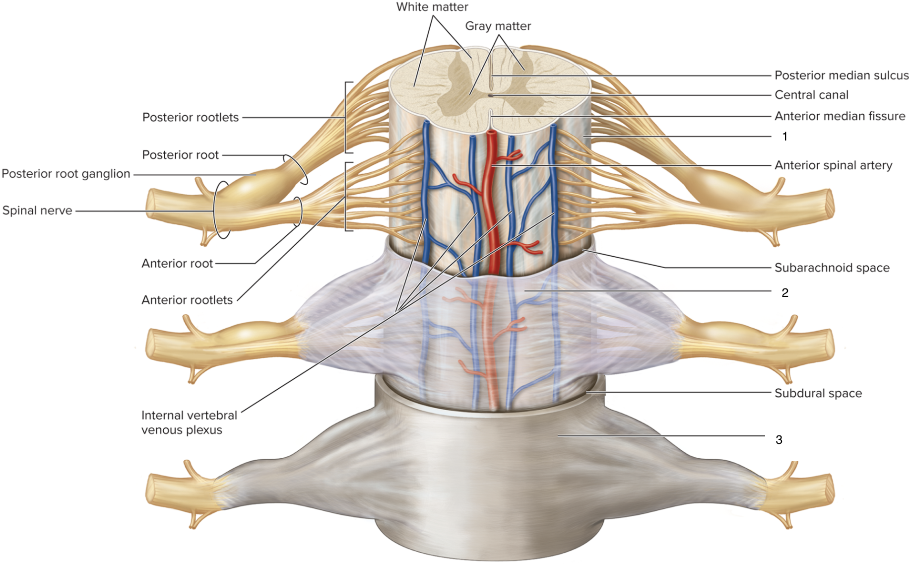
ID 1
Pia mater
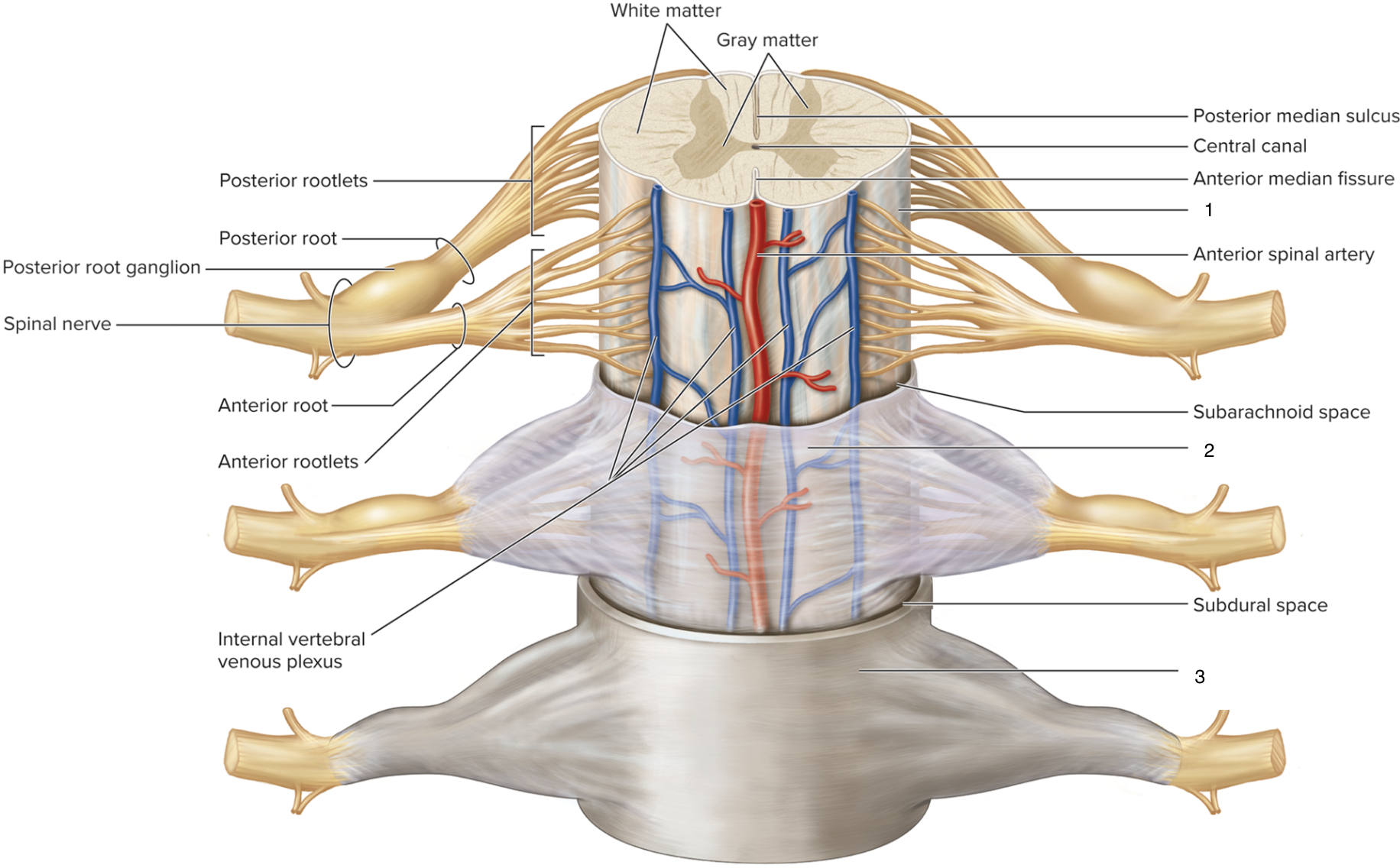
ID 2
Arachnoid mater

ID 3
Dura mater

ID 1
Spinous process of thoracic vertebra

ID 2
Epidural space

ID 3
Dura mater
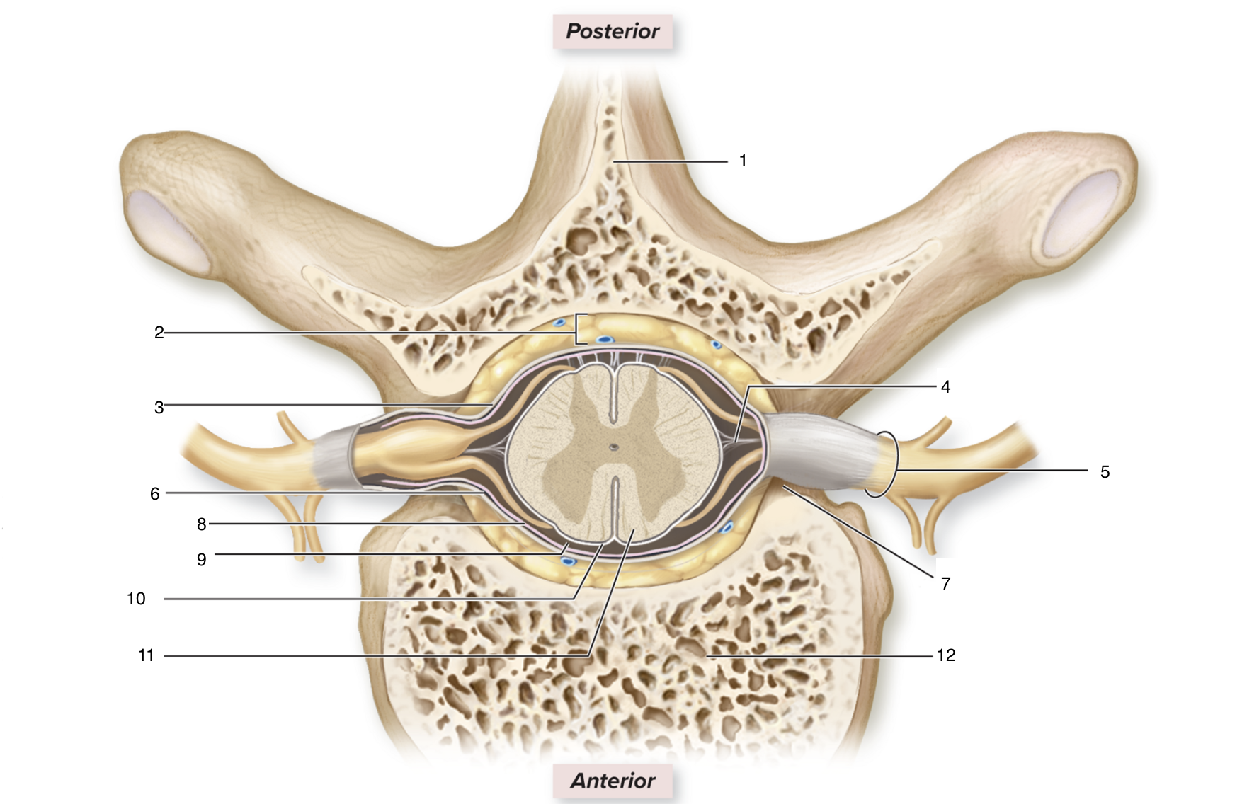
ID 4
Denticulate ligament

ID 5
Spinal nerve

ID 6
Subdural space

ID 7
Intervertebral foramen

ID 8
Arachnoid mater

ID 9
Subarachnoid space

ID 10
Pia mater
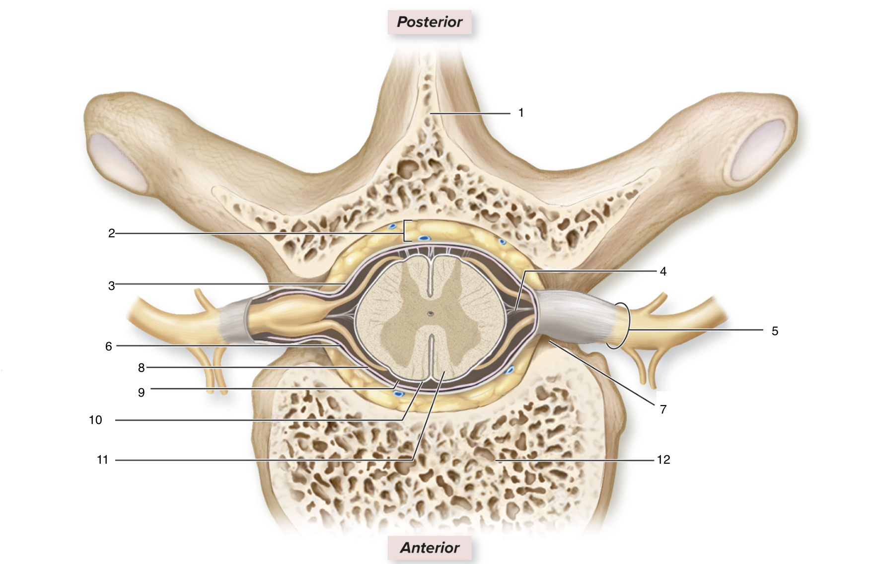
ID 11
Spinal cord

ID 12
Body of thoracic vertebra