Anomalies of the Right Ventricular Outflow Tract & Pulmonary Artery
 Call Kai
 Learn
 Practice Test
 Spaced Repetition
 Match
 Flashcards
 Knowt Play
1/78

There's no tags or description
Looks like no tags are added yet.
79 Terms
List the 5 categories of RV outflow tract anomalies. Which is the Most common type is what?
Pulmonary valve stenosis - valve stenosis is the most common*
Subpulmonary stenosis
Supravalvular pulmonary valve stenosis
Branch PA stenosis
Pulmonary atresia with intact ventricular septum - AKA single ventricle
How common is pulmonary valve stenosis, and is it usually congenital or acquired?
___th most common congenital heart lesion
Almost always _______ and if its the other one what is caused by list 2
4th most common congenital heart lesion
Almost always congenital
May rarely be acquired from carcinoid or rheumatic disease
When can pulmonary valve stenosis present, and what determines when it presents?
It can present from in _____through _____, depending on the degree of ______.
It can present from in utero through adulthood, depending on the degree of stenosis.
Is pulmonary valve stenosis usually isolated, and list the 2 CHDs associated with it.
It is usually isolated.
Associated CHDs:
Tetralogy of Fallot
DORV
List the 3 syndromes/conditions associated with pulmonary valve stenosis.
Noonan syndrome
Rubella
Williams syndrome — associated with branch stenosis
What happens to the measured severity of PV stenosis when the PDA closes, and what may be used before then?
After the PDA closes, the gradient better shows the severity (b/c the PVR drops)
Before that, you may need to rely on the Severity of RV hypertrophy First
Is progression of pulmonary valve stenosis common after the first year of life? & What view is the best to view RVOT obstruction list 3? & what is the best View?
No. Progression after the first year is rare.
What view is the best to view RVOT obstruction: SAX #1 view , PLAX, Subcostal, RVOT view
Pulmonary valve stenosis includes:
What happens when pulmonary valve _________are fused, underdeveloped, or absent?
LIST 2
Pulmonary valve commissures are fused, underdeveloped, or absent
The valve opening / orifice opening becomes limited
The valve may appear dome-shaped
What are dysplastic pulmonary valves associated with?
Sinotubular narrowing.
What abnormal numbers of pulmonary valve cusps may occur List 3, and what are they associated with? list one congential and two leisons
Unicuspid - more stenotic
Bicuspid - more stenotic
Quadricuspid - more regur
They may be associated with Tetralogy of Fallot and with stenosis and regurgitation.
How does the severity of PV stenosis relate to RV hypertrophy?
Mild–moderate PS → normal to mild RV hypertrophy
Moderate RVH → Moderate PS
Severe PS → severe RV hypertrophy (Wall stress)
The degree of stenosis is directly related to the degree of hypertrophy.
What is common with PV stenosis, and does PA size correlate with stenosis severity?
Post-stenotic dilation is common
No, the severity of stenosis does not correlate with the size of the main PA
Simple way to remember:
A large main PA does not automatically mean severe pulmonary stenosis, and a normal-sized main PA does not rule out severe stenosis.
What happens to the RVOT as hypertrophy becomes severe?
Outflow tract is severely affected by degree of
_______and will cause an ________
RVOT becomes _____
Less ______during systole
Decreased ______during diastole
Pressure ________
May eventually produce a ______-to-______shunt
Outflow tract is severely affected by degree of hypertrophy and will cause an obstruction
RVOT becomes stiff
Less contractile during systole
Decreased recoil during diastole
Pressure increases
May eventually produce a right-to-left shunt
(infundiublum becomes thick can lead to subpulmonic obstruction double obstruction)
Decreased recoil during diastole means the right ventricle (RV) cannot relax and spring back to its normal shape as easily after contracting.
Why does this happen?
Pulmonary stenosis causes pressure overload in the RV.
The RV muscle becomes thicker (RV hypertrophy).
The thickened RV becomes stiffer and less compliant.
During diastole, the RV cannot relax and expand as easily to fill with blood.
This causes increased RV filling pressure.
List the 9 clinical findings of pulmonary valve stenosis.
May be symptomatic or asymptomatic ? which one?
What murmur
List 5 clincial presentation
Sudden _____
RV what and ____-axis deviation on EKG
_______and ______with critical PS
May be asymptomatic (b/c PS may be mild)
Systolic ejection murmur
Exertional dyspnea
Fatigue
Chest pain and/or syncope
CHF
Sudden death
RVH and right-axis deviation on EKG
Hypoxemia and cyanosis with critical PS
(S.A.D also applys here)
Hypoxemia means a low level of oxygen in the blood, specifically arterial blood.
Echo Assessment:
List the 4 main things evaluated when imaging the pulmonary valve/RVOT.
____ size and anatomy- using what view _____ parasternal
what finding / shape of the PV leaflets
Level of what
______ through the outflow tract and valve
Valve size and anatomy — high parasternal
Doming of the PV leaflets
Level of obstruction
Gradient through the outflow tract and valve
List the 5 additional findings evaluated on echo with PV stenosis.
List 2 for the PA size and what lesion
List 2 for RV
what TV findings and RA findings
what finding do you see of the septem
and two shunt flow direction
Main and branch PA size and PI
RV hypertrophy and RV function
TR and RA dilation
Septal flattening (b/c of pressure of the RVH)
ASD and PDA flow direction
What additional study may be needed when evaluating an ASD?
Bubble study
What is the primary repair for pulmonary valve stenosis?
Balloon angioplasty/valvuloplasty. - #1 choice
What gradient does the slide give for balloon angioplasty in what severity level PS, and what is its success rate?
Balloon angioplasty is recommended for (what severity level) -PS with gradients up to ____
mmHg
• _____% success rate of relieving the gradient
Mild PS with gradients up to 35 mmHg
>95% success rate in relieving the gradient
What percentage of patients require additional procedures after balloon treatment such as what list 2, and 33% develop significant what lesion afterward
21% require additional procedures such as repeat valvuloplasty or RVOT enlargement
33% develop significant PI afterward
What happens to RV compliance after relief of pulmonary stenosis?
RV compliance improves
When may Surgery or what replacement be required for pulmonary stenosis? List 2 types of stenosis (like there area)
Surgery is required for infundibular stenosis and supravalvular stenosis
Valve replacement may also be required
What is critical pulmonary valve stenosis?
Severe what ? obstruction in which the RV cannot maintain _____saturation above _____% without a what shunt
Severe RV outflow tract obstruction in which the RV cannot maintain oxygen saturation above 90% without a PDA
What direction does blood shunt through the PFO and PDA in critical PV stenosis?
And Blood becomes dependent on what shunt?
PFO: Right → left
PDA: Left → right
Blood becomes dependent on PDA
Why does the patient become dependent on the PDA in critical PS ( the PDA helps supply what blood flow?), and what can result?- extra what load on the which Ventricular ? what about biventricular function
The PDA helps supply pulmonary blood flow.
This can cause:
Extra volume load on the LV
Poor biventricular function
Why does the LV experience volume overload?
In critical pulmonary stenosis, the RV has difficulty pumping blood into the pulmonary artery because the pulmonary valve is severely narrowed.
The PDA stays open, allowing blood to travel from the aorta into the pulmonary artery (left-to-right shunting).
This additional blood travels through the lungs.
The blood returns through the pulmonary veins into the LA, then the LV.
The LV receives extra blood, causing LV volume overload.
Simple way to remember: The PDA sends extra blood into the lungs, which eventually returns to the left side of the heart. This increases the amount of blood the LV must handle.
The important distinction is that the PDA helps supply blood to the lungs in critical pulmonary stenosis, but the additional pulmonary blood flow can overload the LV.
What is another name for subpulmonary stenosis?
Infundibular pulmonary stenosis
List the 5 causes of infundibular pulmonary stenosis.
Discrete ___________obstruction
Ventricular septal ______
Sinus of Valsalva ______
Cardiac ______
RV _________
Discrete fibromuscular obstruction
Ventricular septal aneurysm
Sinus of Valsalva aneurysm
Cardiac tumors
RV hypertrophy
What causes a double-chambered right ventricle?
Anomalous RV muscle bundles
What is a discrete subpulmonary membrane, and where is it located?
A ____with an opening located at or within the ______ostium, usually in the ______portion of the ________
A ring with an opening located at or within the infundibular ostium, usually in the lower portion of the infundibulum
What causes a discrete subpulmonary membrane according to the slide?
Caused by a defect in the ______of the ______
&
Does PS progressive with age?
A defect in the absorption of the infundibulum.
NO PS does not progressive with age!
What is a double-chambered right ventricle?
_______between the RV _____and RV _____caused by abnormal RV what attachments.
Narrowing between the RV inflow and RV outflow caused by abnormal RV muscle-bundle attachments.
What 2 pressure chambers are created in DCRV?
High-pressure RV chamber
Low-pressure infundibular chamber

When is DCRV usually detected, and list the 4 associations given.
Usually detected after the first few _____of life.
4 associations given: What the #1, what membrane finding? list 2 syndromes
Usually detected after the first few months of life.
Associated with:
Membranous VSD***
Subaortic membrane
Down syndrome
Noonan syndrome
How does DCRV change as the patient ages? The muscle bundles become more what as the patient grows & what is the best view ?
Obstruction is progressive with age
Muscle bundles become more prominent as the patient grows
Best view PSAX at the ventricle level
Where does the VSD usually communicate in DCRV, and how does shunt direction change with severity? in less severe the shunt is what and in severe the shunt may what?
The VSD usually communicates with the high-pressure portion of the RV.
Less severe → left-to-right
Severe → shunt may reverse
What may happen if the VSD spontaneously closes in DCRV?
RV dysfunction may occur.
(The VSD acts like the pressure pop -off) Simple way to remember: The VSD closes, but the RV obstruction is still there → continued RV pressure overload → possible RV dysfunction.
Subpulmonary stenosis
How are discrete membranes and DCRV repaired, This repair provides excellent what results list 2? and what happens to RV function afterward with the relief of the what?
Repair is surgical
Surgery gives excellent hemodynamic/functional results
RV function improves after relief of the obstruction
What is the best view for finding subpulmonary obstruction?
Short-axis at the ventricular level.
Where is supravalvular pulmonary stenosis located?
Obstruction _____to the pulmonary valve, and just above the where?
Obstruction Distal to the pulmonary valve, and just above the branch pulmonary arteries.

What 2 forms can supravalvular pulmonary stenosis take?
Discrete membrane
Hypoplastic segment

Where along the PA can supravalvular stenosis occur; Visualized as a discrete membrane or a hypoplastic segment; Located where?
Located Just _____the valve, ____ portion of the PA, or close to the where
Just below the valve region
Mid PA
Near the bifurcation
Etiology
List the 5 syndromes/conditions associated with Supravalvular pulmonary stenosis.
Williams syndrome
Noonan syndrome
Alagille syndrome
Ehlers-Danlos syndrome
Rubella
2-3% will have other CHD and cardiovascular anomalies
What previous repairs/procedures may cause supravalvular pulmonary stenosis?
List 2 (first one what can it produce and 2nd what may be associated with?
Previous repairs can cause it
Pulmonary artery banding can produce a ridge (done for complete AV - canal defect or single ventricle)
LeCompte maneuver may be associated with post-stenotic dilation (done when pt has a D-TGA)

List the 4 defects associated with supravalvular pulmonary stenosis.
List 1 congential defect and 3 shunts
Tetralogy of Fallot
VSD
ASD
PDA
List the 6 management/repair options given for supravalvular pulmonary stenosis.
_____patch angioplasty for main PA
Balloon _______
Intravascular stainless-steel ____
_______graft of the stenosis
______treatment or monitoring or ______Conduit
Dacron patch angioplasty for main PA
Balloon angioplasty
Intravascular stainless-steel stent
Bypass graft of the stenosis
Surgical treatment or monitoring
Rastelli Conduit
Rastelli procedure: includes closing the VSD to the aorta and placing a valved RV to PA conduit for RVOT reconstruction
Dacron patch angioplasty = Widening the narrowed pulmonary artery with a synthetic patch to improve blood flow and reduce the workload on the RV.
What 2 abnormalities can affect the pulmonary artery branches?
Stenosis
Hypoplasia
What congenital heart defects are associated with PA branch stenosis? List 3
Conotruncal defects, including examples such as:
Truncus
TGA (LeCompte maneuver- causes Lt pulmonary branch artery stenosis)
Tetralogy of Fallot
What 2 syndromes are strongly associated with PA branch stenosis and this is the same for Unilateral branch stenosis? which one may be progressive? (%?)
Alagille syndrome — 76%
Williams syndrome — 50%
Williams-associated disease may be progressive
What does Branch PA stenosis do to the RV and PA pressure? List 2 for the RV, and one for the PA
It causes:
RV pressure overload
RV hypertrophy
High PA systolic pressure
What peak PA velocity is associated with branch stenosis,*** and what else may increase PA velocity? List 2
Peak velocity >1.5 m/s
Velocity may also increase from:
Atrial shunts
High cardiac output
Can elevated PA velocity/branch stenosis resolve in infants? if so how long?
&
What is the BEST view for PA Branch stenosis?
Yes. It may resolve within a few months.
(PDA present velocity may be increased b/c of the volume load so make sure its true PS)
BEST VIEW: High PSAX
What may cause unilateral PA branch stenosis, and why can RV pressure remain normal?
____tissue may encircle the _____, similar to a ______
RV pressure may remain normal because the _______ what accommodates the what output
PDA tissue may encircle the branch, similar to a coarctation
RV pressure may remain normal because the opposite PA branch accommodates the cardiac output
How does this happen?
The PDA (patent ductus arteriosus) connects the aorta to the pulmonary artery during fetal development.
The PDA contains tissue that normally constricts after birth to close the ductus.
Sometimes this tissue extends into and encircles (wraps around) part of a branch pulmonary artery, usually the left pulmonary artery (LPA).
When the PDA tissue contracts, it can also narrow that pulmonary artery branch.
This causes branch pulmonary artery stenosis, restricting blood flow to that lung.
Why does your slide say it's similar to coarctation?
Because both involve narrowing of a blood vessel:
Coarctation of the aorta: Narrowing of the aorta.
PDA-related branch PA stenosis: Narrowing of a pulmonary artery branch caused by ductal tissue.
Simple way to remember: PDA tissue wraps around a pulmonary artery branch → PDA tissue contracts → Branch PA narrows → Less blood reaches that lung.
Your slide also mentions that RV pressure may remain normal because the opposite pulmonary artery can accommodate more blood flow.
List the 6 things evaluated on echo for pulmonary branch abnormalities.
Number of PA _____
Level of ______
______through each branch
Flow rate through the ______ - one maybe what than the other?
Branch PA _____
List two of the RV
and what PV lesion
Number of PA branches
Level of obstruction
Gradient through each branch
Flow rate through the branches - One maybe higher than the other
Branch PA size
RV hypertrophy/function and PI
What 2 windows are used to assess branch PA size?
High parasternal
Suprasternal short-axis
What repair options are listed for PA branch stenosis?
Balloon ______
Stainless-steel _____
_____graft of stenosis
____or monitoring, or ______Conduit
Balloon angioplasty
Stainless-steel stent
Bypass graft
Surgery or monitoring
Rastelli Conduit
What is Pulmonary Atresia with an Intact Ventricular Septum consists of?
______pulmonary valve blocks blood flow from the _____into the where
Ventricular septum is ______
Pulmonary blood flow depends on a what shunt?
It is a ________CHD
Atretic pulmonary valve blocks blood flow from the RVOT into the pulmonary arteries
Ventricular septum is intact
Pulmonary blood flow depends on a PDA
It is a cyanotic CHD (PDA isnt large enough to supply blood flow)
What 2 pathways can provide RV outflow in PA/IVS?
Coronary artery sinusoids (like a fistula) connecting the coronary circulation to the RV cavity
Tricuspid regurgitation
How high may RVSP become in PA/IVS?
Up to 200 mmHg.
What direction of flow through the PFO and PDA is necessary for survival in PA/IVS?
PFO: Right → left
PDA: Left → right
BOTH necessary for survival as well*
How common is PA/IVS, and when is it usually diagnosed?
what % diagnosed within the first weeks of life
85% diagnosed within the first weeks of life - b/c the baby is born cyanotic
What happens without treatment and why, and is PA/IVS usually inherited or associated with maternal tetragons?
It may occasionally occur with what abnormalities?
Without treatment it can be fatal due to cyanosis
It may occasionally occur with chromosomal abnormalities
Not associated with inheritance or maternal tetragons
How does pulmonary-valve morphology relate to RV development in PA/IVS?
Completely fused PV causing what→ RV may be what - formed
Very small RV → PV may not have what, and the RVOT may also be what
Completely fused PV causing membranous atresia → RV may be well formed
Very small RV → PV may not have developed, and the RVOT may also be atretic
Simple way to remember: The pulmonary valve can be completely blocked, but the RV can still be well developed.
Well-formed RV means the right ventricle has developed relatively normally in size and structure, rather than being severely underdeveloped (hypoplastic).
RV → RVOT → Pulmonary valve (membranous atresia) → Main pulmonary artery
The membrane is at the normal location of the pulmonary valve, completely preventing blood from passing from the RV into the pulmonary artery.
Simple way to remember: Membranous atresia = The PV is fused shut. Muscular atresia = The muscular outflow tract itself is blocked or underdeveloped.

What are the 2 anatomic types of PA/IVS?
Type 1 — RV hypoplasia
Type 2 — RV dilation
List the 3 findings of Type 1 – RV Hypoplasia PA/IVS.
Small but _______what valve
Hypoplastic ____with coronary artery ____
About 20% have a what shunt**
Small but competent tricuspid valve
Hypoplastic RV with coronary artery fistulae
About 20% have a secundum ASD**
What relationship exists between tricuspid-valve size and RV size in Type 1 RV hypoplasia in PA/IVS?
TV size correlates with RV size.
The amount of RV hypoplasia depends on how large or small the TV is.
List the 3 findings of Type 2 RV Dilation. PA/IVS.
Dilated of what List 3?
what happens with the TV size and what Lesion is found
what happens with the pulmonary valve
Dilated RA, SVC, and IVC
Enlarged tricuspid valve with significant TR
Membranous pulmonary-valve atresia
Simple way to remember: In Type 2, blood cannot exit through the pulmonary valve, but significant TR allows blood to move back and forth between the RV and RA, contributing to RV dilation.
Pathophysiology in PA/IVS, :
Where does the systemic venous blood crosses? - what shunt and is it usually restricted or unrestricted
and why does cyanosis occur?
Systemic venous blood crosses the PFO - Usually unrestricted
Mixing of blood causes cyanosis
Why is PA/IVS called a ductal-dependent lesion?
The _____sends blood to the _____, unless ______collateral arteries are present.
The PDA sends blood to the lungs, unless aortopulmonary collateral arteries are present.
Aortopulmonary collateral arteries are abnormal blood vessels that connect the aorta (systemic circulation) to the pulmonary arteries, allowing blood to reach the lungs through another pathway.
a rare congenital heart defect in which there is an abnormal hole or direct connection between the ascending aorta and the main pulmonary artery.
List the 3 clinical findings of PA/IVS. (what at birth, what is wrong with the heart, and what may occur)
Cyanosis at birth
CHF
Myocardial infarction may occur
List the 5 right-heart/valve findings assessed on echo in PA/IVS.
Tricuspid valve ____and anatomy
what TV and PV lesions
Pulmonary-valve ____if possible
List 3 things about the RV
RV and RA ____
Tricuspid valve size and anatomy
TR and PI
Pulmonary-valve gradient if possible
RV hypertrophy, size, and function
RV and RA size
List the 4 additional things evaluated on echo in PA/IVS.
Main and branch PA ____
Coronary artery _____with color Doppler
Septal _____
what two shunt flow direction
Main and branch PA size
Coronary artery fistulas with color Doppler
Septal flattening
PDA and ASD flow direction
What is the prognosis of PA/IVS without intervention, and what medication keeps the PDA open?
Prognosis is poor without intervention
Prostaglandin (PGE) is used to prevent ductal closure
List the 3 reasons on why a Cardiac catheter procedures that may be used for PA/IVS.
Balloon _______to open/expand the pulmonary valve
Balloon atrial _______to enlarge the ______ovale
PDA ____to keep the ductus open
Balloon valvuloplasty to open/expand the pulmonary valve
Balloon atrial septostomy to enlarge the foramen ovale
PDA stent to keep the ductus open
Pulmonary Atresia with and Intact Ventricular Septum:
What is the purpose of a BT shunt?
To allow more blood to pass through the ____and become _______.
To allow more blood to pass through the lungs and become oxygenated.
Simple way to remember: BT shunt = Subclavian artery or innominate artery connected to right pulmonary artery branch using a graft.
Pulmonary Atresia with and Intact Ventricular Septum:
What may be done if the RV is too small, a what palliation with a what list 2 may be performed , and what other major treatment may be considered?
Single-ventricle palliation with Glenn and Fontan
Heart transplant may be considered
1. Glenn shunt
Connection: Superior vena cava (SVC) → Right pulmonary artery (RPA)
The SVC carries deoxygenated blood from the upper body.
The surgeon connects the SVC directly to the pulmonary artery.
Blood from the upper body flows directly into the lungs, bypassing the RV.
2. Fontan procedure
Connection: Inferior vena cava (IVC) → Pulmonary arteries
The IVC carries deoxygenated blood from the lower body.
The surgeon creates a pathway from the IVC to the pulmonary arteries, usually using a conduit or tunnel.
Blood from the lower body also bypasses the RV.
What long-term survival is given after appropriate treatment for PA/IVS?
About what % of infants alive at age ___ survive beyond age ____
About 65% of infants alive at age 1 survive beyond age 10.
List the 4 things evaluated after PA/IVS repair.
• Evaluate for what PV lesion and Severity
• Evaluate the TV lesions
• Evaluate ___ outflow track and use what doppler for peak and mean gradients
• Evaluate the single ventricle palliation list the 2
PI and its severity
TR
RVOT with CW Doppler for peak and mean gradients
Glenn and Fontan circulation
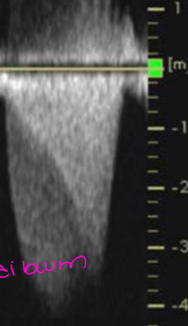
This is when the valve its self is clacified or stenosis what is the wavefrom called? what murmur?
FIXED
Ejection Murmur in systole


This is when there is muscle in the RVH Infundibulm causing obstruction what is this waveform called?
DYNAMIC
(With CW doppler you can see Fixed and PW Doppler usually you can see dynamic / dagger shaped, can show both combintion with each other )
