Outcome 1: Heart Failure, HTN, and PHTN
 Call Kai
 Learn
 Practice Test
 Spaced Repetition
 Match
 Flashcards
 Knowt Play
1/105

Earn XP
Description and Tags
Heart Failure, HTN, and PHTN
106 Terms
what is heart failure
when the heart can’t pump blood well enough to meet the body’s O2 and nutrient needs
types:
left sided (HFrEF, HFpEF)
right sided
high output
left sided heart failure
implies that LV dysfunction is responsible for the heart fail.
systolic heart failure (LV)/heart failure w/ reduced ejection fraction (HRrEF)
LV can’t contract effectively » leading to reduced EF and dec. cardiac output
initially, the extra fluid builds up in the LV and LA bcuz the LV isn’t contracting as forcefully as it should
as this happens, LV and LA begin to stretch and become dilated
blood backs up further into pulmonary circulation » inc. pulmonary pressures
potentially leads to secondary rt-sided heart failure
diastolic heart failure (LV)/heart failure w/ preserved ejection fraction (HFpEF)
LV becomes stiff and non-compliant » preventing normal relaxation and ventricular filling during diastole
LV is limited in its ability to fill, so it remains a normal size and maintains normal sys. function in the early stages of disease
blood accumulates in the LA since it can’t move forward into the LV » causing LA dilatation
as LV diastolic heart fail. progresses, blood backs up further into the pulmonary circulation, as seen w systolic lt-sided heart failure
reduced ventricular filling lowers stroke volume and cardiac output despite a normal LV ejection fraction
right sided heart failure
RV dysfunction is resp. for the heart fail.
decreased RV systolic function
RV can’t contract effectively » leading to dec. output to the lungs
this may occur due to inc. pulmonary pressures w/ primary lung diseases or from too much blood in the pulmonary circulation, often a consequence of lt-sided heart failure (both sys. and dia.)
inc. pulmonary pressures force the RV to work harder » leading to dec. systolic RV function and systemic venous congestion
decreased RV diastolic function
not as common or as freq. identified as LV diastolic HF
RV should be investigated for dia. dys. if LV diastolic HF if suspected
bcuz of intrinsic dysfunction of the RV myocardium (heart muscle) or external restriction from pericardial disease, the RV can’t fill adequately and therefore remains a normal size
as blood backs up from the underfilled RV, it accumulates in the RA » leading to RA dilation
as the diastolic HF progresses, blood backs up into the systemic veins (IVC and SVC) » causing systemic venous congestion
biventricular heart failure
both ventricles have impaired function (systolic and/or diastolic), often due to advanced cardiac disease, longstanding lt-sided HF, or global myocardial dysfunction
pt. exhibits symptoms of both pulmonary congestion from lt-sided HF and systemic venous congestion from rt-sided HF
high output heart failure
cardiac output is normal or elevated, but still insufficient to meet the body’s inc. metabolic demands
causes include..
anemia
hyperthyroidism
arteriovenous malformations or fistulas
beriberi (thiamine deficiency)
pregnancy
Paget’s disease
congestive heart failure (CHF)
used interchangeably w/ heart failure, but it specifically refers to a hype of HF characterized by fluid buildup (congestion) in the body’s tissues
fluid can accumulate in various parts of the body when the heart is unable to pump blood effectively
this is due to poor heart function
leads to symptoms such as..
swelling (edema and ascites)
fluid retention in the lungs (pulmonary edema)
SOB (shortness of breath)
——
can occur in both lt-sided and rt-sided HF
in lt-sided HF:
fluid backs up into lungs » causing pulmonary congestion and breathing difficulties
in rt-sided HF:
fluid accumulates in the body’s peripheral tissues. » leading to swelling in the legs, ankles, and abdomen
systolic vs. diastolic heart failure
systolic HF:
occurs when heart muscle is weakened and can’t pump blood efficiently » leading to reduced cardiac output
diastolic HF:
occurs when the heart muscle becomes stiff and can’t relax properly » impairing its ability to fill w blood
left vs. right sided HF
left:
involves the LV » leading to fluid buildup in the lungs and causes symptoms like SOB
right:
affects the RV » causing fluid retention in the lower extremities and abdomen, often due to PHTN
acute vs. chronic heart failure
acute HF:
a sudden onset of HF symptoms
requires immediate medical attention to prevent severe complications
chronic HF:
a long-term condition where the heart gradually loses its ability to pump blood effectively over time
clinical signs of left-sided heart failure
shortness of breath (SOB)
dec. exercise tolerance
PHTN, pulmonary edema (crackles heard on ausculation)
fatigue and weakness
arrhythmia
specifically atrial fibrillation and atrial flutter (occurs w atrial dilatation)
pulsus alternans
pts. pulse alternates bw strong and weak beats
this indicates dec. LV systolic function » more common w HFrEF and late stage HFpEF
clinical signs of right-sided heart failure
SOB
hepatomegaly
peripheral edema
ascites
jugular venous pressure or distension
arrhythmia
specifically atrial fibrillation and atrial flutter » occurs w atrial dilatation
weight gain due to fluid retention

echo findings of left and right sided heart failure
left sided HF:
LV hypokinesis
LV dilatation
LA dilatation
inc. amount of MR
right sided HF:
RV dilatation (acute and chronic)
RA dilatation
flattening of IVS (D-shaped LV)
paradoxical septal motion
RV hypertrophy (RVH) (chronic)
RV hypokinesis
TV annulus dilatation
IVC and hepatic vein dilatation
heart failure and blood volume
chemoreceptors, baroreceptors, and proprioceptors and involved in regulating blood volume (BV) and blood pressure (BP)
the kidneys and hypothalamus play critical roles through the release of hormones:
renin-angiotensin-adosterone system (RAAS)
antidiuretic hormone (ADH)
——
w/ low cardiac output, the activation of the RAAS and the sympathetic nervous system (SNS) leads to inc. sodium and water retention by the kidneys
can lead to more fluid in the circulation, exacerbating symptoms of congestion and edema
diuretics, such as Lasix, are often prescribed to manage excess fluid volume
heart failure and blood volume cont.
nocturia:
may be experienced during early stages of HF
meaning an inc. urinary output during the night
in a supine position, inc. renal perfusion and subsequent inc. cardiac output and urine production
oliguria:
may be experienced in advanced stages of HF due to significantly reduced renal blood flow, whether supine or not
cardiac output is so low that even a supine position doesn’t sufficiently improve it to enhance urine output
——
urine production is monitored in pts. w HF to help assess renal function and evaluate the need for diuretic therapy
effective management of HF aims to:
alleviate symptoms
optimize cardiac func.
restore hemodynamic stability
through a combo. of lifestyle changes, medication therapy, and in some cases, interventions (ie. device implantation or surgical procedures)
systemic hypertension
term for high BP
hyper- = above/over
-tension = pressure
systemic = refers to systemic arterial vessels (not including arteries in pulm. circulation)
pressure in the systemic arterial circulation is inc.
a condition that can result in structural changes in the heart changes and arteries (hypertensive heart disease)
definitions
hypertension/hypertensive:
sustained high lvls of systemic arterial BP
normotension/normotensive:
normal lvl of systemic arterial BP
hypotension/hypotensive:
sustained low lvls of systemic arterial BP
less than 90/60mmHg would need medication to inc. the BP
white coat hypertension (HTN):
high BP obtained in clinical settings, but at no other location
malignant HTN:
extremely high BP that develops rapidly (180/120mmHg) can lead to organ damage
resistant HTN:
high BP persists even when taking 3 or more medications to control it

pathophysiology of HTN
elevated press. in the systemic arteries creates a press. overload for the LV of the heart
LV is directly connected to the systemic artery circulation via the aorta at the aortic root
when arterial press. is higher than normal, the LV must contract more forcefully to eject blood into the aorta
HTN is an inc. afterload for the LV, and if prolonged and/or severe, it may result in a LV pressure overload (LVPO) state
—
the LV wall undergoes hypertrophy to accommodate this inc. workload
as the LV wall hypertrophies (thickens), the LV dia. func. becomes impaired
the LV sys. function remains preserved
only after prolonged (chronic) uncontrolled HTN, the LV may begin to show signs of reduced sys. func. » eventually leading to dilation of both the LV and LA
What is a potential consequence of prolonged systemic hypertension on the left ventricle?
inc. LV wall thickness
Hypertension increases the ________ for the left ventricle, requiring it to contract more forcefully to eject blood.
afterload
systolic and diastolic components (SBP and DBP) - BP
BP is measured w a blood press. cuff (sphygmomanometer)
a SBP and DBP is obtained
SBP = press. in systole
DBP = press. in diastole
units of measure: mmHg
SBP/DBP mmHg
the average of multiple readings is documented
risk levels according to blood pressure
low risk = 120/80
medium risk = 121-134/80-84
high risk = 135+/85+
pulse pressure - BP
inversely related to blood vessel compliance
directly proportional to the change in BV (blood vol.)
calculated to estimate the probability of a cardiovascular event (myocardial infarction or stroke) and/or to identify if the pt. is in HF w/ dec. cardiac output
normal value = <40mmHg
Pulse Pressure = SBP - DBP
mean arterial pressure (MAP) - BP
avg. BP over the cardiac cycle
body must maintain a minimum of 60mmHg to perfuse the vital organs
—
CALCULATION FROM SBP AND DBP VALUES:
MAP = [2(DBP) + SBP]/3
CALCULATION USING CARDIAC OUTPUT AND SYSTEMIC VASCULAR RESISTANCE:
MAP = Cardiac Output (CO) x Systemic Vascular Resistance (SVR)
CO is influenced by factors such as:
heart rate, preload, afterload, inotropy, contractility
SVR is affected by variables such as:
vessel length, blood viscosity, diameter or radius, vessel stiffness
—
Examples of how MAP can be affected:
Obesity
obese pts. can develop HTN bcuz the inc. length of the vessels will inc. their SVR
Blood Viscosity
certain hematological disorders (polycythemia rubra, anemia, etc.) affect the pts. BP by inc. (polycythemia rubra) or dec. (anemia) the viscosity of pts. blood
inc. blood viscosity = inc. BP (and vice versa)
Atherosclerosis
causes inc. BP bcuz it dec. the diameter and inc. the stiffness of the vasculature in the body
Which of the following factors influence systemic vascular resistance (SVR) and consequently affect the mean arterial pressure (MAP)? Select all that apply.
blood vessel length
blood viscosity
blood vessel diameter
How does an increase in blood vessel length affect mean arterial pressure (MAP)?
it inc. MAP due to higher SVR
etiology vs. risk factors
etiology:
cause of the disease process
risk factors:
things than inc. the likelihood that a disease may develop, but aren’t directly linked to that outcome
primary hypertension
aka essential or idiopathic HTN
contributes to 90-95% of all cases of HTN
etiology:
occurs by itself w/o any other disease process
cause of primary HTN is idiopathic (unknown)
risk factors:
obesity
diabetes mellitus
obstructive sleep apnea
smoking and exposure to smoke, alcohol consumption, physical inactivity, work-related stress, and high salt intake
lifestyle choices
dyslipidemia
inc. age
male sex
race (higher incidence in African Americans)
low socioeconomic/educational status
secondary hypertension
HTN caused by the presence of another disease process
etiology:
a disease process(es) that causes systemic HTN
EXAMPLES:
renal disease » MOST COMMON
sleep apnea
cushing syndrome
coarctation of the aorta**
pheochromocytoma
hypo and hyperthyroidism
vasculitis
pregnancy-induced
atrioventricular fistulas
adrenal hyperplasia
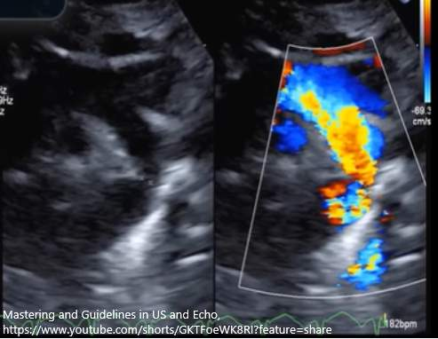
coarctation of the aorta (CoAO)**
the only disease process that causes HTN that can be identified w echocardiography
the narrowing of the desc. Ao, just past the branches of the aortic arch
What is the primary cause of primary hypertension?
idiopathic, no known cause
Which condition that causes hypertension can be identified using echocardiography?
CoAo
why does systemic hypertension matter?
HTN is harmful to the organs and the arterial system:
it creates chronic, mechanical stress on the arterial blood vessels
inc. mechanical stress on vessel walls can lead to aneurysmal changes or even arterial dissection
may also accelerate the development of atherosclerosis and promote thrombus (clot) formation within the arteries
atherosclerosis can impact the amt. of blood flow to the organs
it causes secondary structural and functional changes in diff. organ systems
organs and vessels most affected by HTN
brain:
HTN is the #1 risk factor for having a stroke and transient ischemic attacks (TIAs)
vessel walls throughout the body are weakened or developed plaque due to HTN » making blood vessels in the brain at risk of rupture (hemorrhagic stroke) or blockage from a blood clot (ischemic stroke)
can also lead to hemorrhage and dementia
arteries:
continuous high press. can cause inc. vessel atherosclerosis (blood clots and artery blockages) and damages to the arterial walls (aneurysmal changes and dissections) throughout the body
acute pulm. embolus, stroke, myocardial infarction (MI), and renal disease are complications of HTN
kidneys:
high BP can damage blood vessels and glomeruli in the kidneys, leading to reduced kidney function or renal failure
known as hypertensive nephropathy
eyes/retina (retinopathy):
high BP can cause hypertensive retinopathy, which is characterized by damage to the retina due to narrowed or ruptured blood vessels
impact of hypertension on the heart
HTN can result in the following:
coronary artery disease (CAD)
angina/ischemic heart disease
arrhythmias (ie. atrial fibrillation)
myocardial infraction
heart failure
structural changes in the heart » most significant in concentric LV hypertrophy (LVH), which can result in other complications

concentric left ventricular hypertrophy (LVH)
the most important structural change in the heart in pts. w systemic HTN » impacts diastolic and possibly systolic function of heart
LVPO caused by systemic HTN leads to structural changes in the LV in the form of LVH
—
concentric LVH = inc. # of LV myocytes that are evenly dispersed across all pf the LV walls (concentric)
results in inc. LV wall thickness, inc. relative wall thickness, & inc. LV mass

types of left ventricular hypertrophy (LVH)
concentric:
even distribution of LVH, small LV cavity size w inc. LV wall thickness and LV mass
develops bcuz of LVPO
asymmetric:
uneven distribution, found in hypertrophic cardiomyopathy (HCM)
apical:
focal distribution in LV apex, found in HCM
eccentric:
LV cavity is dilated (inc. volume), LV wall thickness measures normal, LV mass inc.
develops bcuz of LVVO
left ventricular diastolic dysfunction - LVH
pathological LVH is always associated w some degree of diastolic dys.
hypertrophied myocardium becomes stiffer and relaxes more slowly » impairing ventricular filling
—
physiological LVH does not impair diastolic func. despite the inc. wall thickness
an adaptive and reversible response to intensive physical training
ventricle maintains normal compliance and relaxation, which is characterized by inc. myocardial stiffness and impaired dia. filling
once the person stops training, the LV wall thickness gradually returns to normal values
—
LVH due to systemic HTN typically produces at least mild dia. dys. (Grade I)
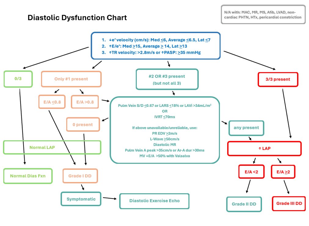
diastolic dysfunction chart

Why does diastolic dysfunction develop as left ventricular hypertrophy (LVH) progresses?
The hypertrophied myocardium becomes stiffer, impairing ventricular filling.
left ventricular systolic dysfunction - LVH
normal LV sys. function in early stage disease, and LV cavity size is small due to hypertrophied LV walls
if HTN remains uncontrolled or becomes severe, the LV wall thickness continues to inc. and may lead to myocardial ischemia and LV systolic dys.
hypertrophy inc. disproportionately to the development of new capillaries in the wall, creating an imbalance bw myocardial O2 supply and demand, esp. during stress
—
systemic HTN is also a major risk factor for coronary artery disease (CAD), ischemia, and arrhythmias
these conditions can further impair sys. function and contribute to the progression of HF
in this context, the resulting HF from hypertensive disease is characterized by reduced LV sys. func. (HFrEF)
What is a primary effect of systemic hypertension on the heart?
It leads to concentric left ventricular hypertrophy.
Which condition is primarily caused by systemic hypertension?
LVPO
conditions that increase the afterload of the LV
blood pressure
aortic stenosis
wall stress (Law of La Place)
clinical findings in hypertensive pts.
asymp. » very common (silent killer)
severe headaches » common
vision changes or vision loss » common
specific ECG changes (inc. ‘R’ wave voltage)
inc. # of myocardial cells of LVH appears on the ECG as an inc. wall diameter
the extra cardiomyocytes cause an inc. voltage of the ‘R"‘ wave
dizziness
chest pain (angina)

treatments for hypertension
treatment of the underlying disease causing the secondary HTN
lifestyle modification (dec. risk factors)
antihypertensive medication
diuretics: removes excess sodium and water from body » reducing blood vol. and BP
ACE inhibitors and ARBs: relaxes blood vessels and lowers BP
calcium channel blockers: relax and widen blood vessels and reduce BP
beta-blockers: dec. heart rate and the force of contraction, to lower BP
Which of the following are common clinical findings in patients with hypertension? Select all that apply.
severe headaches
vision changes or vision loss
inc. ‘R’ wave voltage on ECG

echo findings in hypertension
LOOK AT IMAGE FOR CHARTS
concentric LVH (inc. LV wall thickness)**:
NORMAL
<1.0cm in men, <0.9cm in women
inc. relative wall thickness (RWT):
RWT = [2(PWTd)]/(LVIDd)
RWT doesn’t have to be indexed for pt. body size
NORMAL
0.22/0.24cm - 0.42cm
inc. LV mass**:
NORMAL
<115g/m2 for men
<95g/m2 for women
LV mass is highly predictive of pt. outcomes
—
MORE ECHO FINDINGS..
LV dia. dys:
NORMAL E/A
<0.8
LV sys. function (EF):
NORMAL EF
52-74% for men
54-74% for women
mild LA dilation:
NORMAL (for both men and women)
<34mL/m2
aortopathy:
aortic root and asc. Ao dilation and aneurysmal changes, as well as aortic dissections
effacement of the aorta = ‘bulges’ of the sinus of Valsalva aren’t visible anymore bcuz of aortic dilatations
NORMAL AO ROOT:
<3.7cm for men
<3.3cm for women
NORMAL ASC AO:
<3.4cm for men
<3.1cm for women
mid-cavity LV obliteration/obstruction:
dagger-shaped waveforms with a lower velocity
velocites <2.5m/s
calcifications:
mitral annular calcification (MAC)
AV calcification (cusps of the AV)

simplified version of changes with systemic HTN
diastolic function = mildly dec. (Grade I/Impaired)
LV wall thickness = inc.
LA size = inc.
LV mass = inc.
RWT = inc.
systolic LV function = normal (except in very late stage)
aorta size = inc.
True or False: Increased flow velocity in the left ventricle can be found in hypertensive patients.
true
Which valves can be impacted by systemic hypertension? (Choose one answer or multiple)
AV
MV
differential diagnosis for LVH
HTN (most common)
hypertrophic cardiomyopathy (HCM)
aortic stenosis
chronic renal failure
athlete’s heart
infiltrative diseases like Amyloidosis, Hemochromatosis, and Fabry’s disease
acute myocarditis
coarctation of the aorta (CoAo)
friedreich’s ataxia
Which of the following may be attributed to the presence of hypertension in a patient?
LV wall thickness of 12mm
Which of the following is the normal range or the range of low risk for the patient in terms of the systolic component of blood pressure?
Systolic pressure (mmHg) < 120mmHg
Which of the following can develop if a patient has hypertension that is not controlled?
retinopathy

Which of the following images would align with a hypertensive patient?
grade I DD in the image
The patient's blood pressure was measured from the brachial artery at 114/70. Which of the following is the best description for this value?
normotensive
Which of the following is an effect of hypertension?
concentric LVH
Which of the following affects the mean arterial pressure (MAP)?
SVR
Which of the following is TRUE about mean arterial pressure?
Can be calculated from the systolic and diastolic pressure measurements from the blood pressure cuff
Pulse pressure can be used to identify the probability of which of the following?
HF
The systolic blood pressure can be estimated by which of the following Doppler measurements?
Maximum pressure gradient of the mitral regurgitation
Patient's with chronic hypertension may have mild to moderate mitral annulus calcification.
True
The value of the systolic blood pressure amount is the same as which of the following?
systolic press. of LV
pulmonary hypertension (PHTN)
inc. pressure in the pulmonary arteries
normal values:
mean pulmonary artery pressure (mPAP) = <20mmHg
RV systolic pressure (RVSP) = <40mmHg
pathophysiology of PHTN
1) PHTN is a pressure overload for the RV
PHTN inc. the afterload for the RV
results in dec. stroke volume from the RV into pulmonary circ. + less oxygenated blood
other conditions that inc. afterload of RV include: pulmonary stenosis & any condition that inc. wall stress of RV (Law of La Place)
2) elevated pulm. pressure = higher pressures in rt. heart chambers
high press. in the RA negatively impacts systemic venous return
less blood returning to RA = dec. RV stroke volume & amt. of blood sent for oxygenation
results in lower lvls of circulating oxygen
3) altered structure and function of the rt-sided heart chambers from the high pulm press. known as cor pulmonale
amt. of change depends on severity of RVPO and whether it;s acute or chronic onset
changes include..
dilated RA and ventricle chambers
inc. RV wall thickness
an eventual dec. in RV sys. function
chronic pulmonary hypertension (PHTN)
chronic = has persisted over an extended period of time
longer time duration allows more structural changes to rt. heart chambers to occur
echo should document..
RV size & wall thickness
RA size
RV sys. func.
since lt. sided heart disease and congenital defects also cause PHTN, those structures are assessed as well
acute pulmonary hypertension (PHTN)
acute = happens suddenly
may not see RV hypertrophy (RVH) w/ acute onset since this is a change that takes time to develop
echo will assess…
amt. of dilatation of RV and RA
if there’s RV sys. dys.
most common condition causing acute PHTN: acute pulmonary embolus (APE)
in these pts. assess for any thrombus or residual emboli that may be in the rt. heart or main pulm. artery
cor pulmonale
collective term referring to the dilatation of the RV and RA, anf the RVH that occurs in chronic PHTN
may also imply poor RV sys. func.
What is a primary consequence of pulmonary hypertension on the right ventricle?
dec. stroke volume
What is a primary effect of pulmonary hypertension on the right ventricle?
inc. afterload on the RV
pulmonary arterial pressures become increased when:
pulmonary arterial vessel diameters dec.
blood volume in the pulmonary vasculature inc.
—
elevated pulm. pressures can develop from conditions affecting lung tissue directly or may arise from dysfunction in either side of the heart
etiology/causes of PHTN
pulmonary arterial HTN (PAH)
inc. pulm. arterial press. and vascular resistance in the lung arteries
characterized by vasoconstriction and vascular proliferation in the pulmonary arteries
lung disease
chronic obstructive pulmonary disease (COPD) or conditions causing hypoxia (sleep apnea, APE, asthma)
left-sided heart pathology that causes elevated LA pressure
LV sys. or dia. dys, MV and AV disease
shunts
VSD, PDA (patent ductus arteriosus), ASD
pulmonary artery obstruction
lung cancer, certain autoimmune diseases
chronic blood clots in the lungs can obstruct blood flow » causing inc. press. in pulm. arteries
—
miscellaneous causes
conditions like sarcoidosis or metabolic disorders that indirectly contribute to pulmonary hypertension development
clinical findings with PHTN
asymptomatic patients:
mild PHTN
may experience more symptoms and signs of rt. HF as pulm. press. inc.
clinical signs of rt-sided HF:
SOB
peripheral edema (leg swelling)
jugular venous dystention
reduced venous return to heart due to inc. pressures and columes in the rt. heart » leads for the jugular veins to no longer collapse w inspiration
Kussmaul’s sign: absence of jugular vein collapse w inspiration
fatigue and dizziness
hepatomegaly
ascites
clubbing of fingers & cyanosis
found in pts. w low O2 saturations (pts. w primary lung disease)
treatment for PHTN
medications
oxygen therapy
surgery to correct any underlying cause

right heart catheterization - PHTN
catheter is advanced from either a vein in the groin or the arm into the SVC » into RA, RV, and PA (pulm. art.) to record pressure at each location
can also measure the pulmonary capillary wedge pressure (PCWP) which is equivalent to the LA press. (LAP)
provides a direct measurement of the PA press.
sometimes called a Swan-Ganz or PA catheterization
gold standard for the most accurate measurement of pulmonary pressure
ISSUE: invasive and costly procedure makes it not suitable to use to monitor pts. multiple times during their treatment
so echocardiography is the PREFERRED method to monitor pulmonary pressure’s and pt. response to medication


echocardiography for measuring pulmonary arterial press. - PHTN
doesn’t provide direct measurements, but they provide accurate estimations in a less invasive and costly manner
pulmonary arterial systolic pressure (PASP) is equivalent to RV sys. press. (RVSP) when pulmonary or aortic stenosis isn’t present in pt.
—
reference range values for mPAP and RVSP/PASP in image above
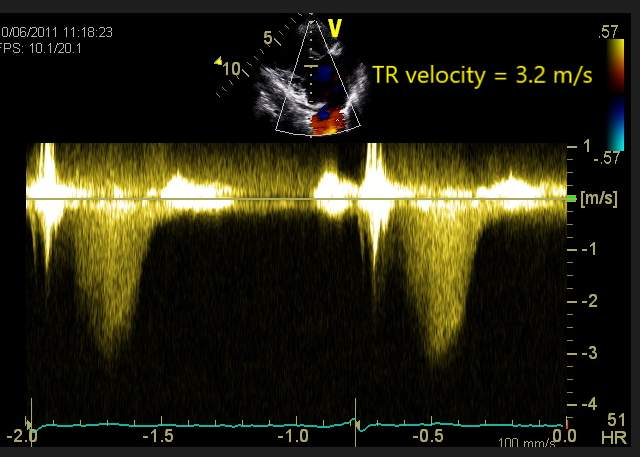
calculating RVSP and/or PASP - tricuspid regurgitation (TR)
waveform most commonly used to measure RVSP
TR is a flow occurring in systole in the rt. heart chambers
TR velo. reflects the press. diff. bw RA and RV in systole
adding the RAP to TR velocity allows us to calculate RVSP
TR velocity is converted to a pressure value in the simplified Bernoulli’s equation
—
TR press. = RV press. - RA press. » RV press. = TR press. + RA press.
RVSP = 4VTR2 + RAP
used when no pulm. stenosis present
RVSP = 4VTR2 + RAP - 4VPS2
used when pulm. stenosis present
—
right atrial pressure (RAP) is an estimated value depending on size and collapsibility of IVC
3mmHg is added when..
IVC =/<2.1cm and collapses w inspiration/sniff
8mmHg is added when..
IVC <2.1cm but doesnt collapse w inspiration
IVC >2.1cm but collapses normally w inspiration
15mmHg is added when..
IVC >2.1cm and doesn’t collapse more than 50% w/ inspiration
calculating RVSP and/or PASP - VSD velocity
ventricular septal defect (VSD) velocity:
not every pt. will have VSD flow
reflects the press. difference bw LV and RV
press. in LV will be the same as pts. SBP amt. if there aren’t any obstructions to flow bw LV and systemic arteries
—
VSD press. = LV press. - RV press.
RVSP = sys. BP (SBP) - 4VVSD2

calculating RVSP and/or PASP - PDA velocity
patent ductus arteriosus (PDA) velocity:
a persistent fetal connection that remains open in adults when it fails to close within the first few days after birth
shunt connects the dec. ao and the PA
excess blood volume from the PDA can cause dilatation of the PA and LA
PDA is an extra volume load for the lt. side of the heart
inc. LV preload » can lead to LVVO if the volume is significant
overtime, if the PDA is large, the LA may struggle to accommodate excess volume, causing it to back up into the lungs » leading to development of PHTN
—
PASP = SBP - 4VPDA2
—
echo:
CW doppler
branches of PA should be clear, if MPA isn’t elongated then PDA flow won’t be seen
bidirectional flow w varying distribution of sys. and dia. components
PDA creates a continuous flow murmur
calculating mPAP - RVOT waveform
RVOT acc. time is useful to estimate RVSP, esp. when a pt. doesn’t have TR, VSD, or PDA
RVOT acceleration time (or PA accel time) Reference ranges:
Normal > 105 ms
Mild PHTN = 80-100 ms
Moderate PHTN = 60-80 ms
Severe PHTN = <60 ms

calculating mPAP - tricuspid regurgitation
mPAP = mean P (TR) + RAP
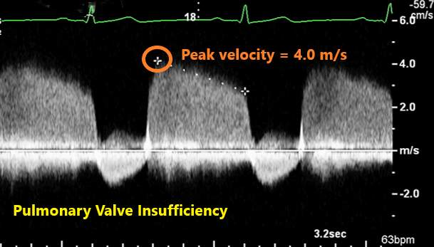
calculating mPAP - pulmonary insufficiency (PI) waveform
peak velo. of pulmonary insufficiency jet at the start of dia
corresponds to the dicrotic notch press, whic is similar to the mPAP value
mPAP = max pressure gradient (PI) + RAP
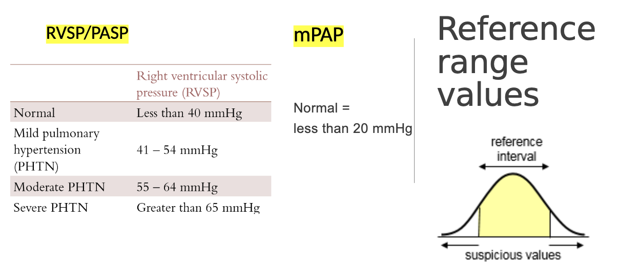
range values for RVSP/PASP and mPAP charts

echo findings in PHTN
signs of inc. pulmonary press. (PHTN):
SPECTRAL MEASUREMENTS
TR velocity - RVSP greater than 40mmHg
RVOT acc. time <100ms
M-MODE & SPECTRAL
RVOT waveform on PW will be less rounded and more angular & may have mid-sys notching or cessation of flow
flying W sign and absent a-dip on PV M-mode
signs of rt heart failure:
dilated RA and RV
RVH in chronic PHTN
D-shaped LV
paradoxical septal motion
dec. RV sys func
secondary TR
IVC plethora (IVC dilatation) and dilated hep. veins, ascites
thrombus or transiting embolus in rt. heart
left sided heart pathology and presence of shunts (ASD, PDA, VSD)

flying 'W sign and absent A-dip in M-Mode
M-mode tracing of pulm. valve
indicated PHTN
PV leaflet creates a ‘w’ shape during sys when its open
‘a-dip’ that is normally present at end-dia disappears when PHTN present
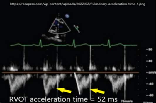
RVOT flow PW dopper w PHTN
less rounded
more angular
may have mid-sys notching

thrombus or transiting embolus in the right heart chambers
APE can cause acute PHTN
positive McConnell’s sign and 60/60 sign are also echo findings for APE
Which of the following echocardiographic findings support decreased right ventricular (RV) systolic function? Select all that apply.
dec. RV fractional area change
TAPSE <17mm
abnormal RV strain
inc. RV wall thickness
Which echocardiographic view is best for accurately measuring the right ventricular (RV) wall thickness?
subcostal 4C view
prognostic indicators for PHTN
dec. RV function
visual EF, TAPSE, RV, MPI, S’ velocity, RV strain, RV FAC
associated w poorer prognosis
presence of pericardial effusion
indicates advanced disease stage
linked to a worse prognosis for pt.
inc. indexed RA volume
correlates w disease severity and poorer outcomes
inc. LV eccentricity index (EI)
indicated leftward IVS shift due to RVPO, worsening prognosis

eccentricity index (EI)
normal LV EI is =/<1
EI = diameter2 (width)/diameter1 (height)

progression of PHTN
RV hypertrophy (RVH):
RV initially thickens its wall to maintain RV cardiac output against inc. afterlpad
RA normal size
apical traction:
RV and RA dilate w weakening RV contractile func, w progressive PHTN
RV cardiac output isn’t maintained
RV apex may be pulled leftward as it dilates
RV-PA uncoupling:
rt heart chambers enlarge significantly
IAS bows into LA
RV becomes more rounded w an abnormal EI
severe stages of PHTN
acute pulmonary embolism (APE)
refers to a sudden blockage of a PA by a thrombus or other material that has traveled/embolized to the lungs
results in rapid inc. in pulm. pressures
serious condition that can be fatal
etiology/cause:
DVT that travelled up to the heart and into lung tissue
embolization from a cardiac mass (vegetation, papillary fibroelastoma)
blockage causes a rapid, significant inc. in pulm. arterial pressures since the blood is unable to move forward into the lungs from the heart
high press. in the lung cause a sudden inc. in afterload for RV
symptoms:
severe chest pain w sudden onset
severe dyspnea (SOB)
tachypnea (rapid breathing)
cyanosis
hypotension
other diagnostic tests:
chest x-ray and ECG = wont indication pulmonary thrombus/embolism
pulmonary angiography (fluoroscopy) and CT pulmonary angiography = GOLD STANDARD exam confirming presence of APE
VQ scan = ~40% of the exams are non-diagnostic for APE
acute pulmonary embolism (APE) - echo findings
McConnel Sign:
normal contractility of apex of RV, but the basal and mid portions will be akinetic or severely hypokinetic
Thrombus in Main Pulm. and/or Branches:
thrombus in the pulm. arteries may be seen on echo, although CT pulm. angiography is the gold standard for detection
thrombus = stationary blood clot
60/60 sign:
RVOT acc. time <60ms
TR max. PG <60mmHg
echo signs of rt. HF:
dilated RV and RA
dec. RV sys. function (abnormal TAPSE)
paradoxical septal motion
D-shaped LV
flying W sign
dilated IVC and hep. veins
acute pulmonary embolism (APE) - treatment
anticoagulant therapy
pulmonary embolectomy
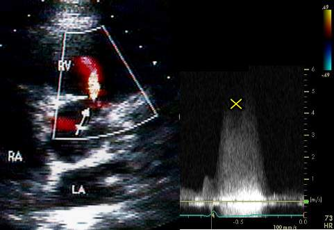
eisenmenger’s syndrome
when the pressures in the rt-sided chambers become higher than the lt-sided chambers due to the presence of a large shunt
right to left flow
deoxygenated blood from the rt. side mixes w/ oxygenated blood in the lt. side
mix. of blood has a lower O2 saturation than normal and is sent out through the aorta to the rest of the body
pt. becomes cyanotic and fatigued, less O2 circulated, and O2 saturation goes down
pt. may have blue tinge to their skin, lips, or fingertips
echo findings:
colour and spectral images/clips document right to left flow
high RVSP
Qp:Qs ratio >1.5:1 correlates w a large shunt
this calc. is used when there’s a shunt present, it determins if the shunt size is sig. or not
compares stroke volume of lt. (Qs) and rt. (Qp) heart
uses LVOT diameter, LVOT VTI, RVOT diameter, RVOT VTI
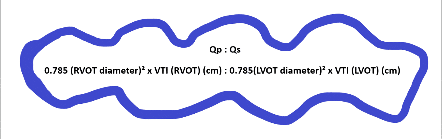
ASD and VSD assessment formula
PDA assessment formula


Looking at the following image, does the patient have pulmonary hypertension?
Don't know. Need spectral Doppler image showing the largest TR jet velocity
Which of the following Doppler measurements estimates the pressure in the pulmonary vasculature?
Acceleration time measured on a PW Doppler of the RVOT flow
Which of the following does 'clubbing' of the fingers and nail bed signify?
cyanotic heart disease