VTMD 206 - Neuro 2
 Call Kai
 Learn
 Practice Test
 Spaced Repetition
 Match
 Flashcards
 Knowt Play
1/110

There's no tags or description
Looks like no tags are added yet.
111 Terms
Cribiform plate
Where does the olfactory nerve (I) exit the skull?
Optic foramen
Where does the optic nerve (II) exit the skull?
Orbital fissure
Where does the oculomotor nerve (III) exit the skull?
Orbital fissue
Where does the trochlear nerve (IV) exit the skull?
Orbital fissure
Where does the trigeminal nerve (V), V1 opthalmic nerve, exit the skull?
Round foramen
Where does the trigeminal nerve (V), V2 maxillary nerve, exit the skull?
Oval foramen/mandibular foramen
Where does the trigeminal nerve (V), V3 mandibular nerve, exit the skull?
Orbital fissure
Where does the Abducens nerve (VI) exit the skull?
Stylomastoid foramen/internal auditory meatus
Where does the facial nerve (VII) exit the skull?
Internal auditory meatus
Where does the Vestibulocochlear nerve (VIII) exit the skull?
Jugular foramen
Where does the Glossopharyngeal nerve (IX) exit the skull?
Jugular foramen
Where does the Vagus nerve (X) exit the skull?
Jugular foramen/foramen magnum
Where does the Accessory nerve (XI) exit the skull?
Hypoglossal canal
Where does the Hypoglossal nerve (XII) exit the skull?
Orange = Orange
Light Green = Parietal
Red (little line) = Interparietal
Pink = Temporal
Dark Green = Sphenoid
Light Blue = Maxillary
White = Nasal
Purple = Incisive
Yellow = Zygomatic
Blue = Palatine
Red = Occitpital
Dark Green (around eye) = Lacrimal
Beige (2) = Ptergoids and Vomer
Orange =
Light Green =
Red (little line) =
Pink =
Dark Green =
Light Blue =
White =
Purple =
Yellow =
Blue =
Red =
Dark Green (around eye) =
Beige (2) =

Infraorbital foramen
Optic canal
Orbital fissure
Rostral alar foramen
Caudal alar foramen
Foramen magnum

Optic canal
Orbital fissure
Rostral alar foramen
Caudal alar foramen
Oval foramen
Foramen magnum

Infraorbital foramen
Cribiform plate
Maxillary foramen
Optic canal
Orbital fissure
Round foramen
Oval foramen
Jugular foramen

Dogs and horses
_____ and _____ have an alar canal
This means that the foramen caudal to the orbital fissure is the rostral alar foramen
Caudally, the next foramen is the caudal alar foramen (and caudal to that is the oval foramen)
Together the alar foramina define the alar canal. WITHIN the alar canal there is a connection to the cranial cavity, this is the round foramen (foramen rotundum = round in latin)
The alar canal is essentially a bony roof covering the round foramen
Foramen orbitorotundum
In ruminants and pigs, there is no alar canal (so no alar foramina), and the round foramen and orbital fissure have fused into the _______ __________
Foramen lacerum
In the horse this is the combination of the oval foramen, carotid foramen and jugular foramen
Intramembranous bone growth
Most skull bones develop through _________ ________ __________
Endochondral
As opposed to the rest of the skull, bones of the cranial base (unpaired) are mostly formed by _________ bone growth (develop from a cartilaginous precursor)
Ethmoid
Sphenoid
Occipital
Pink = Rostral
Red = Middle
Dark Red = Caudal
Label the 3 fossae of the cranial cavity
Pink =
Frontal lobes/olfactory lobes
Red =
Temporal and parietal lobes
Dark Red =
Hindbrain and brainstem

Paranasal sinuses
Cavities between inner and outer layers of specific bones surrounding
Ex. frontal, maxillary, ethmoid, conchal, palatine, sphenopalatine, etc.
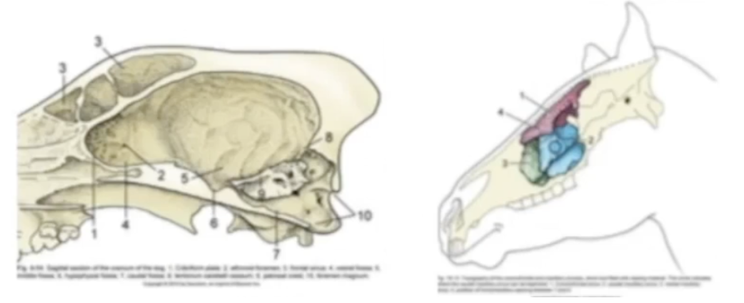
Nasal cavity
Facial part of respiratory passages
Bony margins: nasal aperture to choanae (tube separated by median nasal septum)
Conchae and ethmoidal turbinates (scroll like paper - thin bones) within _____ ______ covered with mucosa to slow down, warm and cleanse inspired air
Defines meatuses

Temporomandibular joint
Synovial joint in the skull of some species
Condylar, with both condyles separated by the head!
Syndochondrosis
Mandibular symphysis
Never get full union in some species, allows for some movement

Afferent
Dorsal root contains _______ nerves
Efferent
Ventral root contains _______ nerves
Somatic afferent
Contained in dorsal root
Voluntary/sensory
Exteroreceptive (pain, heat, tough), proprioception
“Special ______ ______” (vision, hearing, balance)
Visceral afferent
Contained in dorsal root
Autonomic/sensory
Enteroreceptive (stretch, chemical)
“Special ______ ______” (smell, taste)
Visceral efferent
Contained in ventral root
Voluntary/motor
Smooth muscle (ANS)
Somatic efferent
Contained in ventral root
Autonomic/motor
Striated muscle
Afferents, Efferents
In the medulla
Lateral orientation for the _______
Medial orientation for the _______

Afferent, efferent
In the spinal cord, they are slightly reorganized
Dorsal route/horn = _______
Ventral route/horn = _______

SVA
SE, VE, SA, VA, SSA, and SVA?
Olfactory (I)
SSA
SE, VE, SA, VA, SSA, and SVA?
Optic (II)
SE, VE
SE, VE, SA, VA, SSA, and SVA?
Oculomotor (III)
SE
SE, VE, SA, VA, SSA, and SVA?
Trochlear (IV)
SE, SA
SE, VE, SA, VA, SSA, and SVA?
Trigeminal (V)
SE
SE, VE, SA, VA, SSA, and SVA?
Abducent (VI)
SE, VE, VA, SVA
SE, VE, SA, VA, SSA, and SVA?
Facial (VII)
SSA
SE, VE, SA, VA, SSA, and SVA?
Vestibulocochlear (VIII)
SE, VE, VA, SVA
SE, VE, SA, VA, SSA, and SVA?
Glossopharyngeal (IX)
SE, VE, VA, SVA
SE, VE, SA, VA, SSA, and SVA?
Vagus (X)
SE, VE, VA
SE, VE, SA, VA, SSA, and SVA?
Accessory (XI)
SE
SE, VE, SA, VA, SSA, and SVA?
Hypoglossal (XII)
Olfactory (I)
Cranial Nerve
Special visceral afferent
From olfactory cells in nasal epithelium in ethmoid conchae
Short passage through cribriform plate to olfactory bulbs
Meningeal sheathes
Bulb (cranial nerve) size reflects importance of smell
Function:
Transmits smell
Testing:
Concern for loss of smell
Place a covered morsel of food beneath the nose and observe for normal sniffing behaviour
Do not use irritating substances – stimulates trigeminal nerve
Sensory – Special Visceral Afferent
Optic (II)
Cranial Nerve
Special somatic afferent
Brain tract from diencephalon to retina
Cross contralaterally
Optic chiasm
Meningeal extensions
Dura blends with sclera of eye
Optic nerve covered in meningeal sheath
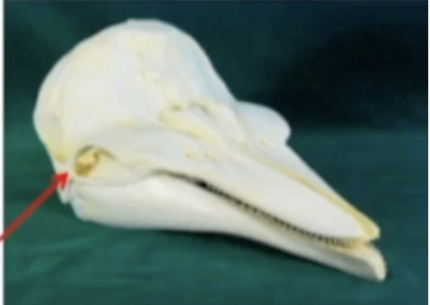
Exit cranial cavity into orbit at optic foramen/canal (red arrow)
Function:
Necessary for vision
Carries the sensory nerves for certain eye reflexes
Closure of the eyelid
Testing:
Menace response: move hand towards patient’s eyes in a threatening manner and watch for blink response
Visual following: drop object and see if patient’s eyes follow
Sensory – Special Somatic Afferent
Oculomotor (III)
Cranial Nerve
Efferent only
Somatic Efferent to remainder of eye muscles (motor)
Dorsal rectus
Medial rectus
Ventral rectus
Ventral oblique
Levator palpebri
Visceral Efferent
Ciliary muscles
Pupil constrictors
Courses with CN IV and VI
Orbital fissure or foramen orbitorotundum
Function:
Carries motor neurons that control most of the eye muscles
Pupillary constriction
Testing:
Pupillary Light Reflex (PLR): see if pupil constricts when you shine light at the eye
Motor – Somatic Efferent, Visceral Efferent
Trochlear (IV)
Cranial Nerve
Somatic efferent to Dorsal oblique (1) of the eye
For axial rotation of the eye
Usually exits through orbital fissure or foramen orbitototundum (in cattle)
Function:
Carries motor neurons that control other muscles of the eye
Testing:
Check up and down movement of eye with head still (adduction)
Motor – Somatic Efferent
Trigeminal (V)
Cranial Nerve
Somatic only (efferent and afferent)
Largest cranial nerve
Three branches (tri-geminal means 3 roots)
Ophthalmic (V-1)
Maxillary (V-2)
Mandibular (V-3)
V1: SA from orbit/frontal area
V2: SA from infraorbital, nasal, and maxillary areas
V3: SA/SE mixed efferent (to mastication muscles), afferent from mandibular area
Function:
Motor nerve to jaw muscles of mastication
Sensory nerves to the face (mouth, nose, and cornea)
Testing:
3 Nerve Branches:
Ophthalmic branch
Palpebral reflex: touch medial canthus of eye and see if it triggers a blink response
Maxillary branch
Touch/pinch upper lip lateral to canine tooth, normal response is wrinkling of face and blink (also depends on facial nerve)
Mandibular branch
Touch/pinch upper lip lateral to canine tooth
Mixed – Somatic Efferent, Somatic Afferent
Abducens (VI)
Cranial Nerve
Somatic efferent to:
Lateral rectus
Retractor bulbi (3)
Also controls eye movements, moves the eye outwards
Usually exits through orbital fissure or foramen orbitototundum
Function:
Carries motor neurons that control other eye muscles
Testing:
Check lateral movement of the eyes with head still (abduction)
Motor – Somatic Efferent
Facial (VII)
Cranial Nerve
SE, VE, VA, SVA
Into internal auditory meatus with VIII - Vestibulocochlear, then through stylomastoid foramen
In petrosal temporal it is mostly
VE (ANS: to lacrimal glands and paranasal sinuses)
SVA (from rostral 2/3 taste buds in the tongue)
SE to stapedius muscle of inner ear
Outside temporal (“free” facial nerve) it is SE to muscles of facial expression. Many branches here join with CN V branches (afferent). Also some VA from oropharynx
Function:
Controls muscles responsible for facial expressions
Also innervates salivary and lacrimal glands
Testing:
Observe face for asymmetrical eyelid closure, widened palpebral fissure, spontaneous blinking, or a drooping ear
Palpebral response: test blinking when touching eye
Evaluate tearing with Schrimer test strips
Mixed – Somatic Efferent, Visceral Efferent, Visceral Afferent, Special Visceral Afferent
Vestibulocochlear (VIII)
Cranial Nerve
Special somatic afferent
SSA nerve fibers from inner ear in petrous portion of the temporal bone
Fibers from vestibule (balance) and cochlea (hearing)
Exit cranial cavity at internal acoustic meatus (in temporal)
Function:
Cochlear nerve – responds to sound
Vestibular nerve – maintains posture and balance
Testing:
Patients should orient head and ears towards loud/unexpected noises (but difficult to test)
Vestibular dysfunction – head tilt, abnormal nystagmus, ataxic, broad stance
Sensory – Special Somatic Afferent
Glossopharyngeal (IX)
Cranial Nerve
SE, VE, VA, SVA
SE to stylopharyngeus and levator palatini mm. (swallowing)
VE to otic ganglion and parotid gland
VA from base of the tongue and carotid body/sinus (blood pressure)
SVA from caudal third of tongue (taste)
Exits via the jugular foramen, common path with CN X to pharyngeal plexus
Function:
Provides sensory and motor control of the throat and vocal cords (pharynx)
Testing:
Touch left/right side of caudal pharyngeal wall w/ applicator stick and observe elevation of the palate and contraction of the pharyngeal muscles (gag reflex)
Mixed – Somatic Efferent, Visceral Efferent, Visceral Afferent, Special Visceral Afferent
Vagus (X)
Cranial Nerve
SE, VE, VA, SVA, SA
The “wanderer”
VA/VE to thorax and gut:
Monitors/Regulates state of internal viscera
VE to most organs between neck and colon (ex. slows heart rate)
SE Pharyngeal, laryngeal, and recurrent laryngeal nerves to muscles of vocalization and some pharyngeal muscles
SA to inner canal or outer ear
SVA to mucosa of oropharynx
Exits through jugular foramen
Function:
Produces sensory and motor control for major internal organs, including the heart and gastrointestinal tract
Motor and sensory innervation to the pharynx
Laryngeal function
Testing:
Touch left/right side of caudal pharyngeal wall w/ applicator stick and observe elevation of the palate and contraction of the pharyngeal muscles (gag reflex)
Mixed – Somatic Efferent, Visceral Efferent, Visceral Afferent, Special Visceral Afferent
Accessory (XI)
Cranial Nerve
SE, VE (?), VA (?)
Cranial part
Really part of CN X? VE/VA
Accessory part: spinal in origin
Runs cranially along the spinal cord between 1st 5 roots, enters through foramen magnum, re-exits through jugular
SE to parts of
Brachiocephalicus
Omotransversarius
Trapezius (dorsal branch of nerve)
Sternocephalicus (ventral branch)
Function:
Carries sensory and motor information for muscles of the head and upper neck
Motor innervation to trapezius muscle
Testing:
Look for trapezius atrophy
Mixed – Somatic Efferent, Visceral Efferent, Visceral Afferent
Hypoglossal (XII)
Cranial Nerve
Somatic efferent to tongue muscles
Hypoglossus
Genioglossus
Styloglossus
Geniohyoidueus
Originates in medulla
Innervates muscles of the tongue
Exits through hypoglossal canal
Function:
Motor nerve to the tongue
Testing:
Inspect tongue for atrophy, asymmetry, deviation
Watch patient drink water to assess tongue function
Motor – Somatic Efferent
Olfactory, Optic, Oculomotor, Trochlear, Trigeminal, Abducens, Facial, Vestibulocochlear, Glossopharyngeal, Vagus, Accessory, Hypoglossal
List Cranial Nerves in Order
S, S, M, M, B, M, B, S, B, B, M, M
List sensory, motor, or both for cranial nerves in order
Organophosphates
_______ inhibit acetylcholinesterase preventing ACh breakdown leading to excess ACh accumulation in synapses
Muscarinic
Increased stimulation of ________ receptors causes:
Salivation
Diarrhea
Miosis
Bronchoconstriction
Difficulty breathing
Nicotinic
Increased stimulation of ________ receptors causes:
Tremors
Fasciculations
Weakness
Difficulty breathing
Atropine, Pralidoxime
2 important drugs treat accumulation of acetylcholine
Continuous depolarization
Continuous neurotransmitter (ACh) stimulation causes ________ at the neuromuscular junction. This inactivates voltage-gated Na+ channels, preventing repolarization and blocking new action potentials resulting in paralysis
Falx cerebri
a fold of the dura mater that extends down into the longitudinal fissure, which separates the two hemispheres of the brain. It helps to stabilize and protect the brain's structure
Tentorium cerebelli
Fold of the dura mater, located between the cerebrum and the cerebellum in the dorsal cranial fossa. It helps to compartmentalize and protect these two brain regions
Cerebellomedullary cistern
A subarachnoid space located at the dorsal aspect of the brainstem and cerebellum. It is a reservoir of cerebrospinal fluid and is important for the circulation and absorption of CSF
2. Dura mater
2’’ Epidural space
4. Arachnoid
5. Arachnoid space
6. Pia mater
7. Membranous tentoreum cerebelli
9. Cerellomedullary cistern
2.
2’’
4.
5.
6.
7.
9.

1. Olfactory bulb
3. Corpus callosum
11. Epiphysis
17. Cerebellar cortex
18. Pons
19. Crus cerebri
20. Mammillary body
21. Hypophysis
22. Infundibulum
25. Interthalamic adhesion
27’ Third ventricle
27’’ Mesencephalic aqueduct
27’’’ Fourth ventricle
1.
3.
11.
17.
18.
19.
20.
21.
22.
25.
27’
27’’
27’’’

Top = Pineal gland
Bottom = Rostral colliculus
Top =
Bottom =

Lateral ventricle
Mesencephalic aqueduct
Fourth ventricle
Infundibular recess
Cavity of olfactory bulb
Third ventricle
Central canal

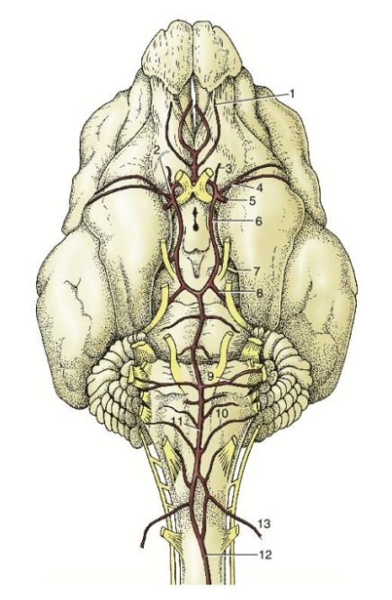
2. Rostral cerebral artery
4. Middle cerebral artery
7. Caudal cerebral artery
8. Rostral cerebellar artery
10. Caudal cerebellar artery
11. Basilar artery
13. Vertebral artery
2.
4.
7.
8.
10.
11.
13.
paired lateral ventricles, third ventricle, fourth ventricle
CSF is produced in the ventricles of the brain
List ventricles
Choroid plexus
Structures found in the ventricles of the brain that produce CSF
Hypophysis/Pituitary Gland
Often called master gland
Produces hormones that affect the activity of other endocrine glands
Acts as relay between nervous and humoral mechanisms
Below hypothalamus
Sits in depression at the base of the skull called the sella turcica
Surrounded by dura
Combination of different types of tissue

Cavernous sinus
Tissues of the venous plexus that surround the pituitary inside the sella turcica
Where venous drainage from pituitary occurs

Epiphysis/Pineal Gland
Located on the caudal end of the roof of the third ventricle
Directly in front of rostral colliculi
Tends to be pigmented/darker

Dorsal sagittal sinus
Straight sinus
Transverse sinus
Basilar sinus
Cavernous sinus
Sinuses return CSF back to circulation

Sematosensation
detection and processing of bodily sensations
Touch, temperature, pain, and proprioception
Thermoreceptors
Somatosensory Receptor
Changes in temperature
Free nerve endings
Superficial, towards epidermis
Associated with small diameter A-delta and C-fibres (similar to nociceptors)
Some unmyelinated
Respond to sudden changes in skin temp; adapt to constant temp
2 types:
Warm
↑ firing rate as temp rises from 30℃ to 45℃
Above 45℃ nociceptors become activated
Cold
↑ firing rate as temp changes from 10℃ to 25℃
↓ firing rate as temp rises 26℃ to 40℃ – ↓ firing rate at temp below 10℃
Can adapt
Temperatures below 15°C or above 43°C evoke thermal sensation accompanied by pain
Transient Receptor Potential (TRP) ion channels
Influx of Na+ and Ca2+
TRP receptors respond to specific temperatures, but also certain foods
Each channel has distinct thermal activation
TRPA1: 12 ̊C – 24 ̊C
TRPM8: 8 ̊C – 26 ̊C
TRPV4: 25 ̊C – 34 ̊C
TRPV3: >33 ̊C
TRPV1: >43 ̊C
TRPV2: >52 ̊C
Mechanoreceptors
Somatosensory Receptor
Physical manipulations (ex. Stretch, indentation, tension of skin)
Low threshold (or high sensitivity)
Don’t need that strong of a stimulus to activate them
Even weak stimulation causes them to fire action potentials
4 major types
Merkel’s Disk
Meissner’s Corpuscle
Ruffini’s Corpuscle
Pacinian Corpuscle
merkel’s disk, meissner’s corpuscle, ruffini’s corpuscle, pacinian corpuscle
4 major types of mechanoreceptors
Nociceptors
Somatosensory Receptor
Extreme temperature, injury, chemicals
Proprioceptors
Somatosensory Receptor
Changes in joint and muscle tension
Signal body position
Tells us where our body/limbs are in space
2 types:
Muscle spindles
Golgi tendon organs
Receptor → First order neuron → Second order neuron → Spinal cord or Brainstem → Third order neuron → Thalamus → 4th order neuron → Cerebral cortex
Somatosensory pathway from receptor to brain
Phasic Receptors
Quick acting
Adapt: stop firing when stimuli are constant (smell)
Turn on/off
Provide info about ‘change’ in stimulus
Reacts strongly when stimulus is changing
Ex. mechanoreceptors

Tonic Receptors
Slow acting, Slow adapting OR
Slow/No adaptation: continue to for impulses as long as the stimulus is there (proprioreceptors)
Provide info about steady state stimulus
Ex. nociceptors

greater
Small receptive field and dense innervation = ______ localization and discrimination of a stimulus for a receptor
Action potentials
Increased intensity of stimulus = Increased number of ______________
Aα
Type of Primary Afferent Axon
Conduct signals from proprioceptors
Largest diameter/Fastest
Aβ
Type of Primary Afferent Axon
Conduct signals from mechanoreceptors
Second largest diameter
Aδ
Type of Primary Afferent Axon
Conduct signals from fast nociceptors and thermoreceptors
Second thinnest in diameter
C
Type of Primary Afferent Axon
Conduct signals from slow nociceptors and thermoreceptors, itch
Thinnest in diameter/Slowest
Merkel’s Disc
Type of Mechanoreceptor
Located in epidermis
Small receptive field – good discrimination
Light Touch & Pressure
‘Sustained light touch’
Vibrations (5-15 Hz)
Slowly adapting
Senses steady state pressure and texture
Dense in fingertips, lips, and genitalia
Located quite superficially, associated with Aβ axons
Vesicles within containing neurotransmitters
Have channels
Critical for tactile acuity
Can help discriminate different patterns in a grating acuity test
More in the fingertips
Meissner’s Corpuscle
Type of Mechanoreceptor
In superficial layers of skin
Small receptive field – good discrimination
Light Touch
Vibration (<50 Hz)
Fast adapting
Main difference from merkel’s disc
40% of sensory innervation of human hand
Detects movement of textures across skin
Layers within ___________, also connected to an Aβ axon
Ruffini’s Corpuscle
Type of Mechanoreceptor
Lie parallel to skin
Deeper than the previous two
More in the dermis
As skin stretches, causes movement of anions, which causes an action potential
Large receptive field
Skin tension
Stretch produced by digit or limb movements
Senses position and movement
Slowly Adapting
Pacinian Corpuscle
Type of Mechanoreceptor
Located in subcutaneous tissue
Much deeper
Large receptive field
Pressure (e.g. poke)
‘Deep touch’
Fast adapting (adapts more rapidly than Meissner’s and lower response threshold)
Discrimination of fine surface textures