physiology quiz (dr. Butcher 1)
 Call Kai
 Learn
 Practice Test
 Spaced Repetition
 Match
 Flashcards
 Knowt Play
1/61

There's no tags or description
Looks like no tags are added yet.
62 Terms
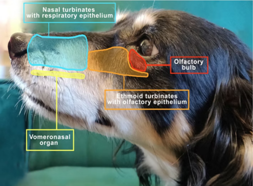
what is the mucosa made of in the nasal cavity? where is it underlying?
epithelium and lamina propria, it underlines submucosa, turbinates (trabecular bone scrolls)
turbinates?

what type of cells do the vestibule (rostal), respiratory portion (middle), and olfactory portion (caudally) have?
vestibule: stratified squamous epithelium that’s continuous with skin
respiratory portion (middle): ciliated pseudostratified columnar epithelium + goblet cells, olfactory cells; tubuloalveolar glands
Olfactory portion (caudally): Similar to respiratory portion but increased olfactory cells


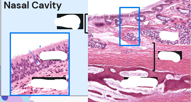

what does the larynx/pharynx look like in terms of tissue/cells?
similar to adjacent cells
part ciliated psuedostratified columnar and parr stratified squamous, depending on location
both contain gland and underlying sheets of skeletal muscle
larynx contains cartilage for support
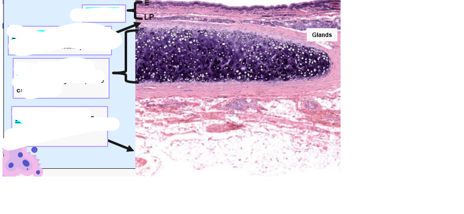
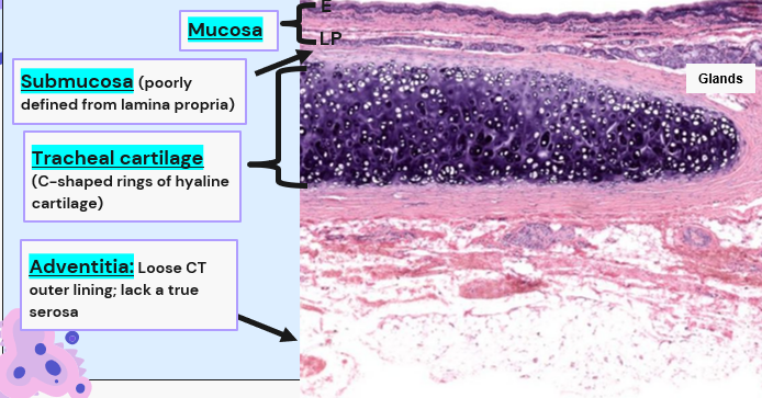
what is the trachea? structure?
it is the passage of air from larynx to the lungs
has C shaped tracheal rings made of hyaline cartilage for structural support
dorsally lined by trachealis m (smooth)
has pseudostrat. columnar epithelium
Mucociliary apparatus/escalator
when does the trachea have opened/closed rings?
closed in non mammals (so like reptiles, birds)
dont have that lined trachealis
open in mammals


Mucocilary apparatus is important why?
for the clearance of dust/dirt and inhaled pathogens from the LRT
bronchi
branch off trachea into mainstem bronchi before lung
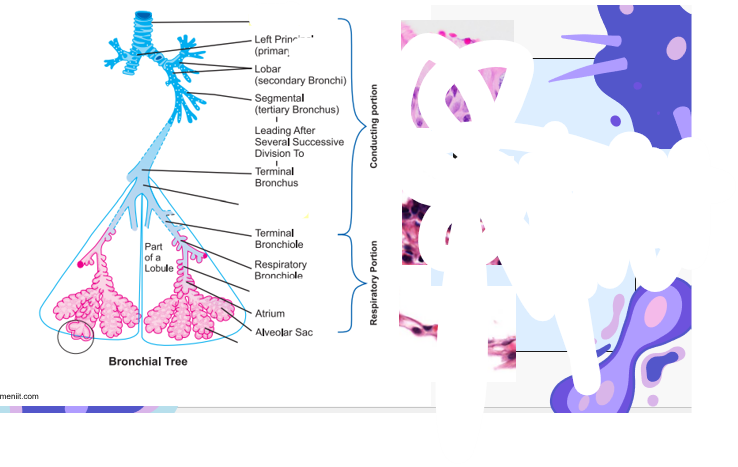
how does tissue transition? what cells to what in the lung?


what is the pleura, bronchi? alveoli


lobe vs lobules?


what is this and the definition?

bronchus
basically same as trachea
1. Ciliated pseudostratified columnar
epithelium + goblet cells
2. Bronchial glands
3. Cartilage
4. Smooth muscle
5. Bronchus associated lymphoid tissue
(BALT)
Bronchiole
Similar to bronchus
tissue: Ciliated pseudostratified columnar epithelium ( smooth muscle)
terminal progress of tissue: Ciliated columnar → ciliated cuboidal→ non-ciliated cuboidal
does the bronchiole have cartilage/ glands?
no

what is the alveolus? epithelial there?
terminal outpouching of bronchioles
simple squamous epithelium (type 1 pneumocytes)
gas exchange
type II pneumocytes: much fewer; cuboidal, produce surfactant
thin layer of supporting collagen (alveolar septum)


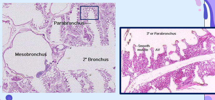
bird anatomy
primary bronchi (mesobronchi): analogous to mammalian bronchi: ciliated pseudostrat. columnar
second bronchi: ciliated columnar
Tertiary bronchi (parabronchi): fenestrated cuboidal epithelium
artia (air vesicles) and air capillaries: analogous to aveloi, simple squamous; site of gas exchange
fish structure
Basically similar to alveolar structure for gas exchange but in Direct contact with environment
secondary lamellae
Gas exchange occurs


main function of respiration
provide oxygen to tissues and remove Carbon dioxide
pulmonary ventilation: in flow and outflow of air between alveoli and atmosphere
diffusion of oxygen and carbon dioxide: between alveoli and blood
transport of oxygen and carbon dioxide: in blood and body fluids
regulation of ventilation
what does the upper respiratory tract contain? the lower?
Upper: Nasal cavity, sinuses, nasopharynx, larynx
lower: Trachea, bronchi, lungs, pleural cavity
what happens in nasal cavity?
air is warmed, humidified, and partially filtered (air-conditioning function)
Large particles trapped in mucous (goblet cells, submucosal glands) and coughed up or swallowed (turbulent precipitation)
Mucociliary elevator
Small particles trapped in mucous
Ciliated epithelium (directionally beat toward pharynx)
Nervous and Local Control of Respiratory Tree: what types function
Parasympathetic nervous system
Branches of Vagus nerve
Secrete Acetylcholine (Ach)
Parasympathetic stimulation increases bronchoconstriction and secretions
Sympathetic nervous system
Mediated by circulating catecholamines (NE, EPI) and B2-adrenergic receptors
Sympathetic stimulation causes bronchodilation
Local secretory factors
Chemical, mechanical, humoral
respiratory pathways: types and what do they do
Conduction
Nasal cavity, trachea, bronchi
Pseudostratified ciliated columnar cells with goblet and serous cells
Cartilage rings and plates keep larger two open, with remaining walls being smooth muscle
Transitional
Primary, secondary, tertiary bronchioles
Progressively less ciliation
Bronchioles are kept open with pressure and mostly smooth muscle, with pulmonary epithelium at the terminal bronchioles
Normal respiratory conditions = low pressure gradient (1-2 cm H2O) or low resistance
• Gas Exchange
Alveolar duct, atrium, alveolar sac, alveolus
what happens without force to keep the lung inflated
lung collapses bc the lung and the chest walls are not attached
how is lung positioned in the body
lung floats in thoracic cavity surrounding by pleural fluid
pleural pressure at rest vs during inspiration
at rest→ slight negative pressure and thus slight suction
during inspiration → expansion of cavity lowers pleural pressure further
pressures reversed during expiration

Where does gas exchange occur in the respiratory system
• Nasal Cavity
• Trachea
• Bronchioles
• Alveoli
• Alveoli
Which receptor predominates in neural control of the respiratory system?
• Mucous receptor
• Acetylcholine
• Alpha receptors
• Beta-2 receptors
• Beta-2 receptors
The mucociliary elevator in the bronchi move large particles downward toward alveoli •
True •
False •
Not enough info given
False •
What happens to the lung when the diaphragm contracts?
• Lung expands
• Lung contracts
• Rib cage movement
• Not enough information given
• Lung expands
At rest, pleural pressure is slightly _______, and then _______ with inspiration.
Negative; decreases
• Negative; increases •
Positive; decreases
Positive; increases
• Negative; decreases
variables that affect gas exchange
the Gas exchange membrane/respiratory exchange membrane ( a thin layer of tissue that separates animal from env. medium)
O2 movement into the lungs via diffusion/convection
rate of diffusion (depends on surface area and thickness of membrane)

active ventilation: what is it? pathway? E? how reliable?
creation of currents of air/water flowing to/from gas exchange membrane
uses E (contracting or beating cilia E)
pathway across membrane: unidirectional, tidal (bidirectional) or non directional
considered more reliable and controllable
passive ventilation
environmental air/water directly or indirectly induce flow to/from a GEM
what is compliance?
a measure of the lungs' ability to stretch and expand
high compliance: lungs expand easily and vise versa
is inspiration work? how does it work
yes, elastin and collagen fibers are interwoven among lung parachyma provide the elastic forces
when u breath in, ur muscles have to generate enough pressure to overcome the forces trying to keep the lungs closed
need 3x the transpleural pressure ( pressure difference to keep lung expanded) to expand air-filled lungs
surface tension: in each alveolus, there is a thin layer of fluid, creating surface tension, which tends to make the alveolus shrink/collapse
respiratory muscles have to work against what?
elastic recoil and surface tension
expiration is what
mostly elastic recoil
surfacant
surface active agent in water
secreted by type 2 alveolar epithelial cells
it interrupts the attraction between the water molecules (decreases surface tension)
increased surfactant = decreased surface tension and vise vera
Neonatal Respiratory Distress Syndrome (RDS) in Pre-term animals
premature animals dont have sufficient mature type 2 alveolar cells- dont produce enough surfacant
Traditional administration through a breathing tube (endotracheal tube) connected to a ventilator
infasurf: a replacement of surfacant, Calfactant - extract of natural surfactant from calf lungs
survanta
CUROSURF
all types of replacement for surfacant
work of inspiration
Expansion of lungs against lung and chest elastic forces (compliance or elastic work)
Overcoming viscosity of the lung and chest wall structures (tissue resistance work)
Resistance to movement of air into the lungs (airway resistance work)
the 4 pulmonary volumes? what happens when u add them all together?
tidal: air inspired or expired
inspiratory reserve: extra volume that can be inspired over tidal
expiratory reserve: max extra volume expired with forceful expiration after tidal
residual: volume remaining after a forceful expiration
added together, lungs can be expanded

correlating the volumes to each other
Inspiratory = Tidal + IRV = how much air can be breathed in
Functional residual = ERV + residual = how much air is in lungs at the end of expiration
Vital capacity = IRV + TV + ERV = max air expelled after max inspiration
Total lung = VC + RV = max volume lungs can be expanded
Alterations to pulmonary volumes and capacity depend on what?
Physical fitness
Age
Height
Sex
Altitude
Pulmonary function test assessed by?
spirometry: used to monitor airway pressures, flows, and volumes
gas exchange areas
alveoli, alveolar air sac, alveolar ducts, and respiratory brinchioles
dead space
air that never reaches gas exchange areas
rate of alveolar ventilation is important for what?
its the new air entering alveolar/min
its a major factor in determining concentrations of oxygen and CO2 int he alveoli
How does oxygen cross a gas-exchange membrane?
Diffusion
Convection
Active transport of ions
diffusion and/or convection
Diffusion
What are force(s) in the alveoli that increase the work of inspiration?
Elastin and collagen stretching
Surface tension
Surfactant
Elastin and collagen stretching and surface tension
Elastin and collagen stretching and surface tension
Tidal volume describes the amount of air inspired or expired at rest •
True
False
Not enough info given
• True
What term(s) describe the max volume lungs can be expanded?
Total lung capacity
Vital capacity
Residual volume
Tidal volume
• Total lung capacity
Air that never reaches gas-exchange areas is termed ______________
Alveolar ventilation
Dead space
Oxygen uptake
Spirometry
Dead space