The Abdomen
 Call Kai
 Learn
 Practice Test
 Spaced Repetition
 Match
 Flashcards
 Knowt Play
1/80

There's no tags or description
Looks like no tags are added yet.
81 Terms
rectus abdominis muscle (ventral)
Tendinous intersections
Interrupts muscle belly to create “abs”— there are 10 sections created
Linea alba
THICK dense irregular CT
Rectus sheath
CT envelope around the abs
Lumbar-vertebral flexion (bending forward)

External oblique muscle - superficial lamina
part 1 of 2
Aponeurosis
flat, sheet-like tendon that covers the abs
Inguinal ligament
bone-to-bone on os coxae
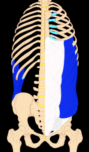
External oblique muscle - deep lamina
part 2 of 2
Doesn’t cover ribs; is in the same plane as ribs, under them
Function:
Bilateral: flex lumbar vertebral column
Unilateral: flexion & contralateral rotation (to opposite sides)

Internal oblique muscle
The aponeurosis splits— half in front of the abs, half behind
function:
flexion of lumbar-vertebral column
flexion & ipsilateral rotation

contralateral
to opposite sides
ipsilateral
to the same side
Transverse abdominis muscle
aponeurosis is posterior to abs
function:
giant abdominal sphincter
core stabilization

Quadratus lumborum muscle
function:
Unilateral: lateral flexion
Bilateral: extends lumbar-vertebral column (holds posture)
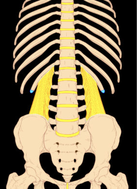
Psoas major muscle
hip flexion

Psoas minor muscle
weak vertebral flexor
only in 40% to 60% of people

dynamic retaining wall
the muscles of the abdomen work together to fight gravity and keep a lot of vital organs in place!
rectus sheath
above the belly button, the rectus sheath is as programmed.
below the belly button, all parts of the rectus sheath switch to in front of the abs to create a stronger retaining wall where there is the most pressure


inferior phrenic arteries
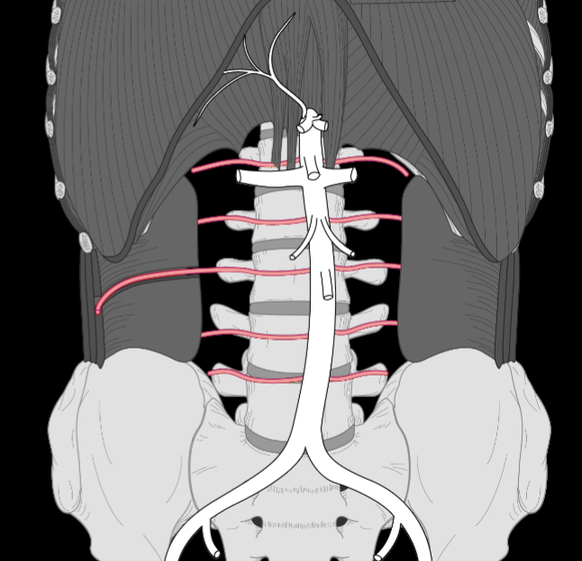
lumbar arteries
where would you find the lumbar arteries?
in between the middle and internal layers

internal thoracic arteries
—> turn into superior epigastric arteries

superior epigastric arteries
down to the belly button

inferior epigastric arteries
up to the belly button
branches from external iliac artery
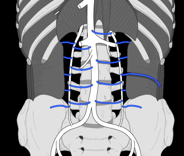
lumbar veins

ascending lumbar veins

venous collateral flow
blood can travel any-which way it wants back to the heart, typically the path of least resistance but can take detours

internal thoracic veins come together from superior epigastric veins

superior epigastric veins
anastomose
to join together
bifurcate
to branch or split

inferior epigastric veins

inferior phrenic veins

thoracic spinal nerves
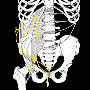
lumbar spinal nerves
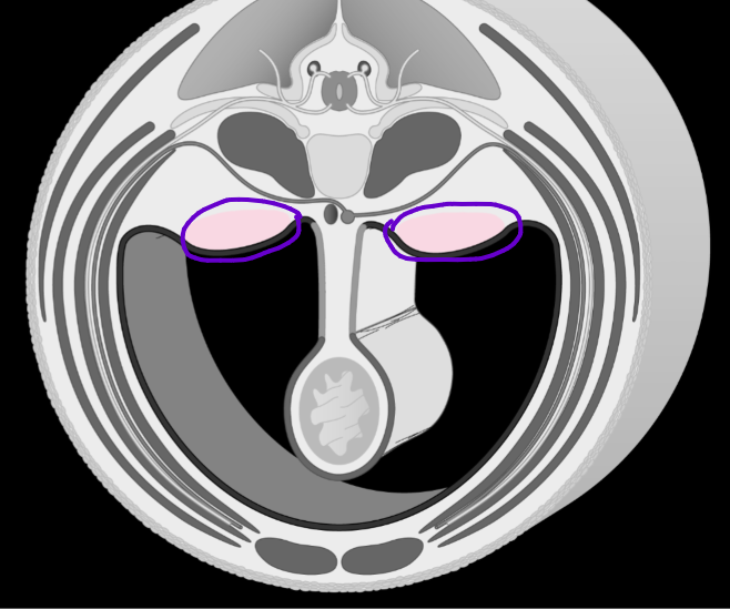
urogenital ridges
develops to be part of the urinary and genital system
Peritoneal organs…
are organs that are in front of the peritoneum
SALT C JISS? (something like that)
Retroperitoneal organs…
are organs towards the posterior cavity wall and are behind the mesenteric layer of the peritoneum
Primary retroperitoneal: has been retroperitoneal since development (not gut tube)
Secondarily retroperitoneal: started peritoneal and migrated to become retro (gut tube)

what kind of organs are visible?
peritoneal

what kind of organs are visible?
retroperitoneal
List peritoneal organs:
stomach, liver, spleen, ileum, jejunum, cecum, appendix, transverse colon, sigmoid colon
List retroperitoneal organs:
pancreas, duodenum, ascending colon, descending colon, rectum
tip for remembering large intestine (retro)peritoneal
the large intestine sews in and out:
when vertical: retroperitoneal
when horizontal: peritoneal

parietal peritoneum
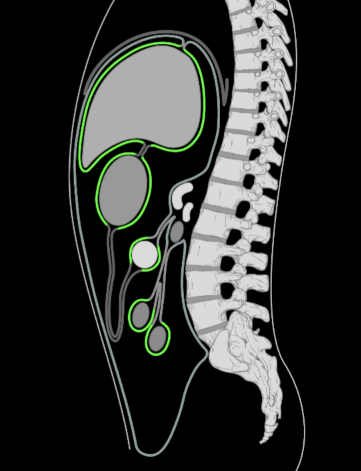
visceral peritoneum

mesentery
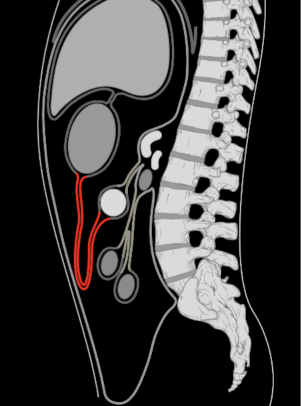
greater omentum:
attaches from the greater curvature of the stomach, covers abdominal organs, and attaches to the transverse colon
omentum
runs visceral to visceral peritoneum (organs to organ)

lesser omentum
runs from the lesser curvature of the stomach to the liver
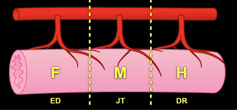
This image represents the arteries that branch off of the aorta; cranial end on the left, caudal on the right. F = foregut, M = midgut, H = hindgut
The celiac trunk supplies the foregut, from the esophagus to the duodenum
The superior mesenteric artery supplies the midgut, from the jejunum to the transverse colon
The inferior mesenteric artery supplies the hindgut, from the descending colon to the rectum

ADoral arteries branch cranially

ABoral arteries branch caudally

what is this showing?
collateral flow between adoral and aboral arteries

celiac trunk —> foregut

superior mesenteric artery —> midgut

inferior mesenteric artery —> hindgut

renal arteries
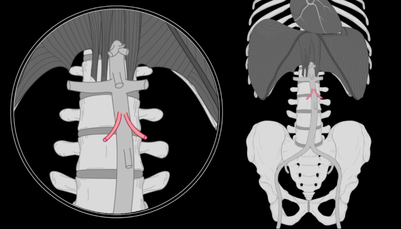
gonadal arteries (up high because gonads start up here and drop with development)
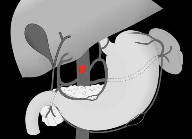
celiac trunk

left gastric artery

esophageal artery (ADoral)

left gastric artery (ABoral)
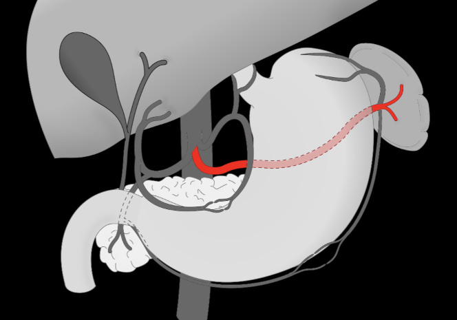
splenic artery

short gastric artery (ADoral)

left gastro-omental artery (ABoral)

hepatic artery

right gastric artery (ADoral)

gastro-duodenal artery (ABoral)

right gastro-omental artery (ADoral)

pancreaticoduodenal artery (ABoral)

hepatic veins: drain the liver

renal veins

gonadal veins
**do NOT match the arteries
right —> IVC
left —> left renal vein

hepatic portal system
first capillary beds are all in the abdominal viscera
second is the liver
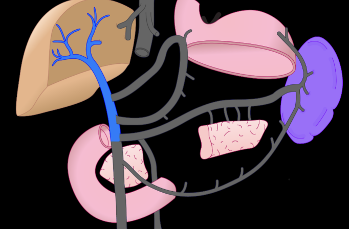
hepatic portal vein
all roads lead to here
**not a hepatic vein— is part of the portal system, not drainage

left gastric vein
left side of lesser curvature of stomach

right gastric vein
right side of lesser curvature of stomach

splenic vein
from the spleen

short gastric vein
off the fundus of the stomach

left gastro-omental vein

inferior mesenteric vein
from hindgut

superior mesenteric vein
from midgut
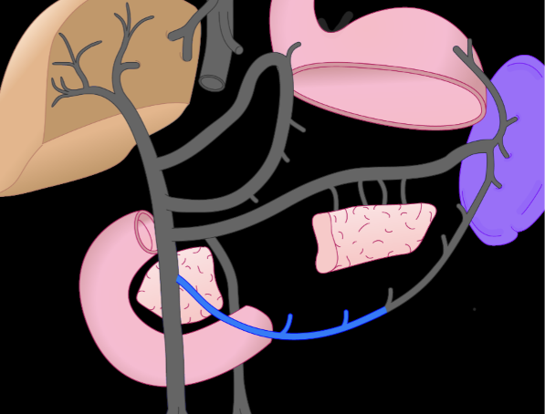
right gastro-omental vein

Hepatic sinusoids
the capillary system inside of the liver

hepatic veins
sympathetic splanchnic nerves…
go to smooth muscle in blood vessels of organs
Parasympathetic nerves…
go to smooth muscle in the wall of organs via either:
Vagus nerve (innervates the foregut & midgut)
Pelvic splanchnic nerves (innervate the pelvis & hindgut)