Anatomy & Physiology Quiz #2
 Call Kai
 Learn
Learn Practice Test
 Spaced Repetition
 Match
Match Flashcards
 Knowt Play
1/42

Earn XP
Description and Tags
Postcranial Skeleton: Appendicular Skeleton
Name | Mastery | Learn | Test | Matching | Spaced | Call with Kai | Chat |
|---|
No analytics yet
Send a link to your students to track their progress
43 Terms
Periosteum
Double-layered connective tissue that covers and nourishes the bone
Diaphysis
Elongated shaft of a long bone
Epiphysis
The end of a long bone, attached to the shaft
Epiphyseal Plate
Plate of hyaline cartilage at the junction of the diaphysis and epiphysis that provides for growth in length of a long bone
Articular Cartilage
Hyaline cartilage covering bone ends at moveable joints
Meniscus (Articular Discs)
Crescent-shaped piece of fibrocartilage in the knee joint that acts as a shock absorber and cushion between the thigh bone (femer) and shin bone (tibia)
Joint Cavity
A fluid-filled space between the connecting bones of a freely moveable synovial joint
Synovial Fluid
Fluid secreted by the synovial membrane; lubricates joint surface and nourishes articular cartilages
Fibrous
Bones joined by collagen fibers of connective tissue
Sutures (Fibrous)
Found only between bones of skull
Syndesmoses (Fibrous)
Junction of bones held together by ligaments
Gomphoses (Fibrous)
Tooth attached to socket with periodontal ligament
Cartilaginous
Bones joined by cartilage
Synchondroses (Cartilaginous)
Joints of hyaline cartilage between the shaft and the epiphyseal plates of growing long bones
Symphyses (Cartiligenous)
Bones are joined by fibrocartilage
Synovial
Articulating bones are separated by a fluid-filled cavity
What are the six cateogires within synovial joints?
Plane, condylar, ball-and-socket, hinge, saddle, pivot
Synarthroses
Immovable joints
Amphiarthroses
Slightly movable joints
Diarthroses
Freely moveable joints (synovial joints)

Temporomandibular Joint: temporal bone of skull & mandible

(Top Purple Dot)
Sternoclavicular Joint: sternum & clavicle

(Second purple dot)
Humeroscapular or Glenohumeral Joint: scapula & humerus

(Second from the bottom)
Metacarpophalangeal (Knuckle): metacarpal & proximal phalanx

(Final dot)
Interphalangeal (Finger): adjacent phalanges

(Second purple dot)
Hip (Coxal): hip bone & femur
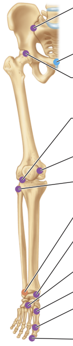
(Fourth dot from the top)
Knee (Tibiofemoral): femur & tibia
![<p>(Fifth from the bottom [purple])</p>](https://assets.knowt.com/user-attachments/599891a7-ea0d-4516-8a79-b112df16df9f.png)
(Fifth from the bottom [purple])
Ankle Joint: tibia & fibula w/ talus
Flexion
Sagittal plane
Decreases the angle between two bones

Extension
Sagittal plane
Increases the angle between two bones

Hyperextension
Extension past the anatomical point

Abduction
Frontal plane
Moves a limb away from the median plane

Adduction
Frontal plane
Moves a limb toward the median plane

Circumduction
Moves the distal end of a limb in a circle

Rotation
Turns a bone around its own long axis

Lateral Rotation
Turns a bone away from the median plane

Medial Rotation
Turns a bone towards the median plane

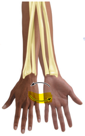
Pronation
Rotates the radius over the ulna

Supination
Rotates the radius and ulna parallel
Dorsiflexion
Moves the superior surface of the foot toward the shin (as in standing on your heel)

Plantar Flexion
Moves the superior surface of the foot farther away from the shin (as in pointing your toes)

Protraction
Moves the mandible forward

Retraction
Moves the mandible back
