STUDY GUIDE
 Call Kai
 Learn
Learn Practice Test
 Spaced Repetition
 Match
Match Flashcards
 Knowt Play
1/298

There's no tags or description
Looks like no tags are added yet.
Name | Mastery | Learn | Test | Matching | Spaced | Call with Kai | Chat |
|---|
No analytics yet
Send a link to your students to track their progress
299 Terms


Ch 6 Q2: What are the medullary cavity and articular cartilage?
The medullary cavity is the marrow cavity inside the shaft. Articular cartilage is hyaline cartilage covering the bone ends at a synovial joint; it reduces friction and absorbs compression.

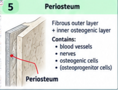
Ch 6 Q3: What is the periosteum, and what does it contain?
A covering on the outside of bone, except at articular cartilage. Its outer fibrous layer contains collagen, blood vessels, lymphatic vessels, and nerves. Its inner cellular layer contains osteogenic cells and bone forming cells. Perforating fibers help anchor it and attached tendons or ligaments.
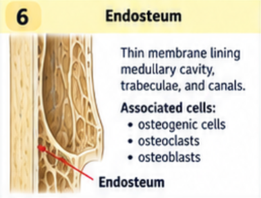
Ch 6 Q3: What is the endosteum, and what cells are associated with it?
A thin cellular lining on internal bone surfaces, including the medullary cavity, trabeculae, and bone canals. Osteogenic cells, osteoblasts, and osteoclasts support growth, repair, and remodeling at these surfaces.
Ch 6 Q4: What are the three main functional bone cell types and their jobs?
Osteoblasts build bone matrix. Osteocytes are mature cells in lacunae that maintain matrix and sense mechanical stress. Osteoclasts break down, or resorb, bone.

Ch 6 Q4: What are osteogenic cells, also called osteoprogenitor cells?
Stemlike precursor cells that divide and produce osteoblasts. The slides include them as a fourth bone cell category, in addition to the three main functional cell types asked for in the guide.

Ch 6 Q5: How do compact bone and spongy bone differ in structure and function?
Compact, or cortical, bone is dense and provides a strong outer shell. Spongy, cancellous, or trabecular bone is a network of thin trabeculae with marrow spaces; it distributes loads while keeping the skeleton light. Both contain living cells and mineralized matrix.

Ch 6 Q5: What is an osteon, also called a Haversian system?
The basic cylindrical organization of much compact bone. Concentric lamellae surround a central canal containing blood vessels and nerves. Osteocytes occupy lacunae between the lamellae and communicate through canaliculi.

Ch 6 Q5: How do central canals, perforating canals, lacunae, and canaliculi differ?
Central or Haversian canals run lengthwise through osteons. Perforating or Volkmann canals connect blood supplies across bone. Lacunae house osteocytes. Canaliculi are tiny connecting channels that allow cell communication and nutrient exchange.

Ch 6 Q5: How are spongy bone trabeculae nourished if they lack typical osteons?
Their thin structure places osteocytes close to marrow spaces. Nutrients diffuse from nearby vessels through canaliculi. Trabeculae contain lamellae and align with common lines of stress.

Ch 6 Q6: Where is compact bone found, and why?
It forms the outer layer of all bones and is especially thick in a long bone's shaft. Its dense organization resists bending, supports loads, and protects internal tissues.

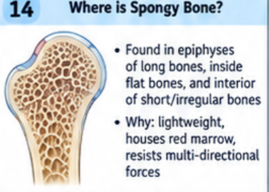
Ch 6 Q6: Where is spongy bone found, and why?
Inside bone, especially in long bone epiphyses and the interiors of flat, short, and irregular bones. Its trabeculae resist stress from several directions, reduce weight, and leave space for marrow.
Ch 6 Q7: What happens first in endochondral ossification?
Mesenchymal cells become chondrocytes and form a hyaline cartilage model. Chondrocytes in the shaft enlarge, the surrounding matrix calcifies, and many chondrocytes die. Osteoblasts form a bone collar as the perichondrium becomes periosteum.

Ch 6 Q7: How does the primary ossification center form?
Blood vessels invade the shaft, bringing osteogenic cells. Osteoblasts deposit bone on remaining cartilage supports. Osteoclasts remove tissue to help form the medullary cavity. The primary center expands along the diaphysis.

Ch 6 Q7: How does endochondral ossification finish in the epiphyses?
Secondary ossification centers develop in the epiphyses. Most cartilage is replaced with bone; articular cartilage remains at joints, and epiphyseal cartilage remains during growth. Spongy bone remains inside the epiphyses.

Ch 6 Q7: What is the order of growth plate zones from epiphysis toward diaphysis?
Reserve or resting cartilage; proliferation, where chondrocytes divide; maturation and hypertrophy, where they enlarge; calcified cartilage, where cells die; ossification, where cartilage is replaced by bone. Division belongs mainly to the proliferative zone.

Ch 6 Q8: What are the four main stages of ordinary fracture repair?
Fracture hematoma; soft fibrocartilaginous callus; hard bony callus; remodeling into mature bone. The callus temporarily bridges and stabilizes the break.

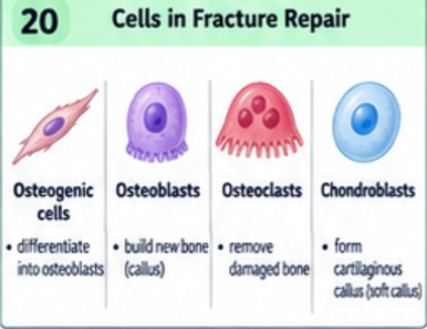
Ch 6 Q8: Which cells participate in fracture repair?
Inflammatory cells clear damaged tissue. Fibroblasts and chondroblasts help form the soft callus. Osteoblasts make new bone. Osteoclasts remove excess and damaged bone during remodeling. New blood vessels support the repair.
Ch 6 Q9: What are the main bone shape classes and examples?
Long: femur, humerus, clavicle, and phalanges. Short: carpals and tarsals. Flat: sternum, ribs, scapula, and many cranial bones. Irregular: vertebrae. Sesamoid: patella. Sutural bones are small extra bones within skull sutures, included as an additional category in the slides.

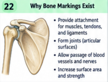
Ch 6 Q9: Why do bone markings exist?
They form joint surfaces, provide attachment sites for muscles and ligaments, or accommodate nerves, vessels, and other tissues. Growth and mechanical loading help shape attachment markings.
Ch 6 Q9: What are a head, condyle, and facet?
Head: a rounded articular projection, often on a neck. Condyle: a rounded joint surface. Facet: a small, smooth, usually flat joint surface. Examples are the femoral head, occipital condyles, and vertebral articular facets.

Ch 6 Q9: What are a tubercle, tuberosity, trochanter, and epicondyle?
Tubercle: a small rounded projection. Tuberosity: a roughened projection. Trochanter: a large projection found on the femur. Epicondyle: a projection above a condyle. These commonly provide attachment sites.

Ch 6 Q9: What are a crest, line, spine, process, and protuberance?
Crest: a prominent ridge. Line: a less prominent ridge. Spine: a sharp or slender projection. Process: a general term for a projection. Protuberance: a projecting bump.

Ch 6 Q9: What are a fossa, fovea, and sulcus?
Fossa: a depression. Fovea: a small pit. Sulcus: a groove. Examples are the olecranon fossa, fovea capitis of the femur, and intertubercular sulcus of the humerus.

Ch 6 Q9: What are a foramen, fissure, canal, meatus, and sinus?
Foramen: an opening through bone. Fissure: a narrow slit. Canal: a passage through bone. Meatus: a canal or its opening. Sinus: an air filled cavity within bone. These accommodate structures or reduce bone weight.

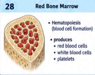
Ch 6 Q10: What does red bone marrow do?
Hematopoiesis: production of red blood cells, white blood cells, and platelets. In adults, much active red marrow is in the axial skeleton and in proximal portions of the humerus and femur.
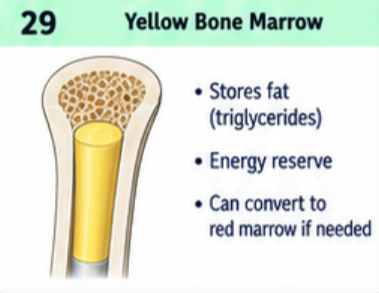
Ch 6 Q10: What does yellow bone marrow do?
It stores triglycerides in adipocytes as an energy reserve. It occupies much of the adult long bone medullary cavity and can convert toward red marrow during severe, prolonged demand for blood cell production.
Ch 6 Q11: What do hydroxyapatite crystals do in bone matrix?
These calcium phosphate crystals make bone hard and resistant to compression. Collagen supplies tensile strength and some flexibility. Bone needs both mineral and collagen for normal strength.

Ch 6 Q12: What is characteristic of a successfully repaired fracture site?
The early callus is bulky and disorganized. Remodeling replaces it with mature bone and restores shape and strength. Bone can heal with true bone instead of a permanent fibrous scar; the site is not automatically stronger forever.

Ch 6 Q13: Why can a bone fracture heal more completely than a torn tendon or ligament?
Bone generally has a better blood supply and strong regenerative capacity. Tendons and ligaments often have poorer blood supply and repair with scar tissue that may not fully restore their original organization. This is a general comparison, not a rule that every fracture is less serious.

Ch 6 Q13: Why is an epiphyseal fracture a concern in a growing child?
Damage to the epiphyseal plate can disrupt lengthwise growth or cause early, uneven plate closure, producing a shortened or angled bone. Risk depends on the fracture and growth remaining. Fractures extending into a joint may also damage articular cartilage.

Ch 7 Q1: Which bones belong to the axial skeleton?
The skull, auditory ossicles, hyoid, vertebral column, ribs, and sternum. This division forms the central axis and contains 80 bones in the typical adult skeleton.

Ch 7 Q1: Which bones belong to the appendicular skeleton?
The pectoral girdles, upper limbs, pelvic girdle, and lower limbs. This division contains 126 bones in the typical adult skeleton and supports limb movement and weight transfer.

Ch 7 Q2: What are the eight cranial bones?
One frontal, two parietal, two temporal, one occipital, one sphenoid, and one ethmoid. The paired bones are parietal and temporal.

Ch 7 Q2: What are the fourteen facial bones?
Two maxillae, two zygomatic, two nasal, two lacrimal, two palatine, two inferior nasal conchae, one vomer, and one mandible. The unpaired facial bones are the vomer and mandible.

Ch 7 Q3: How does a fetal or newborn skull differ from an adult skull?
It has a proportionally larger cranium and smaller face, incompletely ossified bones, unfused regions, and fontanelles. The jaws and paranasal sinuses are less developed. Fontanelles allow molding during birth and room for rapid brain growth.

Ch 7 Q3: What are the major fontanelles?
Anterior, posterior, paired sphenoidal, and paired mastoid fontanelles. They are connective tissue gaps between developing cranial bones that gradually close as the skull grows.

Ch 7 Q4: What does the hyoid bone do, and what makes it unique?
It supports the tongue and provides attachments for muscles involved in swallowing and speech. It does not articulate directly with any other bone; muscles and ligaments suspend it in the neck.

Ch 7 Q5: What are the regions and bone counts of the vertebral column?
Seven cervical, twelve thoracic, and five lumbar vertebrae; the sacrum, formed from five fused vertebrae; and the coccyx, usually formed from four fused vertebrae. This gives about 33 original vertebrae or 26 adult bones.

Ch 7 Q5: What are the four normal spinal curves?
Cervical and lumbar curves are convex anteriorly and are secondary curves that develop after birth. Thoracic and sacral curves are convex posteriorly and are primary curves present during fetal development.

Ch 7 Q5: What are the main parts of a typical vertebra?
A body anteriorly; a vertebral arch made of pedicles and laminae; a vertebral foramen; a spinous process; two transverse processes; and superior and inferior articular processes. Adjacent vertebral foramina form the vertebral canal.

Ch 7 Q6: How can you identify a typical cervical vertebra?
Small body, relatively large triangular vertebral foramen, and transverse foramina. Many have a bifid spinous process. C1, C2, and C7 have important exceptions to the typical pattern.

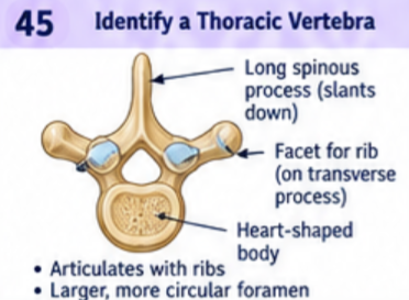
Ch 7 Q6: How can you identify a typical thoracic vertebra?
Costal facets for rib articulation, a generally heart shaped body, and a long spinous process angled inferiorly. Typical thoracic vertebrae also have transverse costal facets; T11 and T12 lack those transverse facets.
Ch 7 Q6: How can you identify a typical lumbar vertebra?
A large, thick body for carrying weight, a triangular vertebral foramen, and a short, broad spinous process. Lumbar vertebrae lack rib facets and transverse foramina.

Ch 7 Q6: What distinguishes the atlas and axis?
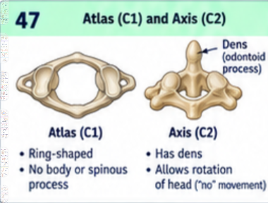
Atlas is C1: a ring without a body or typical spinous process that supports the skull and permits nodding. Axis is C2: its dens, or odontoid process, acts as a pivot for turning the head with the atlas.
Ch 7 Q7: Why is the wedge shape of the sacrum important?
The sacrum fits between the two hip bones like a keystone. Its shape and strong sacroiliac ligaments help stabilize the pelvic ring and transfer upper body weight to the hip bones and lower limbs.

Ch 7 Q8: How many rib pairs are there, and which are true ribs?
Twelve pairs. Ribs 1 through 7 are true, or vertebrosternal, ribs because each reaches the sternum through its own costal cartilage.

Ch 7 Q8: Which ribs are false, vertebrochondral, and floating?
Ribs 8 through 12 are false because they lack an individual direct cartilage attachment to the sternum. Ribs 8 through 10 are vertebrochondral and join cartilage above them. Ribs 11 and 12 are floating and have no anterior attachment to the sternum.

Ch 7 Q9: What does the costal groove contain?
Intercostal vessels and a nerve along the inner inferior border of a rib. Their main superior to inferior order is vein, artery, nerve: VAN.

Ch 7 Q10: Why is the sternal angle an important landmark?
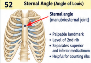
It is the palpable junction of the manubrium and sternal body, where the second costal cartilage attaches. Finding it lets you locate rib 2 and count ribs and intercostal spaces. It lies approximately at the T4 to T5 intervertebral level.
Ch 8 Q1: What bones make up each pectoral girdle?
One clavicle and one scapula. The clavicle connects the upper limb girdle to the axial skeleton at the sternoclavicular joint.

Ch 8 Q2: What are the scapular spine and acromion associated with?
The spine divides the posterior scapula into supraspinous and infraspinous fossae. It continues laterally as the acromion, which articulates with the clavicle. The spine and acromion provide attachment sites for trapezius and deltoid.

Ch 8 Q2: Which muscles attach to the three major scapular fossae?
Supraspinatus originates in the supraspinous fossa; infraspinatus in the infraspinous fossa; subscapularis in the anterior subscapular fossa. Their tendons contribute to the rotator cuff.

Ch 8 Q2: What attaches to the coracoid process?
Pectoralis minor inserts there. The short head of biceps brachii and coracobrachialis originate there. Coracoclavicular and coracoacromial ligaments also attach to it.

Ch 8 Q2: What are the glenoid cavity and the supraglenoid and infraglenoid tubercles?
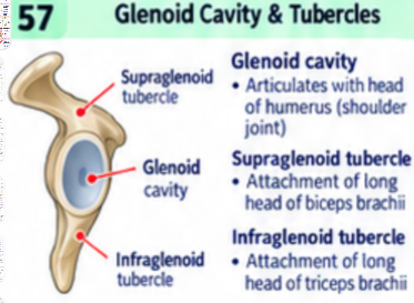
The glenoid cavity articulates with the humeral head and is deepened by the glenoid labrum. The supraglenoid tubercle anchors the long head of biceps brachii; the infraglenoid tubercle anchors the long head of triceps brachii.
Ch 8 Q2: Which major muscles attach along the scapular borders and angles?
Rhomboids and serratus anterior attach along the medial border on different surfaces. Levator scapulae attaches near the superior angle. Teres minor arises along the lateral border; teres major arises near the inferior angle. Also identify the superior border and suprascapular notch.

Ch 8 Q3: What are the proximal humeral landmarks and their associations?
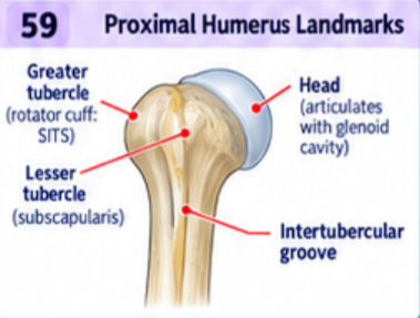
The head articulates with the glenoid cavity. The anatomical neck borders the head; the surgical neck lies below the tubercles. Supraspinatus, infraspinatus, and teres minor insert on the greater tubercle; subscapularis inserts on the lesser tubercle.
Ch 8 Q3: What is the intertubercular groove, and what muscles relate to it?
Also called the bicipital groove, it guides the long head tendon of biceps brachii. Pectoralis major inserts on its lateral lip, latissimus dorsi on its floor, and teres major on its medial lip.

Ch 8 Q3: What are the deltoid tuberosity, radial groove, and humeral epicondyles associated with?
The deltoid inserts on the deltoid tuberosity. The radial nerve runs in the radial groove. Many forearm flexors arise from the medial epicondyle and many extensors from the lateral epicondyle. The ulnar nerve passes behind the medial epicondyle.

Ch 8 Q3: What are the distal humeral joint surfaces and fossae?
The capitulum articulates with the radial head; the trochlea articulates with the ulna. Radial and coronoid fossae receive forearm projections during flexion. The posterior olecranon fossa receives the olecranon during extension.

Ch 8 Q3: What major radial landmarks should you know?
The radius is on the thumb side. Its head articulates with the capitulum and radial notch of the ulna; the neck is just below. Biceps brachii inserts on the radial tuberosity. Brachioradialis inserts near the distal styloid process. The ulnar notch receives the distal ulna.

Ch 8 Q3: What major ulnar landmarks and muscle attachments should you know?
The olecranon receives triceps and forms the elbow point. The trochlear notch fits the humeral trochlea. The coronoid process projects anteriorly. Brachialis inserts on the coronoid process and ulnar tuberosity. The radial notch receives the radial head; a styloid process is distal.

Ch 8 Q3: What connects the shafts of the radius and ulna?
The interosseous membrane, a strong fibrous sheet that stabilizes the forearm, transfers forces, and provides muscle attachment. In anatomical position, the radius is lateral and the ulna is medial.

Ch 8 Q4: Name the proximal carpal row from lateral to medial in anatomical position.
Scaphoid, lunate, triquetrum, pisiform. Lateral means the thumb side. The pisiform sits on the palmar surface of the triquetrum.

Ch 8 Q4: Name the distal carpal row from lateral to medial in anatomical position.
Trapezium, trapezoid, capitate, hamate. The trapezium is on the thumb side and articulates with the first metacarpal.

Ch 8 Q5: How many phalanges are in the pollex and other fingers?
The pollex, or thumb, has two: proximal and distal. Each other finger has three: proximal, middle, and distal. Each hand has fourteen phalanges total.

Ch 8 Q6: What makes up the pelvic girdle, and how is it different from the bony pelvis?
The pelvic girdle is the pair of hip bones, or ossa coxae. The bony pelvis includes both hip bones plus the sacrum and coccyx.

Ch 8 Q6: What three bones form one os coxae?
Ilium, ischium, and pubis. They fuse at the acetabulum, the socket for the femoral head. The ischium and pubis surround most of the obturator foramen.

Ch 8 Q7: What separates the true pelvis from the false pelvis?
The pelvic brim, or boundary of the pelvic inlet. Key landmarks include the sacral promontory and alae, arcuate lines of the ilia, pectineal lines and crests of the pubic bones, and the superior border of the pubic symphysis.

Ch 8 Q7: Where are the true and false pelvis?
The false or greater pelvis lies above the brim and supports lower abdominal contents. The true or lesser pelvis lies below the brim and surrounds the pelvic cavity; it contributes to the birth canal.

Ch 8 Q8: How do typical female and male pelves differ?
A typical female pelvis is wider and shallower, with a larger, more rounded inlet and outlet, a wider subpubic angle, and a shorter, broader sacrum. A typical male pelvis is narrower and deeper, with a more heart shaped inlet and narrower pubic arch. These are general patterns.

Ch 8 Q8: Which pelvic landmarks help distinguish typical female and male pelves?
In the lecture diagram, the female subpubic angle is greater than 90 degrees and the male angle is less than 90 degrees. Female ischial spines generally project less medially, and the greater sciatic notch is wider. Individual anatomy varies.

Ch 8 Q9: What are the major proximal femoral landmarks and attachments?
The head fits the acetabulum; the neck connects it to the shaft. The fovea capitis attaches the ligament of the femoral head. The greater trochanter receives gluteus medius, gluteus minimus, and several lateral rotators; the lesser trochanter receives iliopsoas.

Ch 8 Q9: What important femoral ridges and tubercles should you know?
The anterior intertrochanteric line and posterior intertrochanteric crest connect the trochanters. The linea aspera is a posterior ridge for adductors and other muscles. Gluteus maximus attaches to the gluteal tuberosity. Adductor magnus attaches partly to the adductor tubercle.

Ch 8 Q9: What are the main distal femoral landmarks?
Medial and lateral condyles articulate with the tibia; the anterior patellar surface articulates with the patella. Epicondyles provide ligament attachments. The posterior intercondylar fossa lies between the condyles and contains femoral attachments of the cruciate ligaments.

Ch 8 Q9: What are the main tibial landmarks and attachments?
Medial and lateral condyles form the tibial plateau. The intercondylar region anchors cruciate ligaments and menisci. The tibial tuberosity receives the patellar ligament. The soleal line anchors soleus. The anterior crest forms the shin, and the medial malleolus forms the inner ankle.

Ch 8 Q9: Which muscles attach at the pes anserinus on the proximal tibia?
Sartorius, gracilis, and semitendinosus insert together on the anteromedial proximal tibia. The name means goose foot. These tendons help flex the knee and stabilize its medial side.

Ch 8 Q9: What are the main fibular landmarks and attachments?
The proximal head receives biceps femoris and the fibular collateral ligament. A neck and slender shaft follow; the common fibular nerve passes around the neck. The distal lateral malleolus forms the outer ankle and anchors ankle ligaments. The fibula contributes to stability and muscle attachment.

Ch 8 Q10: What is a sesamoid bone, and which one is in the quadriceps tendon?
A sesamoid bone develops within a tendon. The patella lies in the quadriceps tendon, protects the knee region, and increases the leverage of the quadriceps. The patellar ligament continues from patella to tibial tuberosity.

Ch 8 Q11: What are the seven tarsal bones and their positions?
Talus above calcaneus in the hindfoot; navicular anterior to talus on the medial side; cuboid anterior to calcaneus on the lateral side; medial, intermediate, and lateral cuneiforms anterior to navicular. The cuneiforms align mainly with metatarsals 1, 2, and 3.

Ch 8 Q12: How many phalanges are in the hallux and other toes?
The hallux, or great toe, has proximal and distal phalanges. Each other toe has proximal, middle, and distal phalanges. Each foot has fourteen phalanges total.

Ch 8 Q13: What are the three foot arches and their commonly taught tarsal keystones?
Medial longitudinal arch: talus, . Lateral longitudinal arch: cuboid. Transverse arch: intermediate or middle cuneiform at the tarsal level. The arches distribute weight, absorb forces, and provide spring during walking.

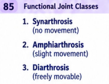
Ch 9 Q1: What are the three functional joint classes based on movement?
Synarthrosis: essentially immovable. Amphiarthrosis: slightly movable. Diarthrosis: freely movable. These describe movement, whereas fibrous, cartilaginous, synovial, and bony describe structure.
Ch 9 Q1: What are examples of synarthroses?
Fibrous sutures between skull bones and gomphoses between teeth and sockets; cartilaginous synchondroses such as a growth plate; and bony synostoses such as fused sacral vertebrae.

Ch 9 Q1: What are examples of amphiarthroses and diarthroses?
Amphiarthroses include cartilaginous symphyses such as the pubic symphysis and joints between vertebral bodies, plus fibrous syndesmoses such as the distal tibiofibular joint. Diarthroses are synovial joints, such as the shoulder, hip, and elbow.

Ch 9 Q2: What is the usual tradeoff between joint strength and mobility?
A joint with a deeper fit and tighter restraints is usually more stable but allows less movement. A looser, shallower joint permits more movement but relies more on muscles and is easier to dislocate. Compare the stable hip with the more mobile shoulder.

Ch 9 Q3: What are the essential structures of a synovial joint?
Articular cartilage on the bone ends; a fluid filled joint cavity; and an articular capsule with an outer fibrous layer and inner synovial membrane. The capsule encloses the joint, and the membrane produces synovial fluid.

Ch 9 Q3: What do articular cartilage, ligaments, blood vessels, and nerves do in a synovial joint?
Articular cartilage reduces friction and distributes compression. Ligaments connect bones and limit unwanted movement. Vessels nourish surrounding tissues; articular cartilage itself is avascular. Nerves provide pain information and position sense, or proprioception.

Ch 9 Q3: What do menisci, articular discs, and labra do?
Menisci and discs are fibrocartilage structures that improve fit and distribute loads; discs can divide a joint cavity. A labrum is a fibrocartilage rim that deepens a socket, as at the shoulder and hip. These structures occur in particular synovial joints, not every joint.

Ch 9 Q3: What are bursae, tendon sheaths, and fat pads?
Bursae are small fluid filled sacs that reduce friction between moving tissues. Tendon sheaths are elongated bursae around tendons. Fat pads cushion spaces and adapt to changes in joint shape during movement.

Ch 9 Q4: What is linear or gliding motion at a joint?
One relatively flat surface slides past another without a large change in bone angle. Examples include small gliding movements between carpal bones and between vertebral articular facets.

Ch 9 Q4: What are flexion, extension, hyperextension, and lateral flexion?
Flexion usually decreases a joint angle; extension increases it. Hyperextension continues beyond anatomical position where permitted. Lateral flexion bends the neck or trunk to the side. Bending and straightening the elbow demonstrate flexion and extension.

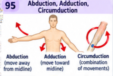
Ch 9 Q4: What are abduction, adduction, and circumduction?
Abduction moves away from the body's midline; adduction moves toward it. For fingers and toes, use the digit's reference axis. Circumduction combines flexion, abduction, extension, and adduction so the distal end traces a circle.
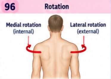
Ch 9 Q4: What are rotation, medial rotation, and lateral rotation?
Rotation turns a bone around its long axis. Medial or internal rotation turns the anterior limb surface toward the midline; lateral or external rotation turns it away. Turning the head at the atlantoaxial joint is rotation.
Ch 9 Q4: What are pronation and supination?
Pronation rotates the forearm so the palm faces posteriorly in anatomical position, or down with the elbow flexed; the radius crosses the ulna. Supination returns the palm anteriorly, or up with the elbow flexed; the bones become parallel.

Ch 9 Q4: What are dorsiflexion, plantar flexion, inversion, and eversion?
Dorsiflexion brings the top of the foot toward the shin. Plantar flexion points the foot downward. Inversion turns the sole medially; eversion turns it laterally.

Ch 9 Q4: What are elevation, depression, protraction, and retraction?
Elevation moves a structure upward; depression downward. Protraction moves it anteriorly; retraction posteriorly. Examples are shrugging the shoulders and pushing the mandible or scapulae forward and backward.

Ch 9 Q4: What are opposition and reposition?
Opposition brings the thumb across the palm toward the fingers, allowing a pinch or grasp. Reposition returns it toward anatomical position. The thumb also rotates during opposition so its pad faces a finger pad.
