A&P Exam II Studyset
 Call Kai
 Learn
Learn Practice Test
 Spaced Repetition
 Match
Match Flashcards
 Knowt Play
1/517

There's no tags or description
Looks like no tags are added yet.
Name | Mastery | Learn | Test | Matching | Spaced | Call with Kai | Chat |
|---|
No analytics yet
Send a link to your students to track their progress
518 Terms
Cutaneous Membrane
Epidermis – superficial epithelium
Dermis – deep connective tissue
accessory features
hair, hair follicles, nails, exocrine glands
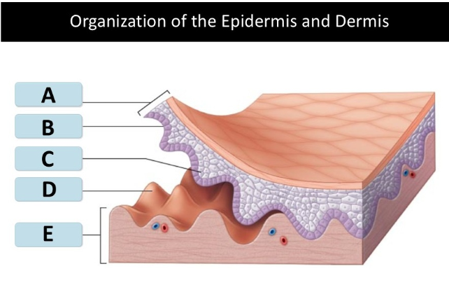
A: dermis
B: epidermis
C:papillary Layer
D: reticular layer
E: subcutaneoud layer
Identify Structures A - E

dermal papilla
Match the cutaneous structure to letter A

epidermal ridge
Match the cutaneous structure to letter B

stratum corneum
Match the cutaneous structure to letter C

stratum lucidum
Match the cutaneous structure to letter D

stratum granulosum
Match the cutaneous structure to letter E

stratum spinosum
Match the cutaneous structure to letter F

stratum basale
Match the cutaneous structure to letter G

basement layer
Match the cutaneous structure to letter H

dermis
Match the cutaneous structure to letter I

A: epidermis
B: basement layer
C: epidermal ridge
D: dermal papilla
E: dermis
Match the letter to the corrseponding structure
integument
contains blood vessels and sensory receptors
subcutaneous layer
(hypodermis) – stabilizes position of the skin and connects the dermis of the skin to underlying tissues
Not part of skin (the integument) Separates the integument from the deep fascia around organs such as muscles and bones
Primarily composed of adipose tissue – subcutaneous fat is for energy storage, insulation and padding
Contains large arteries and veins and acts as reservoir for blood
Site for subcutaneous injections using hypodermic needles
Integumentary system function
– Protection of underlying tissues and organs against impact, abrasion, fluid loss, chemical attack
– Excretion of salts, water, and organic wastes by glands
– Maintenance of normal body temperature through insulation and evaporative cooling
– Production of melanin for protection from UV radiation
– Production of keratin which makes skin tough (protects against abrasion) and repels water
– Synthesis of vitamin which is converted to calcitriol, a hormone important for calcium metabolism
– Storage of lipids in the dermis and subcutaneous layer
– Detection of touch, pressure, pain, vibration, and temperature stimuli
– Coordination of the immune response to pathogens and cancers in the skin
Edpidermis
top layer of the skin
– Composed of stratified squamous epithelium
– Avascular
– Nutrients and oxygen diffuse from capillaries in the dermis
keratinocytes
main cells in the epidermis
– Contain large amounts of keratin – a tough, fibrous protein
stratum basal (stratum germinativum)
1st strata
Attached to basement membrane by hemidesmosomes
Contains basal cells
Contains epidermal ridges, projections which interlock with the dermal papillae in dermis to increase the surface area for attachment of dermis to epidermis
The contour of the skin surface follows the pattern of the epidermal ridges.
▪ In thick skin visible as fingerprints – ridges that increase the surface area for better grip
▪ The pattern is unique and genetically predetermined
stratum spinosum
2nd strata, layer superficial to the stratum basale
– 8−10 layers of keratinocytes bound by desmosomes
– Keratinocytes produced by division of basal cells in stratum basale, some of which continue dividing
– Contains immune cells called dendritic (Langerhans) cells, which defend against microorganisms and skin cancers
stratum granulosum
3rd strata
thin layer superficial to the stratum spinosum
– 3−5 layers of keratinocytes
– Cells from the stratum spinosum get pushed into the stratum granulosum as new cells are produced
– Cells stop dividing and fill with keratin and keratohyalin
– The cells then become thin and flat, become less permeable and become dehydrated
– Nuclei and organelles disintegrate, and the cells die, but remain tightly interlocked
stratum lucidum
4th strata
layer of dead keratinocytes superficial to the stratum granulosum
– Found only in thick skin
stratum corneum
5th strata, keratinization occurs
– top layer, exposed surface of the skin
– 15−30 layers of keratinized cells; most layers
– New cells move from stratum basale toward the stratum corneum and reach it in 7−10 days
– Exposed cells shed after 2 weeks
basal cells
(germinative cells) – stem cells that divide to give rise to new keratinocytes
epidermal ridges
projections which interlock with
the dermal papillae in dermis to increase the surface area
for attachment of dermis to epidermis
Tactile (merkel cells)
together with sensory nerve endings form tactile discs
▪ Found in hairless skin
▪ Sensory receptors for touch
Melanocytes
cells that synthesize the pigment melanin
▪ Distributed through stratum basale and their processes extends into other layer
Keratohyalin
granules promote cell dehydration, aggregation, and cross-linking of keratin fibers
keratinization
the formation of protective layers of cells filled with keratin
insensible perspiration
loss of water which diffuses across the stratum corneum and evaporates
▪ Approximately 500 mL per day
▪ Cannot be felt or prevented
▪ Increase in fluid loss if stratum corneum is damaged
sensible perspiration
– water loss by excretion from sweat glands
▪ Allows for cooling the body
Epidermal growth factor (EGF)
A peptide growth factor which causes epithelial growth, Produced by the salivary glands and glands of the duodenum and Used in laboratories to grow skin grafts in tissue culture
Functions
Promotes division of basal cells
Accelerates keratin production
Stimulates epidermal development and repair
Stimulates secretions from epithelial glands
Dermis
deeper layer of skin, located under the epidermis
– Anchors epidermal accessory structures (e.g., hair follicles and sweat glands)
– Contains a network of blood vessels and nerves
contains:
▪ Superficial papillary layer
▪ Deeper reticular layer
Papillary Layer
top layer of dermis, directly underneath the epidermis
– Consists of areolar tissue, capillaries, lymphatic vessels, and sensory neurons
– Named for dermal papillae that attach epidermal ridges
Dermatitis
inflammation of the dermis
▪ Caused by infection, radiation, mechanical irritation, or chemicals
▪ May produce itching or pain
reticular layer
deeper layer of dermis, above the subcutaneous layer
– Consists of dense irregular connective tissue, rich in collagen & elastic fibers
– Fibers extend and attach the reticular layer to the papillary layer and the subcutaneous layer
Collagen fibers
Strong, can bend but do not stretch
▪ Easily bent or twisted
▪ Add strength and limit flexibility to prevent tissue damage
elastic fibers
stretching and recoil
▪ Provide flexibility
skin turgor
strength and flexibility of skin because of its water content
tension lines
(cleavage lines) – pattern produced by parallel bundles of collagen and elastic fibers in the dermis
– Allow skin to resist applied forces
– A cut made parallel to tension lines heals well with minimal scarring
– A cut made perpendicular to tension lines pulls open and scars more
Cutaneous plexus
deep network of vessels along the border of the reticular layer of the dermis and the subcutaneous layer
subpapillary plexus
network of small vessels in the papillary layer of the dermis
– Contusion (bruise), Caused by damage to blood vessels in the dermis
Nerve fibers
▪ Transmit information from sensory receptors
▪ Control blood flow
▪ Adjust gland secretion rates
sensory receptors
– Tactile (Meissner) corpuscles, in dermal papillae detect light touch
– Lamellar (Pacinian) corpuscles, in the reticular layer detect deep pressure and vibration
carotene
Orange-yellow pigment
▪ Found in orange vegetables
▪ Accumulates in epidermal cells, the dermis, and the subcutaneous layer
▪ Can be converted to vitamin A, required for the maintenance of epithelia and the synthesis of photoreceptor pigments in eye
Melanin
– red-yellow (pheomelanin) or brown-black (eumelanin) pigment produced by melanocytes
▪ Stored in intracellular vesicles (melanosomes), which are then transferred to keratinocytes
▪ Dark-skinned people have larger and more numerous melanosomes
▪ The number of melanocytes and the rate of melanin production can vary in different parts of the body
▪ Melanin protects the skin from ultraviolet (UV) radiation which can damage DNA and cause cancer
▪ Melanin synthesis accelerates when exposed to sun
dermis
match the melanocyte structure to letter B
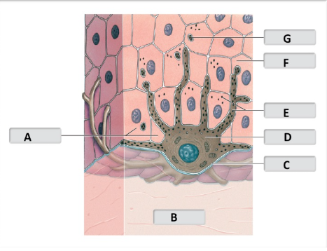
basement layer
match the melanocyte structure to letter C

melanocyte
match the melanocyte structure to letterD
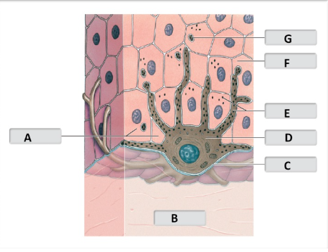
melanin pigment
match the melanocyte structure to letter E

keratinocyte
match the melanocyte structure to letter F
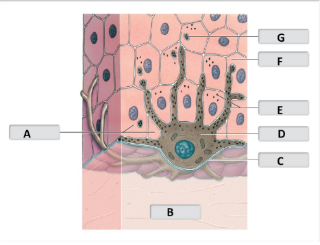
melanosome
match the melanocyte structure to letter G

Hemoglobin
bright red when bound to oxygen
▪ When blood vessels in the dermis dilate, skin reddens
▪ When blood vessels in the dermis constrict, skin pales
turns dark red when not carrying oxygen
cyanosis
bluish coloration of the skin due to reduction in blood supply and oxygen levels
– May be caused by extreme cold, heart failure, severe asthma,
Jaundice
the liver is unable to excrete bile, so a yellow pigment builds in the body
▪ Skin and whites of the eyes may turn yellow
Pituitary tumor
result in excess melanocyte-stimulating hormone (MSH) – skin darkens
Addison’s disease
causes the pituitary gland to release excess adrenocorticotropic hormone (ACT H), which has similar structure and effect to MSH
vitiligo
autoimmune condition that causes loss of melanocytes and results in white patches
basal cell carcinoma
originates in the stratum basale
– Most common type of skin cancer, caused primarily by exposure to UV radiation
– Does not metastasize
squamous cell carcinoma
affect squamous cells in the epidermis of the skin
– Cause exclusively by exposure to UV radiation
– Does not metastasize
malignant melanoma
aggressive cancer of the melanocytes ( most dangerous)
– Metastasis is common
– Signs include asymmetry, irregular border, mottled color, larger than 6 mm, changing size, shape and color
Vitamin D3
(cholecalciferol)
– Produced by epidermal cells in the presence of UV radiation
– The liver and kidneys together convert vitamin into calcitriol
– Calcitriol is essential for the absorption of calcium and phosphate ions by the small intestine
rickets
form of osteomalacia due to insufficient vitamin D3 which results in abnormal bone development
Hair
nonliving, long, cylindrical accessory structure produced in a hair follicle
– Originates in the dermis and projects through the epidermis
– Covers almost all of the body, except the lips, the palms of the hands and the sides of the fingers, the sides and soles of feet, the sides of toes, and portions of external genitalia
Functions
– Protect, cushion, and insulate
– Guard openings from particles and insects
– Serve as sensory receptors
hair follicles
– Originate deep in the dermis
– Wrapped in a dense connective tissue sheath
– Base is surrounded by sensory nerves (root hair plexus)
arrector pili muscle
involuntary smooth muscle
▪ Contraction causes hairs to stand up
▪ Produces “goose bumps”
hair root
portion of hair that anchors it into the skin
▪ Begins at the base of the hair (hair bulb) and extends about halfway to the skin surface
hair shaft
extends from the end of the hair root and forms the protruding, visible portion of the hai
hair structure
-Medulla – central core
– Cortex – intermediate layer
– Cuticle – surface layer
medulla
core of hair containing soft keratin
cortex
comtains thick layers of hard kerayin that give hair thickness
cuticle
thin but tough containg hard keratin
hair follicle
– Internal root sheath – surrounds the hair root and the deeper portion of the shaft
– External root sheath – extends from skin surface to hair matrix
– Glassy membrane – clear layer wrapped in the connective tissue sheath
• Oval hair follicles result in curly hair
• Round hair follicles result in straight hair
hair production
Begins at the hair bulb, at the base of the hair follicle
▪ The hair bulb surrounds the hair papilla – connective tissue containing capillaries and nerves
▪ At base of hair bulb, epithelial cells form a layer known as the hair matrix
– Basal cells in the hair matrix produce hair cells
– As cells divide, hair is pushed up and out of skin
– The cells keratinize and die
hair cycle
the cycle of how hairs grow and shed
– Cells of the hair root absorb nutrients and incorporate them into the hair
– As hair grows, its root is firmly attached to the matrix of the follicle
– At the end of the growth cycle, the follicle becomes inactive, and the hair is now called club hair
– As a new hair growth cycle begins, the follicle produces a new hair and the club hair is shed
– Hair loss result when shedding outpaces production and can be caused by drugs, dietary factors, radiation, stress, or hormones
vellus hair
soft, fine hairs that cover most of body surface
Terminal hairs
– heavy, pigmented hairs on head, eyebrows, and eyelashes
▪ After puberty also the hairs in the pubic area and armpits and sometimes on limbs
sebaceous glands
oil glands) – holocrine glands that produce an oily lipid secretion (sebum) into hair follicles
▪ Simple branched alveolar glands
▪ Sebum lubricates and protects the hair shaft, inhibits bacterial growth and conditions of the surrounding skin
▪ Seborrheic dermatitis – inflammation caused by overactive sebaceous glands

sebaceous follicle
large sebaceous glands that discharge sebum directly onto the surface of the
skin
– Located on the face, back, chest, nipples, and external genitalia

sweat glands
(sudoriferous glands) – glands thatproduce a watery secretion called sweat
▪ Type of sweat glands:
– Apocrine sweat glands
– Eccrine sweat glands
apocrine sweat glands
secrete sweat into hair follicles via merocrine secretion
– Found in armpits, around nipples, and in pubic region
– Begin secreting at puberty
– Produce sticky, cloudy secretions which can be a nutrient source for bacteria, which cause odors
– Surrounded by myoepithelial cells, which squeeze the sweat out of the gland in response to hormonal or nervous signals
Eccrine (merocrine) sweat glands
coiled, tubular glands that secrete sweat directly onto the skin surface
– Widely distributed on the body surface, hands and feet
– Sweat is mostly water plus sodium chloride and other solutes and is called sensible perspiration
– Functions include
▪ Cooling the surface of the skin to reduce body temperature
▪ Excreting water and electrolytes
▪ Providing protection from environmental hazards by diluting them, flushing microorganisms off, and having antibiotic properties
ceruminous glands
produce cerumen (earwax) in the ear canal
▪ Cerumen prevents foreign particles from reaching the eardrum
Autonomic nervous system (ANS)
(AN S) controls sebaceous and apocrine sweat glands
▪ Does not allow for specific regional control
glandular secretions
Eccrine sweat gland secretions are controlled more precisely, and sweating may occur locally
▪ Thermoregulation is the main function of sensible perspiration
▪ Eccrine sweat gland secretions and blood flow regulation allow the body to cool down
nails
accessory structures made of dead, keratinized epidermal cells that protect tips of fingers and toes
• Nail structure
– Nail body – the visible portion of the nail
▪ Covers the nail bed
▪ Bordered by lateral nail grooves and lateral nail folds

hyponychium
thickened stratum corneum beneath the free edge, the distal portion of the nail which continues passed the nail bed
nail root
epidermal fold where nail production occurs
eponychium (cuticle)
stratum corneum of nail root that extends over the exposed nail
lunula
a pale crescent (luna, moon) near the root where blood vessels are obscured
inflammatory phase (1st stage)
(1st stage)
▪ Bleeding, swelling, and pain occur
▪ Mast cells trigger inflammation
migration phase (2nd phase)
▪ A scab (dried blood clot) stabilizes and protects the area
▪ Macrophages clean debris, dead cells and pathogens
▪ Cells from stratum basale migrate around scab to begin epidermis repair
▪ Fibroblasts and endothelial cells migrate into scab and divide to repair dermis and blood supply
▪ Granulation tissue – formed by the blood clot of the scab, the fibroblasts and the capillary network
granulation tissue
formed by the blood clot of the scab, the fibroblasts and the capillary network
proliferation phase (3rd phase)
▪ Scab begins to disintegrate
▪ Fibroblasts produce a collagen fiber meshwork
▪ Epidermal cells migrate over the collagen meshwork
scarring phase (4th phase)
▪ Formation of scar tissue completes the repair process
▪ Scar tissue is less flexible, has more fibers and fewer blood vessels than normal tissue
keloid
a thick raised area covered by shiny smooth epidermis
first-degree burns
partial-thickness burns that damage only the surface of the epidermis
▪ Cause inflammation and minor pain
second-degree burn
partial-thickness burns that damage the entire epidermis and some of the dermis
▪ Cause blistering, swelling, and pain
third-degree
full-thickness burns that destroy the epidermis, the dermis, and extend into the
subcutaneous layer
▪ Destroy hair follicles, nerves, glands
▪ Cannot repair themselves and skin grafts are needed
rule of nines
applies for adults
▪ The head is 9% of body surface area
▪ Each upper limb is 9%
▪ The trunk is 36%
▪ Each lower limb is 18%
▪ Genitalia are 1%