Anatomy of the Heart and the Great Vessels
 Call Kai
 Learn
Learn Practice Test
 Spaced Repetition
 Match
Match Flashcards
 Knowt Play
1/30

There's no tags or description
Looks like no tags are added yet.
Name | Mastery | Learn | Test | Matching | Spaced | Call with Kai | Chat |
|---|
No analytics yet
Send a link to your students to track their progress
31 Terms
Heart overview
Closed system of tubes - carry blood to and from capillary beds
2 separate circulations
Pulmonary circulation
Systemic circulation
Heart location and orientation
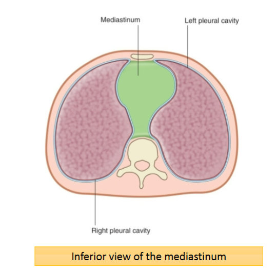
Located in the thoracic cavity, within the mediastinum
Borders of the mediastinum:
Pleural lining of the lungs
Diaphragm
Thoracic vertebrae
Sternum
The mediastinum is divided into a superior mediastinum and inferior mediastinum by a horizontal plane trhough the sternal angle - the heart is located in the inferior mediastinum
The ‘apex’ of the heart projects anteriorly towards the ribs
The ‘base’ of the heart projects posteriorly towards the vertebrae
Pericardium and layers
Pericardium: a fibroserous sac that surrounds thee heart and the roots of the great vessels
Fibrous pericardium: blends with the fibres of the diaphragm, holding the heart in place, prevents overdistension/enlargement of the heart
Serous pericardium: consists of 2 layers and cavoty containing fluid, layers glide over each other
2 layers (parietal and visceral)

Pericarditis - causes, symptoms and treatment
Inflammation of the pericardium
Causes:
Infection, autoimmune disorder, trauma
Symptoms:
Pain, shortness of breath, increased and irregular heartbeat
Pericardium is innervated by the phenic nerve (C3 - C5)
Referred pain to skin of shoulder and chest
Treatment:
NSAIDs, corticosteroids, antibiotics
Pericardiocentesis (removal of excess fluid)
Pericardial Effusion - what is it and treatment
Build up of excess fluid in the pericardial cavity
Can lead to cardiac tamponade (compression of the heart) reducing force of contractions and potentially leading to cardiac arrest
Treatment:
Non-severe: NSAIDs, antibiotics
Severe: pericardiocentesis (removal of excess fluid by needle)
What are the 3 layers of the heart wall?
Endocardium
Myocardium
Epicardium
What are the 2 types of septums?
Interatrial septum
Interventricular septum
Purpose of heart valves and 4 types of heart valves
Prevent regurgitation of blood back
Heart valve disease involves a defect in a heart valve
Valve narrowing (stenosis)
Incomplete valve closure (regurgitation)
Heart valve replacement surgery replaces a faulty heart valve with a new functional heart valve - untreated heart valve disease may lead to blood clots, stroke or heart failure
4 heart valves:
Tricuspid valve - between right atrium and right ventricle
Pulmonary valve - between right ventricle and pulmonary trunk
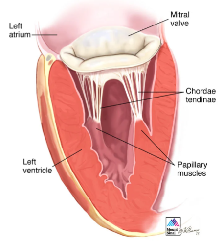
Bicuspid (mitral) valve - between the left atrium and left ventricle
Aortic valve - between the left ventricle and aorta
How many cusps does the tricuspid and bicuspid (mitral) valve have?
Tricuspid valve - 3 cusps
Bicuspid (miral) - 2 cusps
Chordae tendineae
Papillary muscle
Semilunar valves
Aortic and pulmonary valve
The cusps fill with blood forcing them together, causing closure
2 cusps in the aortic valve contain openings for the coronary arteries

Mitral valve stenosis (narrowing) - clinical signs, diagnosis and treatment
Clinical signs
High left atrial pressure
Pulmonary hypertension
Diagnosis
Chest X-ray; echocardiogram
Treatment
Diuretics (congestion)
Beta blockers/ Ca2+ channel blockers (heart rate)
Warfarin (anticoagulant)
Valvuloplasty
Myocardial infarction
Result of blockage of the coronary arteries
Commonly known as a heart attack
Major cause of death due to coronary artery and thrombosis
Causes necrosis in the the wall of the heart
Coronary arteries
Left and right coronary arteries
Don’t overlap their supply or anastomose
Blockage = no blood supply
Cardiac muscle ischamia
Myocardial infection and necrosis

Coronary veins
Cardiac veins drain into the coronary sinus which drains into the right atrium

What are the main anatomical features of the right atrium?
Receives deoxygenated blood
Has relatively thin walls because it only pumps blood into the right ventricle
Contains openings of:
Superior vena cava
Inferior vena cava
Coronary sinus
Communicates with the right ventricle through the tricuspid valve
What is the function of the right atrium?
Receives deoxygenated blood from the systemic circulation
Receives blood from the SVC, IVC and coronary sinus
Passes blood into the right ventricle
Contracts during atrial systole to assist ventricular filling
What are the main features of the right ventricle?
Receives blood from the right atrium through the tricuspid valve
Pumps blood into the pulmonary trunk
Has a thinner wall than the left ventricle
Contains papillary muscles and chordae tendineae which prevent reflux into the right atrium
Contains the moderator band which carries the right bundle branch of the atrioventricular bundle

What is the main anatomical feature of the left atrium?
Receives oxygenated blood from the lungs
Receives blood through the four pulmonary veins
Has a relatively smooth internal surface
Contains pectinate muscles mainly within the left atrial appendage
Communicates with the left ventricle through the mitral valve
What is the function of the left atrium?
Receives oxygenated blood from the pulmonary veins
Stores and transfers this blood to the left ventricle
Contracts during atrial systole to assist filling of the left ventricle
What are the main features of the left ventricle?
Receives oxygenated blood from the left atrium through the mitral valve
Pumps blood into the aorta
Has the thickest muscular wall of all four chambers
Contains trabeculae carneae
Contains larger papillary muscles and chordae tendineae due to increased pressure
Generates the high pressure required for systemic circulation
Why is the left ventricular wall thicker than the right ventricular wall?
The left ventricle must pump blood around the entire systemic circulation
This creates much greater resistance than pumping blood to the nearby lungs
Therefore, the left ventricle must generate a much higher pressure
Its thicker myocardium allows it to produce this pressure
Great vessels of the heart - anterior view diagram
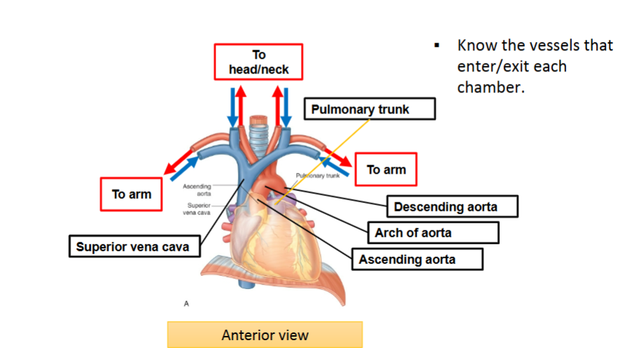
Great vessels of the heart - posterior view diagram

How do the AV valves prevent reflux?
During ventricular systole:
Ventricular pressure rises
AV valves close
Papillary muscles contract
Chordae tendineae become tense
The valve cusps are prevented from inverting into the atria
Blood is therefore directed into the pulmonary trunk/aorta
How do semilunar valves prevent reflux?
During ventricular systole:
Ventricular pressure becomes greater than arterial pressure
Semilunar valves open
Blood is ejected into the pulmonary trunk/aorta
During diastole:
Arterial pressure becomes greater than ventricular pressure
Blood attempts to flow backwards
The cusps fill with blood and close
Electrical conduction system of the heart - ECG

Cardiac conduction system process
Impulse originates from the sinoatrial (SA) node which is located in the right atrium - acts as the heart’s natural pacemaker
The impulse spreads across the right and left atrium causing atrial depolarisation and contraction
Impulse reaches the atrioventricular (AV) node located in the lower part of the right atrium near the interatrial septum
AV node introduces conduction delay allowing time for ventricles to fill with blood before contracting
Impulse then passed into the bundle of His
Bundle of His carries electrical excitation from the AV node through the fibrous skeleton of the heart into the interventricular septum
Purkinje fibres distribute electrical excitation throughout the ventricular myocardium allowing rapid, coordinated ventricular depolarisation and contraction
What is the cardiac plexus formed from?
Parasympathetic fibres from vagus nerve
Sympathetic fibres from sympathetic trunk
Neuronal control of the heart - parasympathetic and sympathetic stimulation
Parasympathetic stimulation:
Decreases heart rate
Reduces force of contraction
Constricts coronary arteries
Sympathetic stimulation:
Increases heart rate
Increases force of contraction
Referred pain
Visceral afferent innervation of the heart corresponds to T1 – T4 spinal cord segments
Skin dermatomes T1 – T4 map to skin of the chest and inner-side of the arm on left side of body
When cardiac cells die during myocardial infarction this results in referred pain in these dermatome
Arrhythmia - what is it and treatment
Irregular heartbeat - atrial fibrillation
‘Episodes’ or ‘burts’ of irregular and rapid beating of atria
Irregular blood flow can lead to clot formation
Treatment:
Rate control medications (beta blockers)
Anticoagulants (warfarin)
Pacemaker implantation
