CNS: Stroke
 Call Kai
 Learn
 Practice Test
 Spaced Repetition
 Match
 Flashcards
 Knowt Play
1/36

There's no tags or description
Looks like no tags are added yet.
37 Terms
What are cerebrovascular diseases and stroke?
Cerebrovascular diseases:
Brain disorders caused by pathological processes involving blood vessels
These processes disrupt normal blood circulation in the brain
Broad category of disorders involving the brain's blood vessels
Stroke: A clinical phenomenon that occurs when there is acute and sudden loss of blood flow and oxygen to the brain
Stroke is a type of cerebrovascular disease
Why does the brain require a continuous supply of blood and oxygen?
The brain is highly dependent on oxygen and requires a continuous supply
Lacks significant energy stores → cannot store enough energy to sustain itself without a continuous supply of glucose and O2 (which provides the main source of ATP → can’t keep that much energy for itself)
Requires continuous supply of glucose and O2 from the blood
These are needed for ATP production, which supports action potentials (needs ATP for ion pumps for depolarization/hyperpolarization) + axonal transport (ATP powers motor proteins that transport materials along axons)
Mechanisms to maintain consistent blood flow over a wide range of blood pressure and intracranial pressure
Stroke: What is hypoxia?
Low levels of oxygen in tissue
Stroke: What is ischemia?
Inadequate blood supply to tissues
Stroke: What is infarction?
Tissue death (necrosis) due to inadequate blood supply
Stroke: What is embolism?
Blocked artery caused by a foreign body, like a blood clot or air bubble, that traveled through the bloodstream
Stroke: What is thrombosis?
Blood clot forms locally and blocks or slows flow of blood
Stroke: What is hemorrhage?
Acute loss of blood from a damaged blood vessel
What are the two ways strokes can be classified?
Ischemic stroke: Blood flow to the brain is blocked (~90% strokes)
Thrombotic occlusion: A blood clot forms locally in the blood vessel, usually where blood flow is slowed or disrupted
Embolic occlusion: Blocks a blood vessel like an artery via a blood clot or air bubble that travels through the bloodstream
Hemorrhagic stroke: Sudden bleeding in the brain (~10% of stroke)
Vascular rupture: Blood vessel that broke or burst
What is the epidemiology of stroke?
5th leading cause of death in the U.S.
In 2021, 1 in 6 deaths from cardiovascular disease was due to strokeq
Every year, more than 795,000 people in the US have a stroke
About 610,000 of these are first or new strokes
A leading cause of disability: reduces mobility in >50% of stroke survivors over 65y
Health disparities:
Risk of having a first stroke is nearly twice as high for non-Hispanic Black adults as for white adults
Environmental factors + status cause these stats (inadequate access to healthcare)
Non-Hispanic Black adults and Pacific Islander adults have the highest rates of death due to stroke
Native American populations have a higher risk than non-Hispanic whites
Age-related increase in risk:
Stroke risk increases with age
In 2014, 38% of people hospitalized for stroke were less than 65 years old
What are the risk factors of stroke?
Age: The incidence of stroke increases with age, double for each decade after 55 years of age
Sex:
Young ages: Women have a high or higher risk of stroke as men
Older ages: Risk is slightly higher for men
Race/ethnicity
What are the genetic factors of stroke?
Mendelian Stroke (single-gene inheritance)
Sickle cell disease: Most common cause of stroke
Autosomal recessive disorder
Familial Amyloid Angiopathy: Autosomal dominant mutation of APP
Sporadic stroke: Shared risk variants with high cholesterol, high blood pressure, other cardiovascular disease
What are the (6) risk factors of stroke?
Hypertension (high blood pressure)
Dyslipidemia (high cholesterol)
Smoking → 15% of stroke deaths per year
Diabetes mellitus
Obesity/metabolic syndrome/diet/sedentary behavior
Inflammation and infection
What is Global Cerebral Ischemia?
A generalized reduction in blood flow throughout the brain, resulting in inadequate oxygen delivery
Global cerebral ischemia → Reduced blood flow or oxygen delivery throughout the brain → Decreased ATP production → Widespread neuronal dysfunction/damage
Global Cerebral Ischemia: How can oxygen delivery to the brain fail?
Reduced cerebral perfusion (blood flow)
Cardiac arrest: Heart stops effectively pumping blood → brain receives insufficient blood and oxygen
Shock or severe hypotension: Very low blood pressure → reduced blood flow to the brain
Reduced oxygen-carrying capacity of blood
Carbon monoxide poisoning (CO): CO binds to hemoglobin → reduces oxygen transport → inadequate oxygen delivery of blood
Global Cerebral Ischemia: What determines the clinical outcome?
Depends on the duration and severity of reduced oxygen delivery:
Shorter/less severe reduction: May cause temporary neurological dysfunction with potential recovery
Longer/more severe reduction: Can cause extensive neuronal damage, permanent disability, or death
Global Cerebral Ischemia: What are the severe outcomes?
Persistent vegetative state: Loss of conscious awareness, although some basic brain functions remain
Brain death: Irreversible loss of all brain function, including brainstem function
Which neurons are most sensitive to hypoxia in global cerebral ischemia? What are the three most sensitive neuronal populations? What are two consequences of severe ischemia?
Neurons vs. Glial Cells
Neurons are more sensitive to hypoxia than glial cells
Neurons have high ATP requirements and depend heavily on continuous oxygen delivery
Three most sensitive neuronal populations:
Pyramidal neurons in the hippocampus
Pyramidal neurons in the neocortex
Purkinje cells in the cerebellum
Consequences of Severe Ischemia
Severe ischemia → decreased oxygen and glucose delivery → ATP depletion
Leads to widespread necrotic neuronal death (irreversible cell injury)
What happens during a brief episode of Global Cerebral Ischemia?
Temporary reduction in blood flow
Reduced blood flow → decrease O2 and glucose delivery to the brain
Causes energy failure (↓ ATP) → Neurons cannot maintain normal electrical activity
Neuronal electrical activity temporarily ceases (b/c action potentials stop ig) → loss of consciousness (fainting)
Restoration of Blood Flow:
If circulation is restored before neurons and glial cells undergo irreversible damage:
ATP production and neuronal electrical activity recover
Neurons and glial cells remain viable
No permanent pathological damage → patient returns to normal
How does brief global cerebral ischemia cause fainting?

What happens during prolonged global cerebral ischemia?
Prolonged ischemia: Blood flow and oxygen delivery to the brain are reduced for too long, resulting in irreversible neuronal damage
Cellular Damage
↓ O₂ and glucose → ↓ ATP production
Cellular metabolism slows and eventually stops
ATP depletion → ion pump failure → loss of cell membrane integrity
Neurons undergo necrosis (irreversible cell death)
Why does the duration matter for prolonged global cerebral ischemia?
4-5 minutes: Severe complete ischemia can permanently damage the most sensitive neurons
Longer periods: Damage extends to additional neurons, including those in the thalamus and brain stem
What is Hypoxic-Ischemic Encephalopathy?
Widespread brain injury caused by inadequate oxygen and blood supply
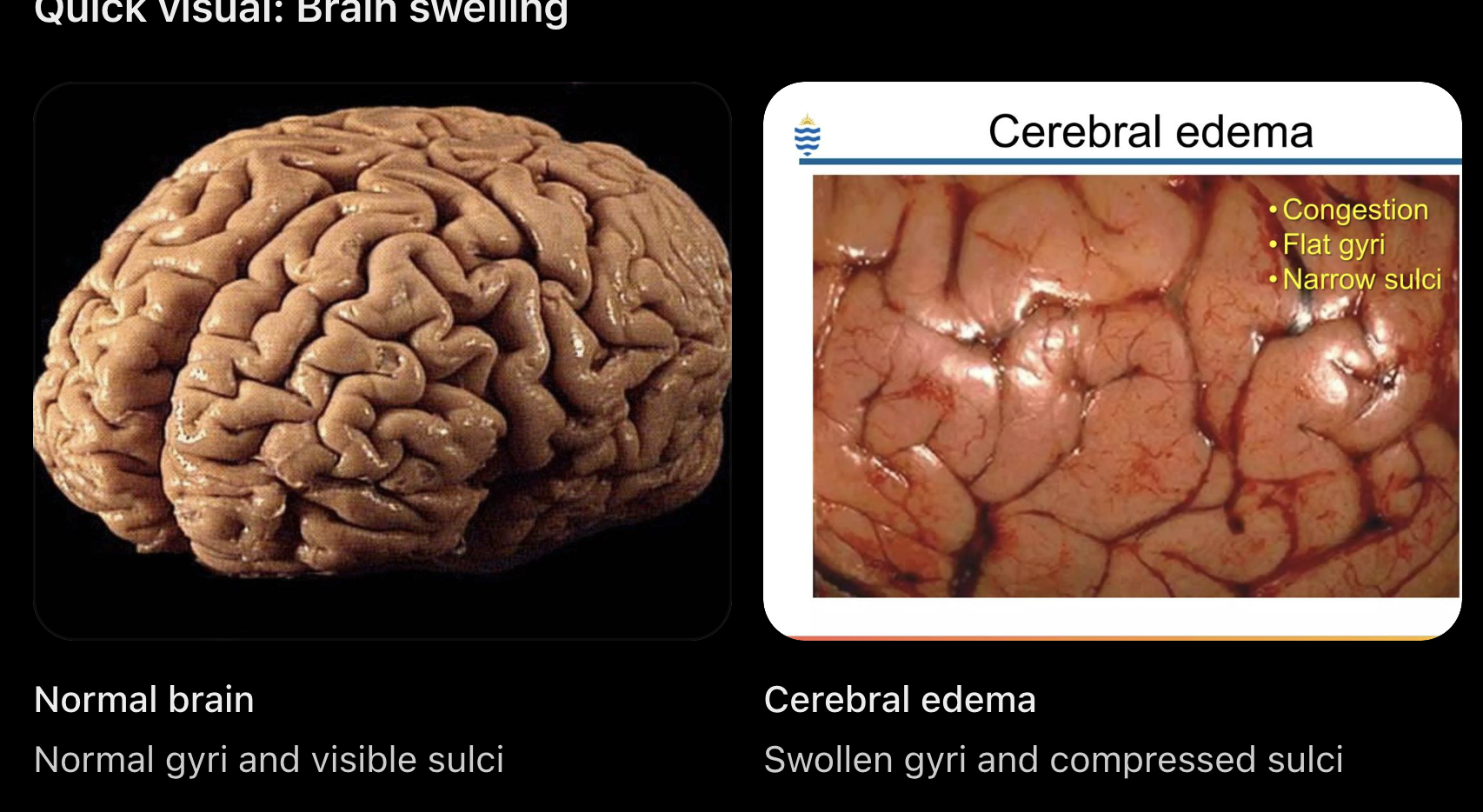
Can cause cerebral edema (brain swelling)
Swelling results in:
Widened gyri: Brain folds become swollen
Narrowed sulci: Grooves between folds become compressed
Why is gray matter more vulnerable than white matter?
Gray Matter: Contains many neuronal cell bodies and synapses → high metabolic activity and ATP demand
White Matter: Primarily contains myelinated axons → generally lower metabolic demand than gray matter
Therefore, gray matter is often more vulnerable to hypoxic-ischemic injury
However, both gray and white matter can still be damaged by severe ischemia.
What are the pathological hallmarks of prolonged global cerebral ischemia? 🟠
Anoxic Neurons (Oxygen-deprived neurons)
Injured neurons shrink
Mitochondria becomes damaged
Nuclei condense (pyknosis)
These changes indicate severe, irreversible neuronal injury
Selective Loss of Sensitive Neurons
The most vulnurable neurons are:
Pyramidal neurons of the hippocampus, neurons of the neocortex, purkinje cells of the cerebellum
Astrocyte Swelling
Astrocytes surrounding injured (anoxic) neurons swell
This occurs partly b/c ATP depletion disrupts ion balance (like the pumps), causing water to enter the cells
Macrophage removal:
Severely damaged neurons eventually undergo cell death
Macrophages (including activated microglia) break down and remove dead neurons and cellular debris
Cortical Atrophy and Gliosis
Cortical Atrophy: Shrinkage of the cerebral cortex due to neuronal loss
Gliosis: Reactive glial cells, especially astrocytes, respond to injury and form glial scars
Prolonged ischemic injury can cause pseudolaminar necrosis
Neurons die preferentially in certain cortical layers
This produces bands-like pattern of necrosis
Over time, neuronal loss and gliosis contribute to cortical atrophy
What are the clinical consequences of Global Cerebral Ischemia? 🟠
Bilateral Hippocampal Damage → Korsakoff’s (severe) Amnesia
Damage to both hippocampi causes significant memory impairment
Anterograde amnesia is more affected than Retrograde amnesia
Anterograde: Difficulty forming new memories
Retrograde: Difficulty recalling previously formed memories
Episodic memory is more affected than Semantic memory impairment
Episodic: Memory of personal experiences/events
Semantic: Memory of general facts and knowledge
Cortical and/or Thalamic Damage (Brainstem Intact)
Can cause dementia-like cognitive impairment or persistent vegetative state
Loss of conscious awareness and emotion
Preserved: Sleep-wake cycles, autonomic functions, and spontaneous breathing
Severe impairment of cerebral function, but brainstem functions remain intact
Damage Extends to Brainstem → Brain Death
Irreversible loss of cerebral and brainstem function
Characterized by:
Complete unresponsiveness
Absence of brainstem reflexes
Apnea: Inability to breathe spontaneously
Electrical silence (flat EEG)
Absence of cerebral perfusion (blood flow)
What is the difference between a persistent vegetative state and brain death?

What are the treatments of Global Cerebral Ischemia?
Neonatal HIE: therapeutic hypothermia
FAST heroes
Face, arm, speech, and time
Tissue plasminogen activator (tPA): Breaks up blood clots blocking blood flow, given within 4.5 hrs of system onset
What is vascular dementia?
Cognitive decline caused by reduced blood supply to the brain, learding to neuronal damage
What is the epidemiology of vascular dementia?
Epidemiology:
2nd most common cause of dementia after Alzheimer’s
Approx 5-10% of people with dementia have vascular dementia
What is the pathogenesis of vascular dementia?
Multiple mini-strokes over several years can cause repeated brain damage
These produce multiple infarcts in the white and gray matter
Infarcts: Area of tissue death due to insufficient blood supply
Accumulation of damage → neuronal loss → cognitive decline
What is the etiology of vascular dementia? [do we really need to know this? it sounds too clinical]
Cerebral atherosclerosis
Vessel thrombosis
Embolization from carotid vessels of the heart
Cerebral arteriolosclerosis from chronic hypertension
Cerebral amyloid angiopathy
What are the pathological changes of vascular dementia?
Cortical atrophy
Hippocampal sclerosis: Scarring and neuronal loss in the hippocampus
These changes may occur after chronic vascular injury
What are the clinical symptoms of vascular dementia? [do i rlly need to know this?]
Dementia
Gait abnormalities
Pseudobulbar signs
Additional symptoms related to specific affected sites
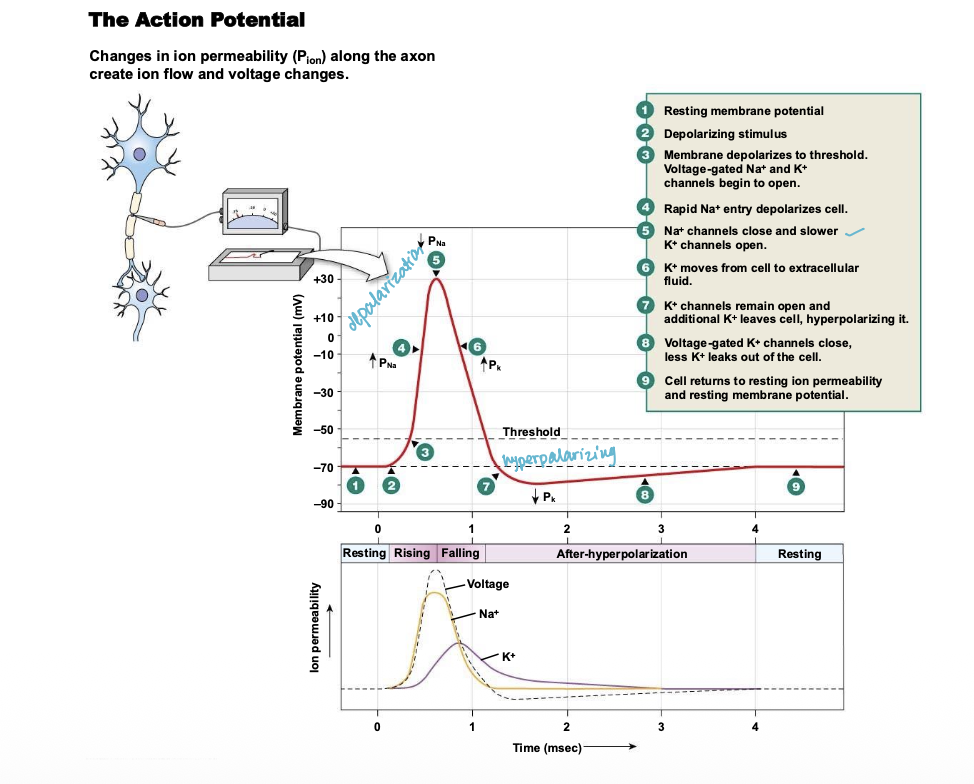
How are action potentials fired?
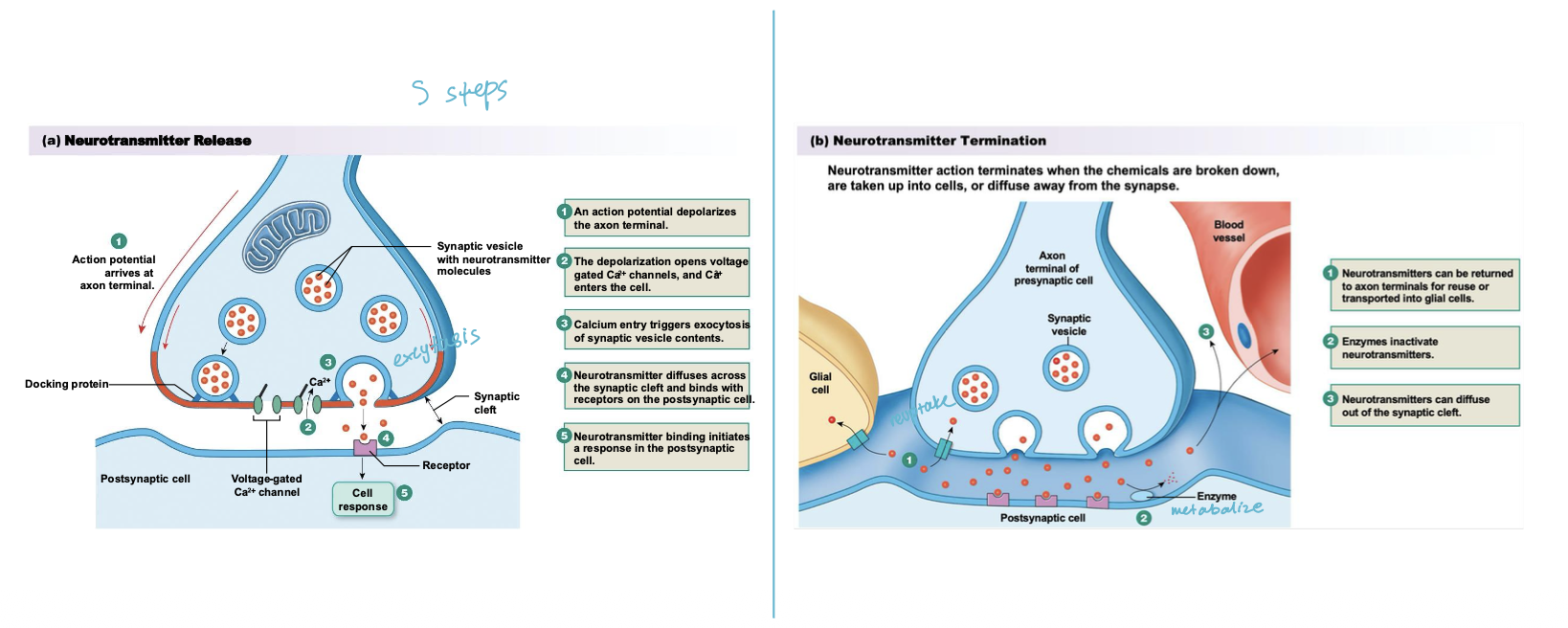
How are neurotransmitters released?
Explain the process of synaptic plasticity or long term potentiation.
Glutamate binds to AMPA and NMDA channels
Net Na+ entry through AMPA channels depolarizes the postsynaptic cell
Depolarization ejects Mg2+ from NMDA receptor-channel and opens the channel
Ca2+ enters cytoplasm through NMDA channel
Ca2+ activates second messenger pathways → cell becomes more sensitive to glutamate
Paracrine from postsynaptic cell enhances glutamate release
