PHGY170 mod2

0.0(0)
Studied by 0 people
call kaiCall Kai
learnLearn
examPractice Test
spaced repetitionSpaced Repetition
heart puzzleMatch
flashcardsFlashcards
GameKnowt Play
Card Sorting

1/37

encourage image

There's no tags or description

Looks like no tags are added yet.

38 Terms

1
New cards

Primary function of nucleus

  • protects cell DNA and allows genes to function by:

    • 1. allows DNA to be replicated and transcribed into mRNA when needed

    • 2. regulates which molecules can access DNA and separate it from other cell compartments

    • organizes DNA, since DNA is fragile and easily damaged

  • Does so through 5 components


2
New cards

what are 5 components of nucleus?

1.

Nucleus subcomponent

Structure

Function

Nuclear envelope

-              Double membrane, in which the outer layer is linked to the endoplasmic reticulum (for protein making) to form part of endomembrane system

-              Protects and creates a barrier between DNA and other cell contents/processes

-              Manages and restricts the types of molecules that can access nucleus

Nuclear pores

-              Has nuclear pore complexes that create access pathways through the nuclear membrane

-              Found on nuclear envelope

-              Generally, manages the entry and exit of molecules from nucleus

-              Small molecules (H2O, O2) pass through membrane freely (no energy needed)

-              Nuclear pore complexes allow and manage movement of molecules (of large molecular size like proteins/mRNA) in or out of nucleus

a) Nucleoplasm/b) nuclear matrix

a)        A viscous fluid mainly composed of water with aqueous ions and molecules, encased by nuclear envelope


b) Made of organized filaments, forming a network, found in nucleoplasm

a)        Contains the aqueous substances required for nucleus activity and helps suspend nuclear parts


b) Organizes chromosomes within nucleus and supports the structural integrity of nucleus

Nucleolus

-              Has machinery for rRNA gene transcription

-              Contains/organizes DNA encoding for rRNA genes

-              Synthesizes rRNA and creates ribosomal subunits for translation

a)Chromatin/ b)chromosomes

a)        Made of DNA and proteins, forms fibres. Found in specific locations in nucleus.


b) The condensed form of chromatin

a)        Condenses and organizes DNA

-              Allows for specific sections to be actively transcribed into mRNA


b)        Found in nucleus during cell division, so DNA is given to new cells


3
New cards

What is purpose of DNA packaging

  • needs to fit lots of DNA into tiny cells

  • allows cell to adjust its organization to access the genes it needs and package away DNA it doesn’t need

  • one of the main ways cell can protect DNA and regulate which genes are being transcribed


4
New cards

what are the 5 levels of DNA packaging?

Level of DNA packaging

Structure

Compression level

1

DNA double helix

-              Contains nucleotides with complementary hydrogen bonds (A-T, C-G) between nitrogenous bases (hydrophobic) in a sugar-phosphate backbone (hydrophilic) with phosphodiester bonds

-              Double stranded, strands run antiparallel

-              DNA is coiled

-              Hydrophilic/hydrophobic regions cause bases to stack in center and backbone remaining outside

o   For stability and due to being in a water-containing environment (a cell)

The least amount of compression

2

Nucleosomes

-              Formed of core DNA (~200 base pairs) wrapped around 8 core histone proteins (octamer) twice + H1 protein (holds core DNA to the core of the histone octamer) + linker DNA (the DNA between sections of core DNA that connects nucleosomes)

-              Nucleosomes are the “beads” on DNA “string”

DNA is shortened by 7x


3

Chromatin fibre

-              The length of nucleosomes is coiled and makes a spiraling fibre

-              Helical shape

-              Diameter ~30-40 nm

DNA is compressed by 42x

4

Chromatin Looped Domains

-              The chromatin fibre (that was 30-40 nm) is arranged into loops

-              Length is ~300 nm


*Levels 1-4 called euchromatin, which is active, can be accessed by proteins for DNA replication/transcription

Again, DNA is compressed, by 750x

5

Heterochromatin

-              The chromatin looped domains are compacted and folded until ~700nm

-              DNA is inaccessible

-              Only found in parts of chromosomes that are inactive (not currently undergoing cell division, interphase)

-              Can be condensed into chromosomes during cell division (mitosis or meiosis)

o   In which the total width of its arms is 1400 nm

The most extreme form of compression, “hyper condensed”


<table style="min-width: 75px;"><colgroup><col style="min-width: 25px;"><col style="min-width: 25px;"><col style="min-width: 25px;"></colgroup><tbody><tr><td colspan="1" rowspan="1" style="width: 155.8pt; border: 1pt solid windowtext; padding: 0cm 5.4pt;"><p class="MsoNormal">Level of DNA packaging</p></td><td colspan="1" rowspan="1" style="width: 155.85pt; border-width: 1pt 1pt 1pt medium; border-style: solid solid solid none; border-color: windowtext windowtext windowtext currentcolor; border-image: none; padding: 0cm 5.4pt;"><p class="MsoNormal">Structure</p></td><td colspan="1" rowspan="1" style="width: 155.85pt; border-width: 1pt 1pt 1pt medium; border-style: solid solid solid none; border-color: windowtext windowtext windowtext currentcolor; border-image: none; padding: 0cm 5.4pt;"><p class="MsoNormal">Compression level</p></td></tr><tr><td colspan="1" rowspan="1" style="width: 155.8pt; border-width: medium 1pt 1pt; border-style: none solid solid; border-color: currentcolor windowtext windowtext; border-image: none; padding: 0cm 5.4pt;"><p class="MsoNormal">1</p></td><td colspan="1" rowspan="1" style="width: 155.85pt; border-width: medium 1pt 1pt medium; border-style: none solid solid none; border-color: currentcolor windowtext windowtext currentcolor; padding: 0cm 5.4pt;"><p class="MsoNormal">DNA double helix</p><p class="MsoListParagraphCxSpFirst"><span>-</span><span style="font-family: &quot;Times New Roman&quot;; line-height: normal; font-size: 7pt;">&nbsp;&nbsp;&nbsp;&nbsp;&nbsp;&nbsp;&nbsp;&nbsp;&nbsp;&nbsp;&nbsp;&nbsp;&nbsp; </span>Contains nucleotides with complementary hydrogen bonds (A-T, C-G) between nitrogenous bases (hydrophobic) in a sugar-phosphate backbone (hydrophilic) with phosphodiester bonds</p><p class="MsoListParagraphCxSpMiddle"><span>-</span><span style="font-family: &quot;Times New Roman&quot;; line-height: normal; font-size: 7pt;">&nbsp;&nbsp;&nbsp;&nbsp;&nbsp;&nbsp;&nbsp;&nbsp;&nbsp;&nbsp;&nbsp;&nbsp;&nbsp; </span>Double stranded, strands run antiparallel</p><p class="MsoListParagraphCxSpMiddle"><span>-</span><span style="font-family: &quot;Times New Roman&quot;; line-height: normal; font-size: 7pt;">&nbsp;&nbsp;&nbsp;&nbsp;&nbsp;&nbsp;&nbsp;&nbsp;&nbsp;&nbsp;&nbsp;&nbsp;&nbsp; </span>DNA is coiled</p><p class="MsoListParagraphCxSpMiddle"><span>-</span><span style="font-family: &quot;Times New Roman&quot;; line-height: normal; font-size: 7pt;">&nbsp;&nbsp;&nbsp;&nbsp;&nbsp;&nbsp;&nbsp;&nbsp;&nbsp;&nbsp;&nbsp;&nbsp;&nbsp; </span>Hydrophilic/hydrophobic regions cause bases to stack in center and backbone remaining outside</p><p class="MsoListParagraphCxSpLast"><span style="font-family: &quot;Courier New&quot;;">o</span><span style="font-family: &quot;Times New Roman&quot;; line-height: normal; font-size: 7pt;">&nbsp;&nbsp; </span>For stability and due to being in a water-containing environment (a cell)</p></td><td colspan="1" rowspan="1" style="width: 155.85pt; border-width: medium 1pt 1pt medium; border-style: none solid solid none; border-color: currentcolor windowtext windowtext currentcolor; padding: 0cm 5.4pt;"><p class="MsoNormal">The least amount of compression</p></td></tr><tr><td colspan="1" rowspan="1" style="width: 155.8pt; border-width: medium 1pt 1pt; border-style: none solid solid; border-color: currentcolor windowtext windowtext; border-image: none; padding: 0cm 5.4pt;"><p class="MsoNormal">2</p></td><td colspan="1" rowspan="1" style="width: 155.85pt; border-width: medium 1pt 1pt medium; border-style: none solid solid none; border-color: currentcolor windowtext windowtext currentcolor; padding: 0cm 5.4pt;"><p class="MsoNormal">Nucleosomes</p><p class="MsoListParagraphCxSpFirst"><span>-</span><span style="font-family: &quot;Times New Roman&quot;; line-height: normal; font-size: 7pt;">&nbsp;&nbsp;&nbsp;&nbsp;&nbsp;&nbsp;&nbsp;&nbsp;&nbsp;&nbsp;&nbsp;&nbsp;&nbsp; </span>Formed of core DNA (~200 base pairs) wrapped around 8 core histone proteins (octamer) twice + H1 protein (holds core DNA to the core of the histone octamer) + linker DNA (the DNA between sections of core DNA that connects nucleosomes)</p><p class="MsoListParagraphCxSpLast"><span>-</span><span style="font-family: &quot;Times New Roman&quot;; line-height: normal; font-size: 7pt;">&nbsp;&nbsp;&nbsp;&nbsp;&nbsp;&nbsp;&nbsp;&nbsp;&nbsp;&nbsp;&nbsp;&nbsp;&nbsp; </span>Nucleosomes are the “beads” on DNA “string”</p></td><td colspan="1" rowspan="1" style="width: 155.85pt; border-width: medium 1pt 1pt medium; border-style: none solid solid none; border-color: currentcolor windowtext windowtext currentcolor; padding: 0cm 5.4pt;"><p class="MsoNormal">DNA is shortened by 7x</p><p class="MsoNormal">&nbsp;</p></td></tr><tr><td colspan="1" rowspan="1" style="width: 155.8pt; border-width: medium 1pt 1pt; border-style: none solid solid; border-color: currentcolor windowtext windowtext; border-image: none; padding: 0cm 5.4pt;"><p class="MsoNormal">3</p></td><td colspan="1" rowspan="1" style="width: 155.85pt; border-width: medium 1pt 1pt medium; border-style: none solid solid none; border-color: currentcolor windowtext windowtext currentcolor; padding: 0cm 5.4pt;"><p class="MsoNormal">Chromatin fibre</p><p class="MsoListParagraphCxSpFirst"><span>-</span><span style="font-family: &quot;Times New Roman&quot;; line-height: normal; font-size: 7pt;">&nbsp;&nbsp;&nbsp;&nbsp;&nbsp;&nbsp;&nbsp;&nbsp;&nbsp;&nbsp;&nbsp;&nbsp;&nbsp; </span>The length of nucleosomes is coiled and makes a spiraling fibre</p><p class="MsoListParagraphCxSpMiddle"><span>-</span><span style="font-family: &quot;Times New Roman&quot;; line-height: normal; font-size: 7pt;">&nbsp;&nbsp;&nbsp;&nbsp;&nbsp;&nbsp;&nbsp;&nbsp;&nbsp;&nbsp;&nbsp;&nbsp;&nbsp; </span>Helical shape</p><p class="MsoListParagraphCxSpLast"><span>-</span><span style="font-family: &quot;Times New Roman&quot;; line-height: normal; font-size: 7pt;">&nbsp;&nbsp;&nbsp;&nbsp;&nbsp;&nbsp;&nbsp;&nbsp;&nbsp;&nbsp;&nbsp;&nbsp;&nbsp; </span>Diameter ~30-40 nm</p></td><td colspan="1" rowspan="1" style="width: 155.85pt; border-width: medium 1pt 1pt medium; border-style: none solid solid none; border-color: currentcolor windowtext windowtext currentcolor; padding: 0cm 5.4pt;"><p class="MsoNormal">DNA is compressed by 42x</p></td></tr><tr><td colspan="1" rowspan="1" style="width: 155.8pt; border-width: medium 1pt 1pt; border-style: none solid solid; border-color: currentcolor windowtext windowtext; border-image: none; padding: 0cm 5.4pt;"><p class="MsoNormal">4</p></td><td colspan="1" rowspan="1" style="width: 155.85pt; border-width: medium 1pt 1pt medium; border-style: none solid solid none; border-color: currentcolor windowtext windowtext currentcolor; padding: 0cm 5.4pt;"><p class="MsoNormal">Chromatin Looped Domains</p><p class="MsoListParagraphCxSpFirst"><span>-</span><span style="font-family: &quot;Times New Roman&quot;; line-height: normal; font-size: 7pt;">&nbsp;&nbsp;&nbsp;&nbsp;&nbsp;&nbsp;&nbsp;&nbsp;&nbsp;&nbsp;&nbsp;&nbsp;&nbsp; </span>The chromatin fibre (that was 30-40 nm) is arranged into loops</p><p class="MsoListParagraphCxSpLast"><span>-</span><span style="font-family: &quot;Times New Roman&quot;; line-height: normal; font-size: 7pt;">&nbsp;&nbsp;&nbsp;&nbsp;&nbsp;&nbsp;&nbsp;&nbsp;&nbsp;&nbsp;&nbsp;&nbsp;&nbsp; </span>Length is ~300 nm</p><p class="MsoNormal">&nbsp;</p><p class="MsoNormal">*Levels 1-4 called euchromatin, which is active, can be accessed by proteins for DNA replication/transcription</p></td><td colspan="1" rowspan="1" style="width: 155.85pt; border-width: medium 1pt 1pt medium; border-style: none solid solid none; border-color: currentcolor windowtext windowtext currentcolor; padding: 0cm 5.4pt;"><p class="MsoNormal">Again, DNA is compressed, by 750x</p></td></tr><tr><td colspan="1" rowspan="1" style="width: 155.8pt; border-width: medium 1pt 1pt; border-style: none solid solid; border-color: currentcolor windowtext windowtext; border-image: none; padding: 0cm 5.4pt;"><p class="MsoNormal">5</p></td><td colspan="1" rowspan="1" style="width: 155.85pt; border-width: medium 1pt 1pt medium; border-style: none solid solid none; border-color: currentcolor windowtext windowtext currentcolor; padding: 0cm 5.4pt;"><p class="MsoNormal">Heterochromatin</p><p class="MsoListParagraphCxSpFirst"><span>-</span><span style="font-family: &quot;Times New Roman&quot;; line-height: normal; font-size: 7pt;">&nbsp;&nbsp;&nbsp;&nbsp;&nbsp;&nbsp;&nbsp;&nbsp;&nbsp;&nbsp;&nbsp;&nbsp;&nbsp; </span>The chromatin looped domains are compacted and folded until ~700nm</p><p class="MsoListParagraphCxSpMiddle"><span>-</span><span style="font-family: &quot;Times New Roman&quot;; line-height: normal; font-size: 7pt;">&nbsp;&nbsp;&nbsp;&nbsp;&nbsp;&nbsp;&nbsp;&nbsp;&nbsp;&nbsp;&nbsp;&nbsp;&nbsp; </span>DNA is inaccessible</p><p class="MsoListParagraphCxSpMiddle"><span>-</span><span style="font-family: &quot;Times New Roman&quot;; line-height: normal; font-size: 7pt;">&nbsp;&nbsp;&nbsp;&nbsp;&nbsp;&nbsp;&nbsp;&nbsp;&nbsp;&nbsp;&nbsp;&nbsp;&nbsp; </span>Only found in parts of chromosomes that are inactive (not currently undergoing cell division, interphase)</p><p class="MsoListParagraphCxSpMiddle"><span>-</span><span style="font-family: &quot;Times New Roman&quot;; line-height: normal; font-size: 7pt;">&nbsp;&nbsp;&nbsp;&nbsp;&nbsp;&nbsp;&nbsp;&nbsp;&nbsp;&nbsp;&nbsp;&nbsp;&nbsp; </span>Can be condensed into chromosomes during cell division (mitosis or meiosis)</p><p class="MsoListParagraphCxSpLast"><span style="font-family: &quot;Courier New&quot;;">o</span><span style="font-family: &quot;Times New Roman&quot;; line-height: normal; font-size: 7pt;">&nbsp;&nbsp; </span>In which the total width of its arms is 1400 nm</p></td><td colspan="1" rowspan="1" style="width: 155.85pt; border-width: medium 1pt 1pt medium; border-style: none solid solid none; border-color: currentcolor windowtext windowtext currentcolor; padding: 0cm 5.4pt;"><p class="MsoNormal">The most extreme form of compression, “hyper condensed”</p><p class="MsoNormal">&nbsp;</p></td></tr></tbody></table><p></p>
5
New cards

what is the endomembrane system?

  • includes RER, SER, Golgi

  • The linked machinery that processes the transport of cargo throughout the cell

  • exocytosis = moves cargo out of cell using exocytic pathway (directs contents of secretory vesicles to PM, includes ER, Golgi, exocytic vesicles)

  • endocytosis = transports cargo into cell using endocytic pathway (cells absorb molecules by engulfing them, involving early and late endosomes and lysosomes)


6
New cards

Briefly describe the parts of the endomembrane system

  1. nucleus

  • connected to ER cisternae by outer layer of nuclear envelope

  • some nuclear pores connect nuclear envelope with ER membranes so molecules can pass through these structures freely

  1. RER

  • has ribosomes

  • site for protein translation/modifications

  1. SER

  • no ribosomes

  • for lipid processing, carb metabolism, stores calcium

  1. transport vesicles

  • proteins made in ER are shuttled in transport vesicles to Golgi for further modification

  • also used to deliver cargo between organelles

  1. Golgi

  • involved in protein modification and transport

  • labels proteins/molecules with signals to go to correct ddestination


7
New cards

how are larger molecules transported throughout the cell?

  • needs the endomembrane system

  • only small molecules can easily move around/out of the cell


8
New cards

Describe functions of RER and smooth ER

  • RER

    • has attached ribosomes, which assemble here to produce proteins that must go to another organelle

    • *free ribosomes in cytosol only make proteins that stay in cytosol

    • responsible for final stages of assembling polypeptide chains into protein and packaging it

    • made of cisternae, flattened membrane disks, connected to outer nuclear envelope

  • SER

    • Creates lipids (phospholipids for

      plasma membrane, steroids)

      - Site of carbohydrate metabolism

      (G6P—>C6H12O6)

      - In muscle cells, manages Ca2+

      concentration for muscle

      contraction

      - Does not have ribosomes for protein

      synthesis


9
New cards

What are transport vesicles? what is vesicle-mediated transport?

  • used to shuttle materials between organelles in endomembrane system using vesicle-mediated transport

  • small vesicles bud off lipid membranes of organelles/the cell to transport cargo (e.g. soluble proteins) —> they transport cargo in/out of cell or to a different location of the cell

    • once arriving at final destination, they fuse with lipid membranes


10
New cards

What are the different parts of the Golgi?

Cis

  • Accepts proteins from the ER that

are currently in the endomembrane

pathway

  • The start point for the Golgi

  • First receives vesicles and begins

working on them


Medial

  • Center/middle region of Golgi

  • Adds oligosaccharides/sugars (e.g.

M6P) to proteins or modifies current

oligosaccharides

  • Can add sugar groups to lipids


Trans

  • Responsible for the last packaging

stage as well as ensures cargo is

sent to its proper, destined

organelles/locations


11
New cards

In detail, what does the trans golgi do?


  • since its the final region of Golgi before cargo departs, its important for sorting and packaging

  1. other organelles

  • Based on cell type, various cell organelles need different combination of proteins

  • Each cell has specialized requirements needed for trans Golgi

network

  1. Retrograde transport:

  • Some proteins coming from the ER have an ER retention signal

  • Once processed by Golgi, they’re packed with COPI coating proteins and sent back to the ER

  1. Lysosome:

  • The medial Golgi can add M6P (an example of a sugar modification)

  • M6P receptors in trans Golgi network identifies proteins with M6P tags—>go to lysosome for

degradation

  1. Cargo transport:

  • Exocytosis allows vesicles to be secreted outside the cell


12
New cards

What are endosomes, lysosomes, peroxisomes used for?

  • all are membrane-bound organelles responsible for breakdown of protein/macromolecules

  1. endosomes

  • hold content that comes into the cell from EC space through endocytosis

  • sorts and send cargo to correct final locations

  1. lysosomes

  • break down proteins/lipids/sugars into building blocks

  1. peroxisomes

  • breaks down molecules, specifically those generating H2O2 as byproduct

  • thus, cells can neutralize H2O2 to prevent cell damage


13
New cards

Give 3 examples of targeting proteins

  • when signal sequences/lack of them are used to target proteins to different areas of the cell

  1. proteins targeted to endomembrane system (ER, golgi, endosomes/lysosomes, PM, etc)

  • proteins would contain target signal sequences or tagged within endomembrane system

  • they would usually end up in other parts of the cell, like as transmembrane proteins

  1. proteins destined for cytosol

  • these proteins lack signal peptides, since free floating ribosomes translate them into the cytosol

  1. proteins that need to be secreted from cell (via exocytosis)

  • have distinct signal sequences

  • essential for body functions like hormones or forming the ECM


14
New cards

How does a protein enter the endomembrane system

  1. protein enters the endomembrane system by entering ER lumen

  • Proteins translated in ribosomes directly attached to RER can be translated directly into the lumen


15
New cards

Describe the steps of translocation into the ER (4)

  • def: the process which proteins go from being translated to transported into the ER

  • for translocating soluble proteins into the ER

  1. signal sequence

  • transport of proteins in the ER begins during translation, since the ER signal sequence is translated as part of the protein

  1. SRP binding

  • this sequence interacts with a receptor (the SRP, signal recognition particle) which binds to the ribosome that’s translating the protein

  • pauses translation

  • needs GTP

  1. ribosome docking

  • while translation is paused, ribosome docks onto ER membrane by SRP interacting with SRP receptor + translocon complex

  • allows the translocation of the growing protein into the ER

  1. translocation

  • translation restarts

  • once protein has translocated into the ER, the signal peptide sequence is cleaved off the protein

  • translation finishes

  • protein folds inside ER lumen

  • the finished protein = meant to be soluble and not attached to a membrane


<ul><li><p>def: the process which proteins go from being translated to transported into the ER</p></li><li><p>for translocating soluble proteins into the ER</p></li></ul><ol><li><p>signal sequence</p></li></ol><ul><li><p>transport of proteins in the ER begins during translation, since the <strong>ER signal sequence</strong> is translated as part of the protein</p></li></ul><ol start="2"><li><p>SRP binding</p></li></ol><ul><li><p>this sequence interacts with a receptor (the SRP, signal recognition particle) which binds to the ribosome that’s translating the protein</p></li><li><p>pauses translation</p></li><li><p>needs GTP</p></li></ul><ol start="3"><li><p>ribosome docking</p></li></ol><ul><li><p>while translation is paused, ribosome docks onto ER membrane by SRP interacting with SRP receptor + translocon complex</p></li><li><p>allows the translocation of the growing protein into the ER</p></li></ul><ol start="4"><li><p>translocation</p></li></ol><ul><li><p>translation restarts</p></li><li><p>once protein has translocated into the ER, the signal peptide sequence is cleaved off the protein</p></li><li><p>translation finishes</p></li><li><p>protein folds inside ER lumen</p></li><li><p>the finished protein = meant to be soluble and not attached to a membrane</p></li></ul><p></p>
16
New cards

How are proteins destined to be embedded into lipid membrane translocated?

  1. transmembrane domain

  • the translocon for transmembrane proteins recognizes a signal (transmembrane signal anchor sequences)

  • initiates process for protein to be embedded in membrane

  1. protein enters lipid bilayer

  • transmembrane signal sequences are embedded in lipid bilayer

    • will become the transmembrane domain of the protein once its a functional, mature protein


<ol><li><p>transmembrane domain</p></li></ol><ul><li><p>the translocon for transmembrane proteins recognizes a signal (transmembrane signal anchor sequences)</p></li><li><p>initiates process for protein to be embedded in membrane</p></li></ul><ol start="2"><li><p>protein enters lipid bilayer</p></li></ol><ul><li><p>transmembrane signal sequences are embedded in lipid bilayer</p><ul><li><p>will become the transmembrane domain of the protein once its a functional, mature protein</p></li></ul></li></ul><p></p>
17
New cards

How are proteins post-translationally modified?

  • Occurs once the proteins are in the ER and folded

  • definition: the covalent modification of the protein after translation

  • examples:

    • adding proteins/sugars/lipids/new functional groups that can change the final target location/structure/fx of protein

    • cleaving of peptide bonds in the protein

    • the ER signal sequence (at N-terminus) is removed


18
New cards

How does the ER fold proteins?

  • ER facilitates protein folding in addition to post-translation mods

  • 2 specific enzymes used:

    • 1. PDI (protein disulphide isomerase)

      • folding and stabilization of a protein is facilitated by a disulfide bond forming between cysteine AAs

      • PDIs help form this disulfide bond between thiol groups in cysteine’s side chains

    • 2. binding protein

      • Chaperonins (type of protein) helps fold polypeptide by binding hydrophobic patches in newly translated proteins

      • BIP is an example of a chaperonin

        • brings patches together so they fold into the hydrophobic interior of mature proteins

        • once buried in protein folds, BiP can no longer access hydrophobic patches


<ul><li><p>ER facilitates protein folding in addition to post-translation mods</p></li><li><p>2 specific enzymes used:</p><ul><li><p>1. PDI (protein disulphide isomerase)</p><ul><li><p>folding and stabilization of a protein is facilitated by a disulfide bond forming between cysteine AAs</p></li><li><p>PDIs help form this disulfide bond between thiol groups in cysteine’s side chains</p></li></ul></li><li><p>2. binding protein</p><ul><li><p>Chaperonins (type of protein) helps fold polypeptide by binding hydrophobic patches in newly translated proteins</p></li><li><p>BIP is an example of a chaperonin</p><ul><li><p>brings patches together so they fold into the hydrophobic interior of mature proteins</p></li><li><p>once buried in protein folds, BiP can no longer access hydrophobic patches</p></li></ul></li></ul></li></ul></li></ul><p></p>
19
New cards

How is a peptide bond formed

  • The carboxylic acid group of one AA and the amino group of another AA undergo a dehydration reaction to form peptide bond

  • peptide bonds only occur between these 2 groups in the AA backbones, not in the R groups of the AAs

  • the AAs can rotate around the peptide bond (a rigid plane)


<ul><li><p>The carboxylic acid group of one AA and the amino group of another AA undergo a dehydration reaction to form peptide bond</p></li><li><p>peptide bonds only occur between these 2 groups in the AA backbones, not in the R groups of the AAs</p></li><li><p>the AAs can rotate around the peptide bond (a rigid plane)</p></li></ul><p></p>
20
New cards

WHat are the 2 ends of an AA

N-terminus

  • the end with an amino acid group free

C-terminus

  • the end with the carboxylic acid group free


21
New cards

What is primary structure of proteins?

  • Simplest: linear chain of amino acids/peptide sequence containing peptide bonds (between carboxyl/amino group) through dehydration reaction (releases water)

  • Described as three or single amino acid codes

  • Numbered from N-terminus (amino group free end) to C-terminus (carboxyl group free end)

  • E.g. Gly-Leu-Lys


<ul><li><p>Simplest: linear chain of amino acids/peptide sequence containing peptide bonds (between carboxyl/amino group) through dehydration reaction (releases water)</p></li></ul><ul><li><p>Described as three or single amino acid codes </p></li><li><p>Numbered from N-terminus (amino group free end) to C-terminus (carboxyl group free end)</p></li><li><p>E.g. Gly-Leu-Lys</p></li></ul><p></p>
22
New cards

What is the secondary structure of proteins?

  • Refers to specific parts of the peptide arranged into alpha helix and/or beta sheets

  • Both form due to specific hydrogen bonding (between hydrogen in amino group of one amino acid + oxygen in carboxyl group of another) for stability

  • Alpha helix: coils, hydrogen bonds form in the backbones of each fourth amino acid

  • Hydrogen bonds (in beta sheets) form in the backbones between the amino acids arranged in rows

  • Parallel (both rows run N—>C) or antiparallel (peptide bonds change direction in the rows)

  • Multiple secondary structure units form domains
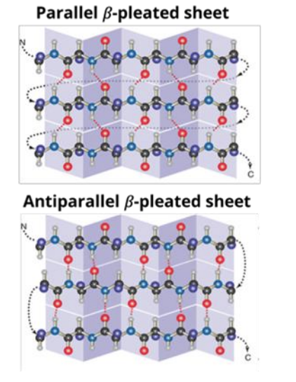

<ul><li><p class="MsoListParagraphCxSpFirst">Refers to specific parts of the peptide arranged into alpha helix and/or beta sheets</p></li><li><p class="MsoListParagraphCxSpFirst">Both form due to specific hydrogen bonding (between hydrogen in amino group of one amino acid + oxygen in carboxyl group of another) for stability</p></li><li><p class="MsoListParagraphCxSpFirst">Alpha helix: coils, hydrogen bonds form in the backbones of each fourth amino acid</p></li><li><p class="MsoListParagraphCxSpFirst">Hydrogen bonds (in beta sheets) form in the backbones between the amino acids arranged in rows</p></li><li><p class="MsoListParagraphCxSpFirst">Parallel (both rows run N<span>—&gt;</span>C) or antiparallel (peptide bonds change direction in the rows)</p></li><li><p class="MsoListParagraphCxSpFirst">Multiple secondary structure units form domains</p></li></ul><p></p>
23
New cards

What is tertiary protein structure?

  • A complete protein’s 3D shape

  • Globular unit (folded amino acid chain), each held by disulfide bridges/ionic bonds

  • E.g. Ribonuclease A

  • Chaperone proteins for correct protein folding

  • Has secondary structures and (usually multiple) domain regions

  • has disulfide bonds


<ul><li><p>A complete protein’s 3D shape</p></li><li><p class="MsoListParagraphCxSpMiddle">Globular unit (folded amino acid chain), each held by disulfide bridges/ionic bonds</p></li><li><p class="MsoListParagraphCxSpMiddle">E.g. Ribonuclease A</p></li><li><p class="MsoListParagraphCxSpMiddle">Chaperone proteins for correct protein folding</p></li><li><p class="MsoListParagraphCxSpMiddle">Has secondary structures and (usually multiple) domain regions</p></li><li><p class="MsoListParagraphCxSpMiddle">has disulfide bonds</p></li></ul><p></p>
24
New cards

What are domains?

Domains: a fundamental/independent unit, creates proteins

  • Can have specific functions (like enzymes)

  • Typically continue functioning during evolution (conserved)

  • Function is possible, folds independently

  • 20 to hundreds of amino acid residues

E.g. Zinc fingers/SH2/Kinase

25
New cards

What is quaternary protein structure?

  • Several proteins are arranged to form a complex

o   If they cannot function independent from complex, called “subunits”

E.g. Hemoglobin (4 subunits)
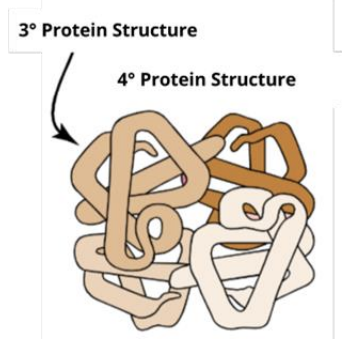
<ul><li><p>Several proteins are arranged to form a complex</p></li></ul><p class="MsoListParagraphCxSpMiddle"><span style="font-family: &quot;Courier New&quot;;">o</span><span style="font-family: &quot;Times New Roman&quot;; line-height: normal; font-size: 7pt;">&nbsp;&nbsp; </span>If they cannot function independent from complex, called “subunits”</p><p class="MsoListParagraphCxSpMiddle">E.g. Hemoglobin (4 subunits)</p>
26
New cards

High temperatures of change in pH result in protein…

  • denaturation or unfolding


smaller, subtle changes occur when proteins are modified/bind to other proteins/other chemicals


27
New cards

What happens for sickle cell anemia?

  • a single AA change in hemoglobin

  • glutamate —> valine

  • the valine sticks to the oxygen binding site of another hemoglobin


28
New cards

What are 2 major types of modifications a cell makes?

  1. covalent

  • relatively long lasting

  • examples: disulfide bonds, adding lipids/sugars

  • adding PO4, CH3, acetyl groups change protein shape, which can activate or inactivate proteins

  1. noncovalent

  • short lived

  • examples: proteins interacting with eachother in binding sites, or small molecules like O2, Ca, Mg, binding briefly


29
New cards

How are proteins transported from the ER to another organelle?

  • a vesicle is needed, cargo can’t freely float

  • a small vesicle buds off ER membrane, move through cytosol, and fuse with Golgi membrane

  • *Vesicles can easily dock/embed into destination organelles membranes since they are formed of the same lipid membranes as organelles


30
New cards

What are the 9 steps of vesicle mediated transport?

Vesicle trafficking is the nine-step endomembrane process where cargo is transported (from the initialacceptor) with vesicles.  

1.        A) Cargo selection

  • The necessary/appropriate cargo is guided to the section of a membrane destined for vesicle formation

    • Uses signal sequences/membrane receptors

  • Ensures accuracy/productivity

1.        B) Coat proteins (cytosolic)

  • Binds to membrane (destined to form vesicle’s exterior)

  • Causes receptors/proteins (tagged with signal sequences) to gather on membrane surface

    • For later budding of vesicle

-              Coat proteins: COPII (ER to move materials to Golgi), COPI (coat vesicles for return to ER), Clathrin (used by Golgi for transport/exocytosis/endocytosis)

2.        Budding

  • Coat proteins work with (cytosolic) adaptor proteins for budding of membrane

    • Pulls membrane into concave shape

3.        Scission

  • Dynamin proteins pinch vesicle off membrane (which enters cytoplasm)

4.        Uncoating

  • The vesicle’s coat/adaptor proteins deconstruct then recycle to make other vesicles

5.        Transport

  • Motor proteins (Kinesin) in cytosol connect to vesicles for transportation to the correct location (using cytoskeleton’s microtubules)

6.        7. Tethering/docking

  • Tether protein joins vesicle to acceptor membrane’s receptor, shortening distance

  • Vesicle precisely attaches to appropriate acceptor target proteins

  • After positioning itself on the acceptor membrane’s surface, fusion (of vesicle and this membrane) can occur

8.        Fusion

  • Of lipid and acceptor membrane

  • Requires two pairs of v-SNAREs and t-SNAREs

    • Vesicle’s contents can be released into acceptor organelle

9.        Disassembly

  • Leftover (tether/fusion proteins/receptors) deconstruct and return to original organelle

  • Vesicle’s contents enter acceptor organelle


31
New cards

What is exocytosis vs. endocytosis?

Exocytosis

  • process releasing intracellular molecules within vesicles by fusing with PM

  • for several reasons: expulsion of waste, secretion of intracellular signals

endocytosis

  • process used to bring materials from EC space into the cell

  • can import multiple types of cargo, to small macromolecules to entire cells (phagocytosis)


32
New cards

What are the stages of exocytosis?

  • the expulsion of cargo out of the cell

  • the endomembrane system does it through:

  1. Proteins are translated in the ER

  2. Post-translational modifications and protein folding in the ER

  3. Vesicle-mediated transport between the ERand Golgi apparatus. Other macromolecules may be produced in their respective cellular locations and sent to the Golgi apparatus.

  4. Further post-translational protein modifications and packaging by the Golgi apparatus in preparation for exocytosis from the cell.

  5. At this point, cargo may be constitutively secreted or undergo regulated secretion.


33
New cards

What are 2 ways exocytosis is regulated?

  • its regulated during the transportation process from vesicles coming from TGN to the PM

  1. constitutive secretion

  • uncontrolled

  • always happening

  • example: ECM proteins like collagen, produced and excreted continuously

  1. reglulated

  • requires a signal to stimulate the cell

  • cells often have specialized storage granules released in response to specific signals

  • example: neurotransmitters, insulin


<ul><li><p>its regulated during the transportation process from vesicles coming from TGN to the PM</p></li></ul><ol><li><p>constitutive secretion</p></li></ol><ul><li><p>uncontrolled</p></li><li><p>always happening</p></li><li><p>example: ECM proteins like collagen, produced and excreted continuously</p></li></ul><ol start="2"><li><p>reglulated</p></li></ol><ul><li><p>requires a signal to stimulate the cell</p></li><li><p>cells often have specialized storage granules released in response to specific signals</p></li><li><p>example: neurotransmitters, insulin</p></li></ul><p></p>
34
New cards

Describe the endocytic pathway

4 steps

  1. formation of endosome

  2. the early endosome

  3. the late endosome

  4. the lysosome


35
New cards

decsribe step 1 of endocytic pathway

  1. formation of endosome

  • cargo that is being imported is transported into the cell via vesicles

  • clathrin is the coat protein involved in vesicle formation during endocytosis

    • initiates the invagination of the PM to form the vesicle that will take cargo to the early endosome

    • clathrin coat is disassembled by cytosolic proteins (auxilin and Hsc70), causing the vesicle to transport and fuse w/early endosome


<ol><li><p>formation of endosome</p></li></ol><ul><li><p>cargo that is being imported is transported into the cell via vesicles</p></li><li><p>clathrin is the coat protein involved in vesicle formation during endocytosis</p><ul><li><p>initiates the invagination of the PM to form the vesicle that will take cargo to the early endosome</p></li><li><p>clathrin coat is disassembled by cytosolic proteins (auxilin and Hsc70), causing the vesicle to transport and fuse w/early endosome</p></li></ul></li></ul><p></p>
36
New cards

describe step 2 of the endocytic pathway and examples of cargo destinations

  1. the early endosome

  • is made of vesicles from PM and trans golgi network

  • a) endocytic vesicles containing cargo and cargo receptors from the PM form

  • b) early endosome fusion

    • vesicles with M6P receptors and their cargo (H+ pumps, LIMPs (hydrolases with M6P modifications)) fuse w/endocytic vesicle, forming the early endosome

  • c) receptors release cargo

    • the slight acidity of the early endosome (pH 6.4-6.8) cause cargo receptors and M6P receptors to change shape, releasing their cargo)


  • once vesicles fuse together, cargo and receptors dissociate in the slightly acidic environment

    • its acidic because of H+ pumps that add H+ to the interior of the endosome, lowering pH and initiating dissociation of proteins

    • cargo still remains in endosome, while dissociated receptors are recycled back to PM

    • some cargo/receptors can be transported elsewhere in the cell, depending on their signal sequences


examples of cargo destinations

  1. storage granules

  • cells can take in cargo and store for later

  • e.g. neurons recycle vesicles that store neurotransmitters

  1. across the cell

  • some cells have specific functions for different sides of the cell, and will transport cargo across the cell for this reason

  • e.g. epithelial cells that line intestines will transport cargo from the side next to the intestine to the interior

  1. into the cell

  • endocytosis can take in signalling molecules that have a downstream fx

  • e.g. the molecule can be taken into the nucleus to turn on gene transcription


<ol start="2"><li><p>the early endosome</p></li></ol><ul><li><p>is made of vesicles from PM and trans golgi network</p></li><li><p>a) endocytic vesicles containing <strong>cargo and cargo receptors </strong>from the PM form</p></li><li><p>b) early endosome fusion</p><ul><li><p>vesicles with M6P receptors and their cargo (H+ pumps, LIMPs (hydrolases with M6P modifications)) fuse w/endocytic vesicle, forming the early endosome</p></li></ul></li><li><p>c) receptors release cargo</p><ul><li><p>the slight acidity of the early endosome (pH 6.4-6.8) cause cargo receptors and M6P receptors to change shape, releasing their cargo)</p></li></ul></li></ul><p></p><ul><li><p>once vesicles fuse together, cargo and receptors dissociate in the slightly acidic environment</p><ul><li><p>its acidic because of H+ pumps that add H+ to the interior of the endosome, lowering pH and initiating dissociation of proteins</p></li><li><p>cargo still remains in endosome, while dissociated receptors are recycled back to PM</p></li><li><p>some cargo/receptors can be transported elsewhere in the cell, depending on their signal sequences</p></li></ul></li></ul><p></p><p>examples of cargo destinations</p><ol><li><p>storage granules</p></li></ol><ul><li><p>cells can take in cargo and store for later</p></li><li><p>e.g. neurons recycle vesicles that store neurotransmitters</p></li></ul><ol start="2"><li><p>across the cell</p></li></ol><ul><li><p>some cells have specific functions for different sides of the cell, and will transport cargo across the cell for this reason</p></li><li><p>e.g. epithelial cells that line intestines will transport cargo from the side next to the intestine to the interior</p></li></ul><ol start="3"><li><p>into the cell</p></li></ol><ul><li><p>endocytosis can take in signalling molecules that have a downstream fx</p></li><li><p>e.g. the molecule can be taken into the nucleus to turn on gene transcription</p></li></ul><p></p>
37
New cards

what is step 3 of the endocytic pathway?

  1. the late endosome

  • the early endosome matures to become late endosome

  • key features:

    • a) lowered pH

      • protein pumps arriving from vesicles from TGN contribute to acidity of interior of endosome

      • lowers pH more (pH 5-6)

    • b) recycling

      • continued recycling of receptors and cargo

    • c) cargo degradation

      • cargo is degraded as pH continues dropping

      • this unfolds proteins

      • degradation of cargo here (or in lysosome) recycles building blocks


38
New cards

what is step 4 of the endocytic pathway?

  1. the lysosome

  • when pH becomes more acidic (pH<5), enzymes like proteases are activated

  • converts the late endosome to lysosome

  • a protective glycocalyx layer of LIMPs and glycosylated proteins forms to prevent lysosome from degrading itself
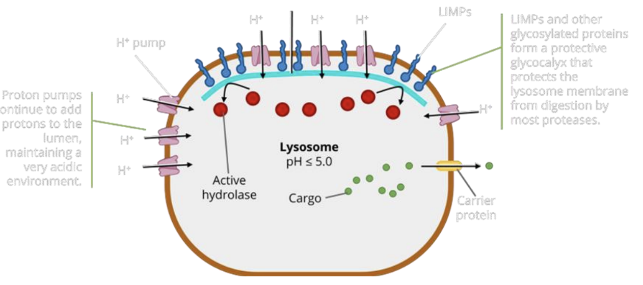

<ol start="4"><li><p>the lysosome</p></li></ol><ul><li><p>when pH becomes more acidic (pH&lt;5), enzymes like proteases are activated</p></li><li><p>converts the late endosome to lysosome</p></li><li><p>a protective glycocalyx layer of LIMPs and glycosylated proteins forms to prevent lysosome from degrading itself</p></li></ul><p></p>



