general terms for regions and cranial skeleton (coyote)
 Call Kai
 Learn
Learn Practice Test
 Spaced Repetition
 Match
Match Flashcards
 Knowt Play
1/84

There's no tags or description
Looks like no tags are added yet.
Name | Mastery | Learn | Test | Matching | Spaced | Call with Kai | Chat |
|---|
No analytics yet
Send a link to your students to track their progress
85 Terms
cranium
the skull; comprises braincase and rostrum
anterior/rostrum
front part of skull
posterior/occipital
back of skull
dorsal
view of the top
ventral
underside
lateral
from the side
fossa
wide depression
canal
bony tunnel
foramen
large hole in the bone
condyle
rounded elevation
process
localized projection of bone
suture
rigid joints
temporal opening
lateral opening of the skull immediately posterior to the orbit; evolutionarily derived from the temporal fenestra of synapsids
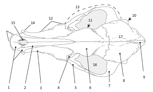
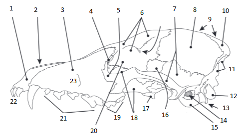
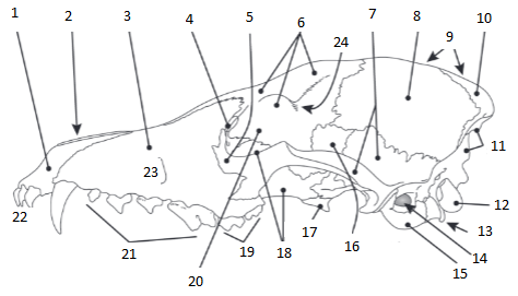
1
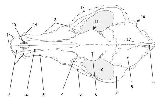
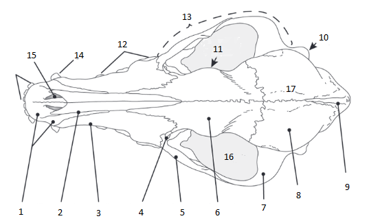
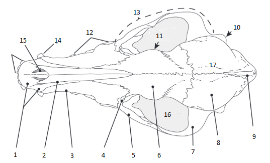
premaxilla: paired bones forming the lower margin of the outer nasal openings and the anteriormost portion of the palate; holds incisor teeth
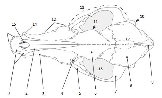
2
nasal: paired bones forming the anteriormost roof of the nasal cavities
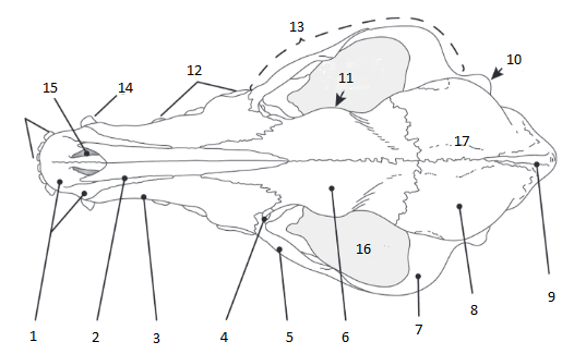
3
maxilla: paired bones that bear all upper teeth except the incisors
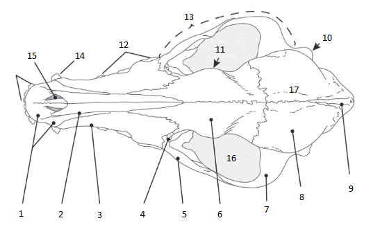
4
lacrimal: paired bones located on or adjacent to the anteriodorsal base of the zygomatic arch; perforated by the lacrimal canal (or tear duct)

5
jugal: paired bones that form the central portion of the zygomatic arch
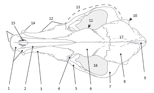
6
frontal: paired bones located posterior to the nasals and dorsal to maxillae; support antlers and horns in taxa with these structures

7
squamosal: paired bones located lateral and ventral to the parietals; part of the zygomatic arch

8
parietal: paired bones located posterior to the frontals, forming the majority of the roof of the braincase; the suture that joins the frontal and parietal bones
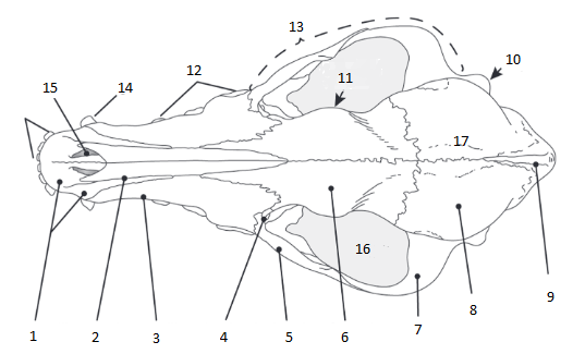
9
interparietal: often indistinct or obliterated as it is a diagnostic trait; when visible, located at the posterior border of the parietals

10
mastoid process: ventrally projecting small process of the mastoid bone
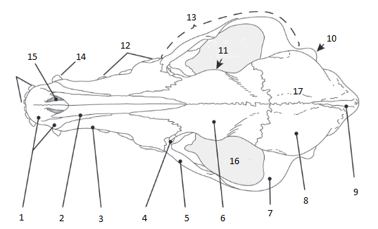
11
postorbital process (forms postorbital bar in some species when process connect across orbit, such as in slow loris skull)
postorbital plate
separates the orbits from the temporal fossa and opening; all higher primates have one
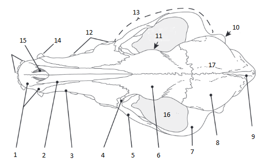
12
upper premolar

13
zygomatic arch: the conspicuous arch of bone extending laterally on either side of the cranium; contains maxilla, jugal, and squamosal bones
14
upper canine

15
incisive foramen: two little holes on premaxilla
16
orbit: socket-like depressions in which the eyes are housed; the eye sockets
17
sagittal crest: a ridge of bone located along the midline of the posteriordorsal aspect of the skull; serves for additional (temporal) muscle attachment and is therefore most prominent in taxa requiring large temporal muscles
occipital (lamboidal) crest
important place for attachment of neck muscles
occipital bone
posterior wall of the braincase; formed by the fusion of the supraoccipital, basiooccipital, and two exoccipitals (does not include auditory bullae)
1
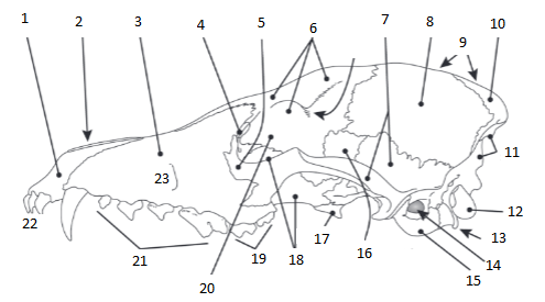
premaxilla

2
nasal
3
maxilla
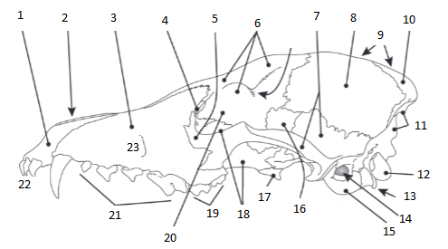
4
lacrimal

5
jugal

6
frontal
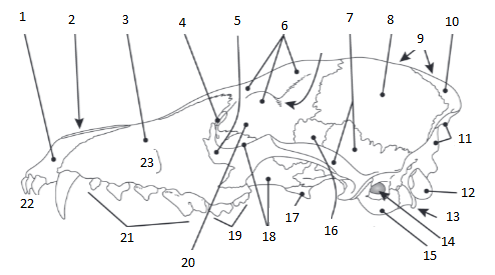
7
squamosal
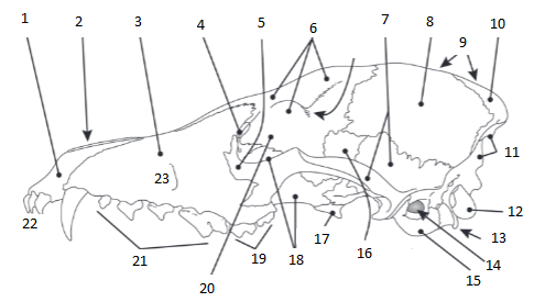
8
parietal
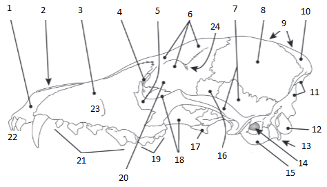
9
sagittal crest

10
interparietal
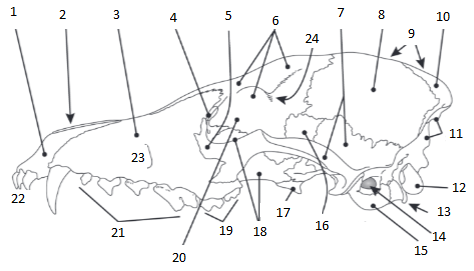
11
supraoccipital: above foramen magnum (“supra” means above)
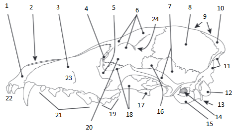
12
occipital condyle (on exoccipital bones): articulation points for the skull, setting into depressions of the atlas (the first vertebra)
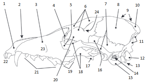
13
paraoccipital process: serves for insertion of certain chewing muscles and for muscles that open the jaw
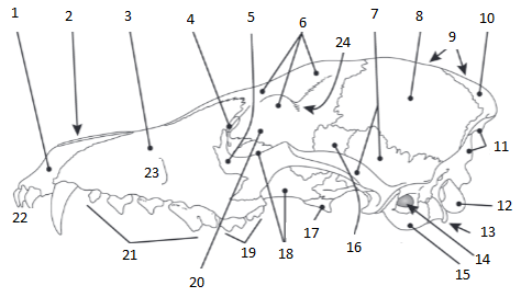
14
external auditory meatus
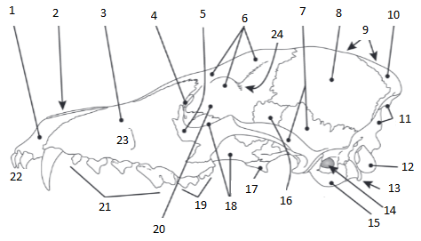
15
auditory bulla: encloses parts of the middle and inner ear; bulges
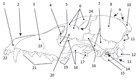
16
alisphenoid

17
pterygoid: pair of wing-like bones
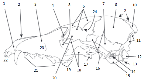
18
palatine: paired bones forming the posterior portion of the palate
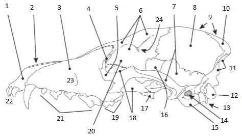
19
molars 1-2
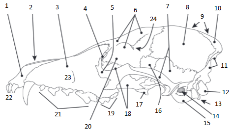
20
orbitosphenoid
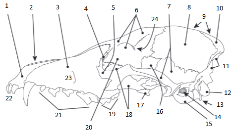
21
premolars 1-4

22
incisors 1-3
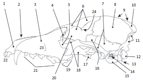
23
infraorbital process
24
postorbital process
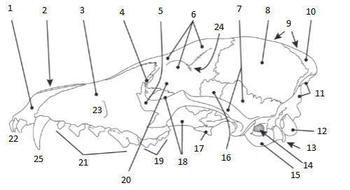
25
canine

1
premaxilla (ventral)
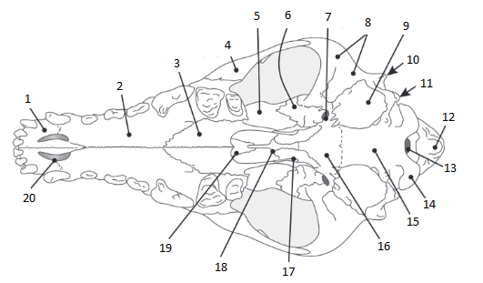
2
maxilla (ventral)
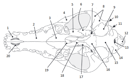
3 & 5
palatine (ventral): paired bones forming the posterior of the palate

4
jugal (ventral)
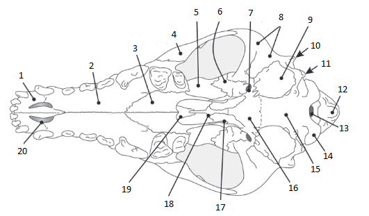
6
alisphenoid (ventral): perforated by alisphenoid canal; wing-like

7
foramen ovale (ventral) (a canal)
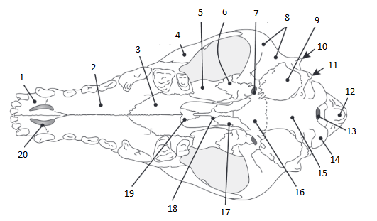
8
squamosal (ventral)

9
auditory bulla (ventral): encloses parts of the middle and inner ear; bulges

10
mastoid process (ventral): ventrally projecting small process of the mastoid bone

11
postorbital process (ventral)
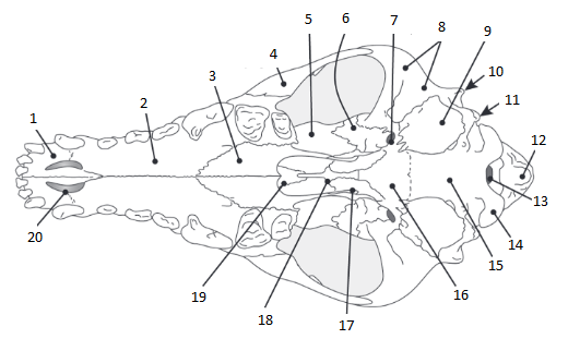
12
supraoccipital (ventral)
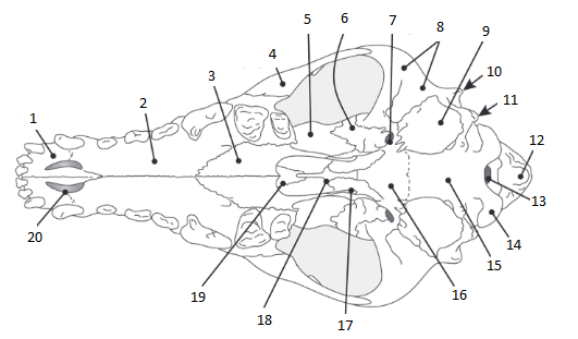
13
foramen magnum (ventral); where the spinal cord attatches

14
occipital condyle (ventral): articulation points for the skull, setting into depressions of the atlas (the first vertebra)
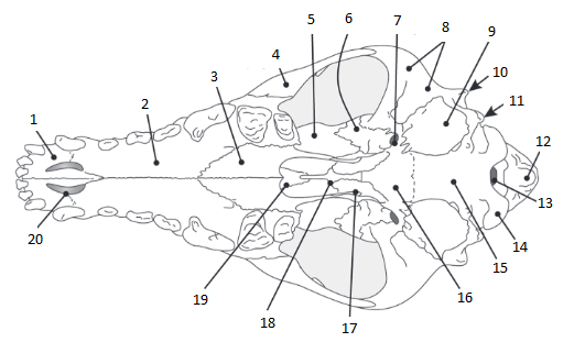
15
basioccipital (ventral); where occipital bone ends
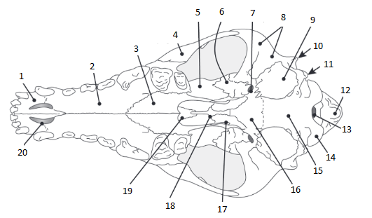
16
basiosphenoid (ventral)
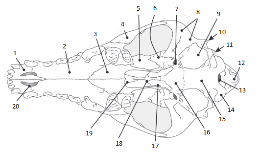
17
pterygoid (ventral): pair of wing-like bones
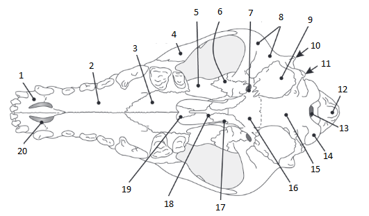
18
presphenoid (ventral): long little bone

19
vomer (ventral): separates the two sides of the nasal passages
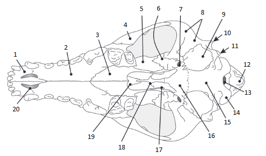
20
incisive foramen (ventral)
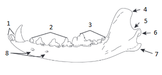
1
incisors 1-3
2
premolars 1-4
3
molars 1-3
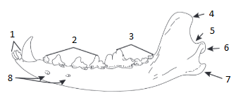
4
coronoid process; inserts into the temporal fossa; serves for attachment for portions of the temporal muscle
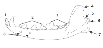
5
sigmoid notch
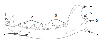
6
condyloid process: bears the mandibular condyle, which articulates with the cranium in the mandibular fossa
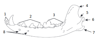
7
angular process: may be enlarged for insertion of powerful masseter and other jaw musculature
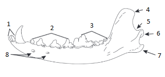
8
mental foraminae/canal: on mandible