Antibody Based Immunotherapy
 Call Kai
 Learn
 Practice Test
 Spaced Repetition
 Match
 Flashcards
 Knowt Play
1/23

There's no tags or description
Looks like no tags are added yet.
24 Terms
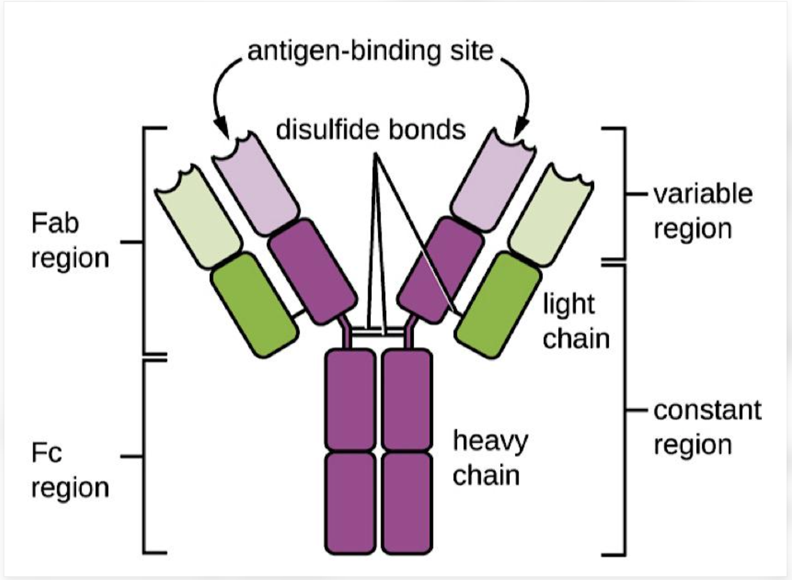
Draw an IgG based antibody. explain each part functions. What are the 2 main roles of the antibody
Two heavy chains and two light chains: form the antibody.
Variable regions: at the tips of the Y; determine what antigen the antibody recognises.
Fab regions: the two arms that bind the antigen.
Fc region: the stem, which interacts with immune cells and other immune-system components.
Hinge region: provides flexibility.
An antibody has two broadly different jobs:
Recognise a particular target using its Fab regions.
Produce biological effects through its Fc region.
How do you make a monoclonal antibody?/ Explain Hybridoma technique. Why wont this classical hybridoma technique work?
Step 1: Obtain antibody-producing B cells.
A mouse is immunised with an antigen, stimulating B cells to produce antibodies against it.
Step 2: Fuse B cells with myeloma cells.
B cells produce antibodies but have limited lifespans. Myeloma cells can proliferate indefinitely.
Step 3: Select and clone hybridomas.
The resulting hybrid cells combine antibody production with sustained growth. Select the cells producing the desired antibody and clone them.
The traditional method produces mouse antibodies. Giving these to humans can cause anti-drug immune response, potentially reducing efficacy and causing adverse reactions.
3 strategies for Production of human antibodies
Phage display: selects an antibody based on its ability to bind a target.
Humanization: modifies an existing non-human antibody to make it more human-like (Preervind CDR from mice while the antibody structure is from human)
HuMab mice: use genetic engineering to generate antibodies with human immunoglobulin sequences.
Where does an antibody bind?
complementarity-determining regions (CDRs): make the most important contacts with the antigen and determine much of the binding specificity.
Framework regions: provide structural support for the CDR domains.
4 types of therapeutic antibody

In caner cells, what are the 3 broad areas the antibodies could target
Tumour-associated surface antigens, such as CD20.
Growth-promoting or environmental targets, such as EGFR and VEGF.
Immune checkpoints, such as PD-1 and PD-L1.
What is anti-idiotypic antibody. How is it significant in tummour immunology
The immune system can produce a second antibody that binds to the unique binding region of the original antibody. This second antibody is called an anti-idiotype antibody.
Anti-idiotype antibodies can sometimes mimic the shape of the original antigen. Because of this, researchers can investigate their use as vaccines to stimulate an immune response against a tumour-associated antigen. (However it doesnt happen with every antigen - disadvantage- needs to be tailored for every tumour)
Three mechanisms of monoclonal antibody action
I. Direct effects
The antibody blocks a growth signal or receptor, which can inhibit proliferation or promote cell death.
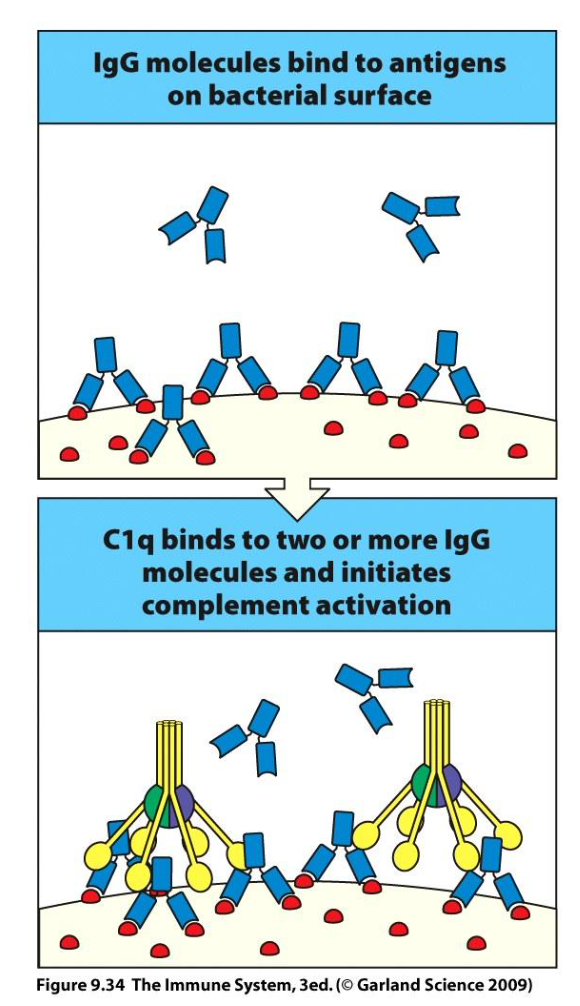
II. Complement-dependent cytotoxicity (CDC)
Antibodies bound to the target recruit complement proteins. Complement activation can damage and lyse the target cell.
III. Fc-receptor-mediated effector functions
Immune cells recognise the antibody's Fc region and attack, engulf or otherwise damage the antibody-coated target.

Explain this graph
The combination leaves a smaller fraction of surviving cells than radiation alone.
Some antibodies can make tumour cells more vulnerable to other treatments, including radiation or chemotherapy.
The antibody is not necessarily replacing the other treatment; it can enhance its effect.
Why molecular biomarkers, including RAS mutation status, are important when selecting patients for anti-EGFR therapy
Make sure there is no downstream mutation
How do you improve efficacy for antibodies based tumour therapy

Antibody–drug conjugates (ADCs) components and how it works
An ADC consists of three main components:
Antibody: recognises the target antigen.
Linker: connects the antibody to the drug.
Payload: a potent cytotoxic compound that damages the cancer cell.
The usual sequence is:
The antibody binds its target.
The antibody–target complex is internalised by the cell.
The payload is released, depending on the ADC design.
The payload damages the cell.
Complement dependent cytotoxicity. how does it work
Fc-receptor-mediated effector functions - 3 types
NK cells: can kill antibody-coated cells through ADCC. (APOPTOSIS)
Macrophages: can engulf antibody-coated cells through antibody-dependent cellular phagocytosis (ADCP). (PHAGOCYTOSIS)
Neutrophils: can kill antibody-coated cells through Fc-receptor-mediated mechanisms. (TROGOCYTOSIS)
How are fc receptors involved in relation to B Cell lymphoma
It leands to antibody dependent cellular cytotoxicity

The balance of activating and inhibitory signals determines whether an NK cell kills its target. Explain / 3 inhibitory signals for nk
Tolerance: inhibitory signals dominate, so the NK cell does not kill a healthy cell.
Missing self: reduced expression of MHC class I can remove inhibitory signalling and favour NK-cell activation.
Stress-induced signals: increased activating ligands on a stressed or tumour cell can favour killing.
How do Macrophages act as effector cells
Macrophages can recognise antibody-coated tumour cells through Fc receptors and engulf them.
The key mechanism is ADCP: antibody-dependent cellular phagocytosis.
Fc mediated neutrophil killing function and advantage
They can mediate effective antibody-dependent killing.
They can be mobilised using G-CSF.
They can release chemotactic signals that recruit other immune cells.
Trogocytosis: ?
Trogoptosis: ?
Where is it seen
Trogocytosis: one cell removes pieces of another cell's membrane.
Trogoptosis: target-cell death associated with trogocytosis.
Neutrophil based killing
IgA–FcαRI interactions can amplify neutrophilic inflammation and worsen tissue damage. True or false
True
What is the disadvantage associated with Iga mediated neutrophil killing
Short Half life of IGA
How does recruiting t cells work with antibodies
Bispecific antibodies have two different antigen-binding sites (Fab regions): one binds the tumour antigen, and the other binds CD3 on the T cell, bringing them together.
BsAb formats can be used to target different effector cells. Give an example
T cells.
One binding region recognises a tumour-associated antigen, while another binds a molecule on the T cell, often CD3.
The antibody acts as a physical bridge, bringing the T cell close enough to activate tumour-cell killing.
Checkpoint inhibition on myeloid cells (e.g. CD47-SIRPalpha interaction) enhances tumor cell killing. How does this work
CD47 is a protein on the surface of many cells, including cancer cells.
SIRPα is a receptor found on myeloid cells, especially macrophages.
When CD47 binds to SIRPα, it sends an inhibitory signal to the macrophage.
The message is essentially: “Don't eat me!”
This prevents the macrophage from engulfing the cell.
In cancer, high expression of CD47 happens. Checkpoint inhibition on myeloid cells helps in phagocytosis by macrophages.