All things HEART
 Call Kai
 Learn
Learn Practice Test
 Spaced Repetition
 Match
Match Flashcards
 Knowt Play
1/32

There's no tags or description
Looks like no tags are added yet.
Name | Mastery | Learn | Test | Matching | Spaced | Call with Kai | Chat |
|---|
No analytics yet
Send a link to your students to track their progress
33 Terms
What day does the heart starts to beat? | What happens after heart looping?
Day 22 | Septum begins to form

True or False: By 8 weeks after fertilization, the heart has completed its major morphogenetic development, but it continues to mature, grow, undergo metabolic changes, and adapt to changes in blood flow after birth.
True
What are the 13 process of the the Embryonic Heart Development ?
1. Heart fields form
The heart begins from two symmetrical heart fields derived from the lateral plate mesoderm.
These cells migrate through the primitive streak and settle between the ectoderm and endoderm.
2. Heart tube forms
During body folding, the two heart fields fuse together to form a primitive heart tube.
The tube has three main layers:
Endocardium → inner layer
Extracellular matrix → middle layer
Myocardium → outer muscular layer
3. The heart tube begins to beat and develop chambers
The primitive heart tube begins rhythmic contractions.
Different parts grow at different rates, producing:
Dilations → future heart chambers
Constrictions → future partitions/septa
4. Second heart field adds structures
The second heart field adds cells to the ends of the heart tube.
These contribute to the:
Outflow tract
Sinus venosus
5. Heart tube loops
The heart tube undergoes rightward (dextral) looping.
This rearranges the positions of the future venous and arterial portions.
Septation of the heart begins around this stage.
🫀 Septation: Dividing the Heart into Chambers6. Atrial septation
The primitive atrium is divided into right and left atria:
Septum primum grows downward and initially separates the atria.
Openings develop in the septum primum and combine to form the foramen secundum.
A second septum, the septum secundum, develops to the right of the septum primum.
Together, they create the foramen ovale.
7. Foramen ovale allows fetal blood flow
In the fetus, the foramen ovale acts as a one-way passage for blood from the right atrium → left atrium.
This allows fetal blood to bypass the non-functioning lungs.
Shortly after birth, it closes permanently, completing functional atrial septation.
💪 Ventricular Septation8. Ventricular septum develops
Ventricular septation occurs after/alongside atrial septation and is not complete when the primary atrial septum forms.
The ventricular septum develops through:
Growth and remodeling of trabecular sheets
Expansion of the ventricular chambers
Fusion of several tissues, including the endocardial cushions and muscular septum
9. Final interventricular septum
These processes produce the:
Muscular interventricular septum
Membranous interventricular septum
Together, they separate the right and left ventricles.
🩸 Outflow Tract Development10. Outflow tract divides
The outflow tract develops spiraling ridges.
These ridges grow and fuse together.
This divides the primitive truncus into two major pathways:
Aorta
Pulmonary tract
This ensures that blood leaving the ventricles is directed into the correct vessels.
🩸 Development of Blood Vessels11. Vessels remodel
The embryonic venous and arterial vessels undergo major remodeling through:
Incorporation into the developing heart
Degeneration of some vessels
Fusion of vessels
Growth of other vessels
The aortic arches are particularly important in this remodeling and contribute to the mature arterial circulation.
❤ Epicardium12. Epicardium develops
The epicardium develops from tissue near the atrioventricular (AV) junction.
It spreads over the outside of the myocardium.
It becomes a layer of squamous epithelial cells and connective tissue.
13. Epicardium helps form coronary structures
The epicardium is important for the development of:
Coronary blood vessels
Cardiac fibroblasts
Q: Why is understanding the embryonic origin of heart cells important for understanding congenital heart disease?
A: Because where a heart cell originates determines what structure it will eventually form. If a group of cells has abnormal migration, development, or signaling, the structures derived from those cells can develop abnormally, producing specific congenital heart defects.
How do precardiac cells become the heart fields?
A: During gastrulation, precardiac cells from the epiblast migrate through the primitive streak and then move laterally. They form the left and right heart fields, which organize into the first and second heart fields.
What is the difference between the first and second heart fields?
A: They contribute to different parts of the heart:
First heart field (FHF) → primarily forms the primitive heart tube, left ventricle, and atria.
Second heart field (SHF) → adds cells to the developing heart tube and contributes to the right ventricle, outflow tract, valves, and parts of the atria.
Key idea: The heart is built by multiple cell populations, not from one uniform group of cells.
Why must cells from the second heart field migrate to the primitive heart tube?
The primitive heart tube needs additional cells to grow and expand into the mature four-chambered heart. Second-heart-field cells migrate to the developing tube and contribute structures such as the right ventricle and outflow tract.
How can abnormal cell migration lead to congenital heart defects?
A: If cells fail to migrate to the correct location, the structures they are supposed to form may be missing, malformed, or incorrectly connected. Therefore:
Abnormal migration → abnormal development → congenital heart defect
Why can a single developmental problem cause multiple congenital heart defects?
A: Because one embryonic cell population can contribute to multiple structures. If something disrupts that population early in development, several structures derived from it may be affected at the same time.
This is why understanding cell lineage helps explain why certain heart malformations occur together
What is the role of cardiac neural crest cells in heart development?
A: Cardiac neural crest cells interact with the second heart field and are particularly important for development of the outflow tract and great vessels. Abnormalities affecting these cells can therefore contribute to congenital heart defects and are associated with certain genetic syndromes

What differentiated the myocardium from the endoocardium?
The myocardium secretes a thick layer of acellular extracellular matrix, also known as cardiac jelly, which creates a distinct layer between the myocardium and the endocardium
What are the three steps the heart has to go through to the normal cardiac septation?
Looping
Convergence
Wedging
What is looping and what is the right way for the heart to loop and the wrong way?
Looping is the first obvious structural left-right lateralization in the developing embryo
The heart tube elongates and folds into an “S” shape to the right. The direction of looping will determine the ultimate ventricular positions.
Looping to the right will give the normal dextro, or “D,” looping.
If the primitive tube loops to the left, this will result in levo, or “L,” looping, with the left ventricle on the right side and the right ventricle on the left side
What are some rare genetics condition known to have dextrocardia ?
Kartagener syndrome
Kartagener syndrome is a rare genetic disorder characterized by the classic triad of situs inversus (mirror-image placement of internal organs), chronic sinusitis, and bronchiectasisHeterotaxy syndrome is a rare congenital condition where internal organs in the chest and abdomen are abnormally arranged, misplaced, or duplicated across the left-right axis of the body.
What about the second step to normal heart septation? what is it and what does abnormality in this process called?
Convergence is the process of proper orientation of the inflow and outflow tracts
Ideally, the inflow tract rises cranially behind the outflow tract such that the inflow and outflow tracts are aligned, with the outflow tract ventral to the inflow tract.
Abnormalities can lead to irregularities in atrial and ventricular septa and outflow tracts.
Convergence and looping are dependent on lengthening of the polar ends of the heart tube, which is achieved by the addition of cells from the second heart field.
Since cardiac neural crest cells influences the second heart field, any neural crest cell–associated diseases such as 22q11 deletion syndromes and velocardiofacial syndromes will occur as well as abnormalities of these cardiovascular structure (which makes sense of why most Trisomies babies have some heart conditions?)
Elaborate on wedging. What is it and what are the abnormalities if it doesn’t occur?
Wedging
It is when the outflow tract rotates counterclockwise approximately 45°so the future aorta could sit behind the future pulmonary artery
Basically the aortic portion shortens, and the future site of the aortic valve “wedges” between the future tricuspid and mitral valves.
This separates the mitral valve from the septum, which is why the mitral valve has no connections to the septal wall.
If convergence and wedging do not occur correctly, then the outflow tract, the ventricular septum, and the AV endocardial cushion tissue do not meet. This results in conotruncal defects such as double inlet left ventricle, double outlet right ventricle, and tetralogy of Fallot.
Once the heart has gone through the three steps of cardiac septation (looping, converging and wedging) what happens next?
Once the primitive heart has done the above, then Septation of the heart continues by the swelling of the extracellular matrix between the endocardium and the myocardium at specific regions of the heart tube.
Basically the existence / positioning of the
- AV junction
- The outflow tract
- The primary atrial septum
- The ridge of the interventricular septum
- Valves
As chamber differentiation proceeds, endocardial cushions grow into the lumen of the tubular heart and act as primitive valves to promote unidirectional blood flow.
What are some neural crest cells Contributions to Cardiogenesis?
The neural crest cells from the neural tube migrate through the third, fourth, and sixth aortic arches to contribute to the walls of the aortic arch arteries, including
The proximal aorta
The stems of the proximal coronary arteries
The sympathetic and parasympathetic innervation of the heart (including the cardiac ganglia).
A subset enters the heart within the endocardium and is involved in septation of the aortic and pulmonary trunk.
Another subset also appears to enter the heart and regulates the formation and maturation of the cardiac conduction system.
What would disruption in neural crest cells cause?
Removal or disruption of neural crest cells causes cardiac inflow anomalies
1. Double inlet left ventricle
Tricuspid atresia
Straddling tricuspid valve
Cardiac outflow anomalies (persistent truncus arteriosus)
Abnormalities in the aortic arch arteries (interruption of the aorta, double aortic arch, variable absence of the carotid arteries, and left aortic arch)
22q11 deletion and related syndromes that include craniofacial and outflow tract abnormalities
22q11 deletion syndromes are associated with abnormalities in cardiac neural crest cells and the second heart field - can you elaborate of some of these defects are?
Mostly conotruncal defects such as
Tetralogy of Fallot
Pulmonary atresia with ventricular septal defect
Truncus arteriosus
Interrupted aortic arch
Isolated aortic arch anomalies
Ventricular septal defects.
What is the role of the epicardium in the cardiogenesis?
The epicardium, the outer layer of the heart, forms by the growth of epithelial tissue from the dorsal mesothelium of the sinus venosus region. This single-cell layer spreads in a stereotyped pattern over the myocardium.
It provides the bulk of the non-cardiomyocyte cellular components, including smooth muscle cells, fibroblasts, and endothelial cells
The cells within the epicardium invade the myocardium and the endocardium, giving rise to valve cells, fibroblasts, smooth muscle cells, and other components of the coronary vasculature.
What are some genetics conditions that afttect the heart development and how?
T21
- (Down syndrome) is the presence of three copies of chromosome 21 or a translocation of an extra piece of chromosome 21
- CHDs detected in T21 infants include AV septal defect (a defect of endocardial cushions), ventricular septal defect, persistent ductus arteriosus, and tetralogy of Fallot.
- Likely to have persistent pulmonary hypertension, both with and without CHD.
T18
- Edwards syndrome, is characterized by slow prenatal growth resulting in low birth weight, small craniofacial features, anomalies of the extremities, and CHDs.
- CHDs seen in T18 include ventricular septal defect, persistent/patent ductus arteriosus, pulmonary stenosis, coarctation of the aorta, and abnormal heart valves.
T13
- Patau syndrome, is characterized by prenatal death or death soon after birth, slow growth and low birth weight, craniofacial anomalies, holoprosencephaly, exophthalmos, or omphalocele
- Ventricular septal defects are the most common.
22q11 deletions.
- (DiGeorge/velocardiofacial syndrome) is considered a neurocristopathy, or a disease caused by disturbances in the generation, migration, or differentiation of neural crest cells.
- It is also categorized as a RASopathy because it results in abnormalities in signaling of the RAS pathway.
Most of these patients have a microdeletion of the region 22q11.2 that contains many genes (30–40)
- A large proportion of individuals with this syndrome (75%) have CHDs that include conotruncal defects such as truncus arteriosus, interrupted aortic arch B, and tetralogy of Fallot.
What are the categories for congenital heart defects


How would it be decided for level 2 vs 3 delivery of CHD?


How would you know if tetrallogy of fallot is prostin dependent in utero?
There are markers on fetal echocardiograms that can help predict whether a fetus with tetralogy of Fallot will require PGE or an intervention in the neonatal period. Morphologic markers including the pulmonary valve (PV) annulus size, the PV to aortic valve ratio, and the main pulmonary artery to PV ratio have been shown to predict both ductal dependence and need for neonatal procedures.
In TGA in utero, what would be predictive of the need for urgent atrial septostomy?
In fetal life, the size of the foramen ovale versus the septal length, a hypermobile primum atrial septum, and reversal of flow in the ductus arteriosus have been shown to be predictive of the need for urgent postnatal balloon atrial septostomy to ensure adequate atrial level communication.
What about in the case of HLHS? How would we know the need for immediate intervention in utero?
Urgent decompression of the left atrium after birth can improve survival. Fetal assessment of the pulmonary venous inflow and the vasoreactivity of the pulmonary vascular bed
Responsiveness of the fetal pulmonary vasculature to maternal hyperoxygenation (exposure of the mother to 60% inspired oxygen for 10 minutes) can also help identify fetuses that will require urgent postnatal intervention.
What is hydrops fetalis?
It’s the result of fetal heart failure is associated with placental edema (increased right heart preload) and resultant hypoxia. The response of the mature heart to hypoxia is to increase the heart rate, but the fetus decreases its heart rate in response to hypoxia.
Expand on placental edema and it’s connection with Hydrops fetalis
This can be seen in any condition that volume loads the right ventricle, including large AV malformations, sacrococcygeal teratomas, and TTTS. 7 There is a resultant increase in the combined cardiac output (CCO). The CCO increase reflects the amount of preload increase. Most infants do well without evidence of hydrops until the CCO reaches 700 to 800 mL/kg/min.

What are some Differential Diagnosis of Causes of Hydrops Fetalis?
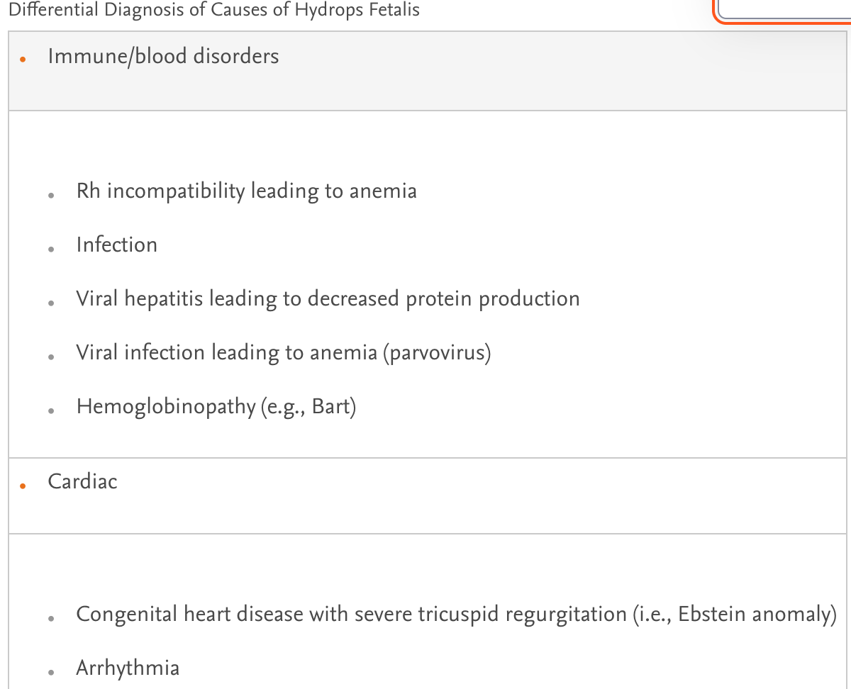
What is the pathophysiology pattern of poetential cause of increased preload and connection to hydrops fetalis?
The donor twin often exhibits volume depletion and nephrosclerosis. This results in elevated ET-1 and angiotensin II. Doppler of the umbilical artery shows decreased diastolic flow and increased resistance.
The increased resistance rarely results in ventricular dysfunction; however, postnatal studies have shown abnormalities in vascular compliance and increased pulse wave velocities in the donor twin.
Donor hormones from the response to hypovolemia are transferred across the placenta to the recipient twin.
The recipient is thus exposed to increased volume and vasoconstrictors. Angiotensin II is a potent stimulant for hypertrophy.
Thus the recipient twin often presents with cardiomyopathy—dilated, hypertrophic, or a combination of the two. Many fetuses demonstrate marked ventricular hypertrophy, which may result in right ventricular outflow tract obstruction, pulmonary stenosis, and tricuspid valve regurgitation.
What are some fetal echocardiographic prognostic indicators that can predict hydrops fetalis outcomes? - what are these indicators called? What do we do about these findings?
Cardiac function
Cardiac size
Systemic venous Doppler profile of the ductus venosus or umbilical vein
Presence of AV valve regurgitation
Presence of pericardial effusions, pleural effusions, ascites, or skin edema.
These indicators can be compiled to obtain a cardiovascular profile scoreTreatment should be directed toward underlying pathology, including
- resolving abnormal peripheral impedance
- treating high output failure, myocardial dysfunction, arrhythmia, anemia, or twin-twin transfusion syndrome.