Bioc 307 - Exam 1
 Call Kai
 Learn
Learn Practice Test
 Spaced Repetition
 Match
Match Flashcards
 Knowt Play
1/112

There's no tags or description
Looks like no tags are added yet.
Name | Mastery | Learn | Test | Matching | Spaced | Call with Kai | Chat |
|---|
No analytics yet
Send a link to your students to track their progress
113 Terms
four types of noncovalent interactions
ionic interactions
hydrogen bonds
van der Waals forces
hydrophobic interactions
ionic interaction (charge-charge interaction)
(aka salt bridges) electrostatic attraction/repulsion between charged groups
effect of distance between two charged groups on energy of their interaction
energy most optimal when two atoms approach one another within 4-5 A

relative dielectric constants for nonpolar vs polar solvents
nonpolar = 1-2
polar = 50+
effect of solvent polarity on ionic interactions
more polar → greater interference/dielectric constant → weakens ionic interaction
what type of solvent is water?
super polar
solvent that makes ionic interaction stronger
lower dielectric constant - vacuum, hexanes
H bond donor vs acceptor
H bond donor = the molecule that supplies H
H bond acceptor = the electronegative atom with a lone pair that attracts H
groups that serve as H bond donors
H bonded to an EN atom
groups that serve as H bond acceptors
EN atom w/ lone pair - O, N, F
importance of van der Waals interactions in biochem
weak + nonspecific individually but sum of many can be strong + important for shape
hydrophobic effect
association of nonpolar groups w/ each other in aqueous systems - want to minimize their interaction w/ water
amphipathic
compound w/ both nonpolar and polar aspects - ex: phospholipids
properties of water
polar, bent structure, can H-bond with tetrahedral network
polarity determined by….
difference in EN > 0.4 between two atoms sharing covalent bond
asymmetric dipole arrangement
coulomb’s law
E = k * [ (Q1 * Q2) / Dr)
clathrate structures
water caged around nonpolar solute - low entropy → unfavorable

water when surrounding nonpolar solutes
highly ordered and low entropy → nonpolar solutes form cluster to increase entropy
basic α-amino acid structure
central C bonded to carboxyl, amino, H, and R group - L-isomer
α-COOH group ionization
weak acid (pKa 2) → donates proton
α-amino group ionization
weak base (pKa 9) → accepts proton
pH
acidity of a sol’n - lower pH → incr [H]
pKa
how tightly molecule holds onto proton - lower pKa → gives up H+
when pH = pKa…
half of the molecules are protonated and half deprotonated
pH < pKa
sol’n has higher [H+] → molecule keeps proton
pH > pKa
sol’n has lower [H+] → molecule kicks out proton
zwitterion
neutral molecule where the charges cancel out to zero
isoelectric point (pI)
pH where zwitterionic form is most likely to be found
pI equation
(pKa 1 + pKa 2) / 2
nonpolar amino acids
ala, val, leu, ile, met, pro, phe, trp
polar, uncharged amino acids
ser, thr, asn, gln, tyr, cys
positively charged amino acids
lys, arg, his
negatively charged amino acids
asp, glu
glycine (g)
achiral, small, flexible, nonpolar, alpha C

alanine (ala)
small and boring, R group = methyl, nonpolar

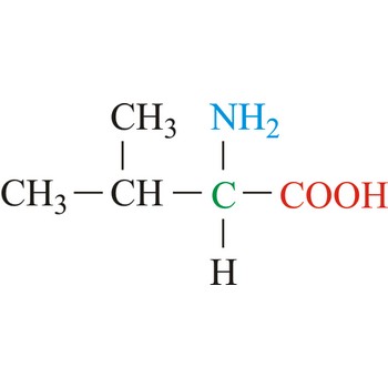
valine (v)
bulky, hydrophobic, nonpolar
leucine (l)
more bulky/hydrophobic, isobutyl

isoleucine (i)
bulky, hydrophobic, side chain = chiral

methionine (m)
nonpolar, start codon, contains S

proline (p)
nonpolar, connects back to amino group → rigid

phenylalanine (f)
nonpolar, aromatic, hydrophobic - phenyl ring connected to B C

tryptophan (w)
nonpolar, indole group that H bonds

serine (s)
OH group - polar, can H bond and be phosphorylated

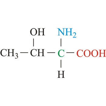
threonine (t)
polar w/ secondary alcohol
asparagine (n)
polar w/ amide group - not titratable

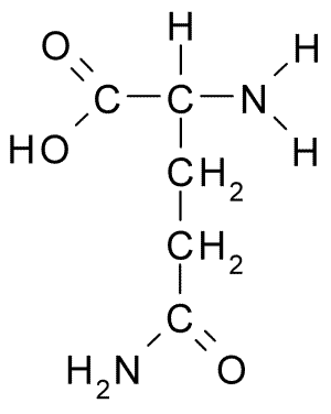
glutamine (q)
polar w/ amide group - not titratable
tyrosine (y)
polar phenol group - can H bond, titratable when basic (pKa = 10)

cysteine (c)
thiol group - can’t H bond but titratable to S- (pKa = 8)

lysine (k)
polar, contains amino group (pKa = 11)

arginine (r)
contains guanidino group (pKa = 12) - always polar + positive

histidine (h)
contains polar imidazole group (pKq = 6) - proton pulled off if pH > 7 → neutral

aspartic acid (d)
beta carboxylate group (pKa = 4) - always polar, negatively charged
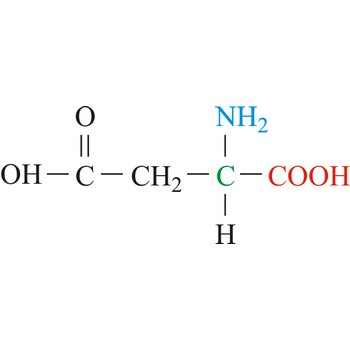
glutamic acid (e)
gamma carboxylate group (pKa = 4) - always polar, negatively charged

hydropathy of amino acids
if it moves to bilayer → hydrophobic + favorable (-ΔG) - determines protein folding
kinase
enzymes that add a phosphate - specific ones for Ser/Thr and Tyr
phosphatase
enzymes that remove a phosphate
effect of kinases
adds big negative charge → amino acid becomes polar → changes shape + function
disulfide bonds
strong covalent bonds between two cysteines - can only occur in oxidizing environments
if pH = 7 & pI < 7
protein has more acidic residues → overall negative charge
examples of protein secondary structure
alpha-helices and beta-sheets
α-helix structure
secondary structure stabilized by H-bonds in backbone four residues apart
H bond donor/acceptor in α-helix
backbone carbonyl = acceptor
amide’s hydrogen = donor
chirality of α-helix
typically right-handed - (-60, -50)
α-helix # of residues per turn
3.6 residues/turn
orientation of R groups relative to axis of α-helix
R groups facing outside - determines protein’s properties
packing density of atoms in α-helix
distance for 1 full turn pitch = 5.4 A/turn
distance per residue rise = 1.5 A/residue
β-conformation structure
pleated zig-zag shape
H bond donors/acceptors in β-sheets
orientation of R groups in β-pleated sheet
alternatively above and below plane of strand

parallel β conformation
sheets have same directionality - weaker b/c H bond geometry less than ideal

antiparallel β conformation
sheets run in opposite directions - more stable/linear structure + more common
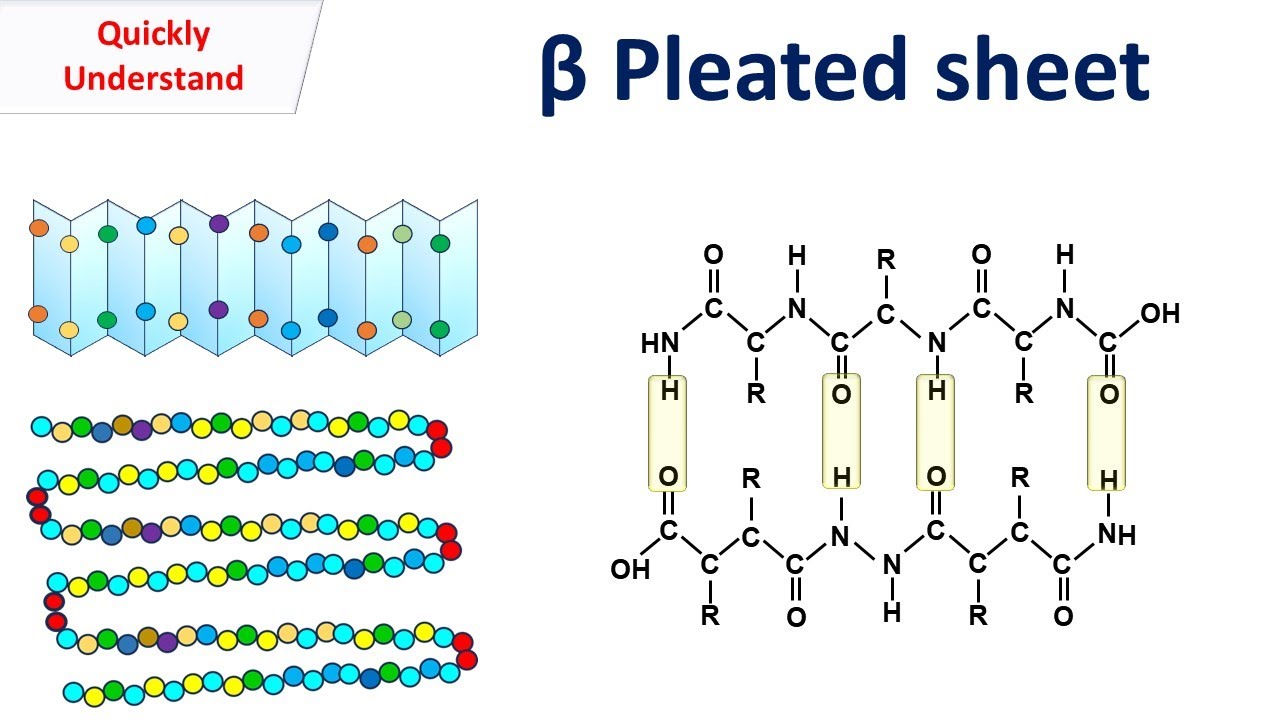
non-covalent interaction stabilizing a-helix and β conformations
hydrogen bonds
tertiary protein structure
overall 3-d conformation of whole polypeptide chain in its folded state
structural domain of protein
structurally independently folded region of a protein - have different functions

6-atom planar peptide group
resonance structure of peptide bond restricts rotation of atoms around omega bond

phi vs psi angles
phi: counter-clockwise between N and α-C
psi: clockwise between α-C and carboxyl-C
Ramachandran plots
shows possible phi and psi values for protein conformations
location of phi angles in Ramachandran plot
fourth quadrant: (-120, +120)
location of psi angles in Ramachandran plot
3rd quadrant (-100, -60) for right handed α-helix and 1st quadrant (60, 60) for left-handed α-helix
conformation
spatial arrangement of atoms/groups that can change by bond rotation with no covalent bond breaking
configuration
spatial arrangement of atoms/groups that cannot change without breaking covalent bonds
peptide bond
amide bond - condensation of acid and amine (catalyzed by ribosome + not spontaneous)
amino acids not found in α-helices
glycine: more energetically stable to be flexible than in ordered conformation
proline: rigid ring + has no proton on N to be H bond acceptor
protein motif
(aka super-secondary structures) smaller, recurring patterns of secondary structures - cannot usually fold on their own, building blocks of protein folding

amino acids most likely buried inside proteins
cysteine, isoleucine, tryptophan, phenylalanine
amino acids most likely facing outside of protein
aspartic acid, glutamic acid, lysine, glycine
protein folding
spontaneously arriving at the correct combo of phi and psi angles for every residue in the polypeptide sequence - becomes functional protein
quaternary structure
multiple independent polypeptide chains coming together to form a functional complex
free energy equation
ΔG = ΔH - TΔS
direction + source of enthalpy change (ΔH) for protein folding
negative b/c of internal interactions
breaking bonds with water to form stronger bonds
protein core has lower dielectric constant than water
sources of entropy change (ΔS) for protein folding
positive: conformational constraints - limiting phi and psi angles
negative: hydrophobic effect - collapse of hydrophobic core → liberation of water
under native conditions, how is free energy state of folded vs unfolded form of protein?
overall (-) ΔG so protein folding is spontaneous
free energy funnel
depicts thermodynamics of protein folding - highest energy states has most possibilities but heads towards energy minimum of native structure

significance of lymphotactin
exists as two conformations in equilibrium with two different functions - exception to the one protein = one function rule
other factors beside hydrophobic collapse in protein folding
salt bridge = internal electrostatic interactions that brig opposite charges together inside protein
zinc finger = non-protein cofactor that holds structure together
effects of urea on proteins structure
breaks noncovalent interactions + changes solvent properties of water - more important b/c prevents collapse of hydrophobic core (breaks 2’ and 3’ structures)

effects of B-mercaptoethanol on protein structure
reduces covalent interactions (disulfide bonds) - breaks 3’ and 4’ structures

Anfinsen’s experimental setup
denature native ribonuclease A in a reversible way using urea + B-mercaptoethanol
Anfinsen’s experimental conclusions
reversibly denatured protein was able to spontaneously refold and regain activity → evidence that primary sequence has all the info needed to find it’s tertiary native structure

globular proteins
compact, roughly spherical proteins
water-soluble - often help w/ transport