Outcome 6: Chromosomal Abnormalities, Fetal Hydrops, and Multiples
 Call Kai
 Learn
 Practice Test
 Spaced Repetition
 Match
 Flashcards
 Knowt Play
1/23

There's no tags or description
Looks like no tags are added yet.
24 Terms
medical screening for chromosomal abnormalities
method for detecting disease or body dysfunction before an individual would normally seek medical care
in OB u/s, both the pt. (maternal) and baby (fetal) have screening options that assess the likelihood of developing a variety of conditions
not every pt. pursues every screening option
typically, pts. will pursue screening options if they have inc. risk for specific conditions
examples of screening tests:
QUAD screening test
OB ultrasound
nuchal translucency
fetal anatomy assessment (including soft markers)
chorionic villus sampling (CVS)
amniocentesis

2nd trimester prenatal risk assessment (QUAD screen test)
performed 14-20wks GA
sample of maternal blood acquired
measures..
alpha fetoprotein (AFP)
high AFP could indicate neural tube defect, abdo wall defect, multiples
low AFP would indicate T21
free BhCG (or total hCG)
unconjugated estriol
inhibin A
tests for aneuploidy, neural tube defects, & other conditions
QUAD screening doesn’t diagnose, it means further testing necessary
common soft markers
choroid plexus cysts
ventriculomegaly (lat. ventricle >10mm)
mega cisterna magna (CM >10mm)
hypoplastic/absent nasal bone
enlarged nuchal fold (NF >6mm)
echogenic intracardiac focus
aberrant rt. subclavian artery
echogenic bowel
fetal renal pelvic dilatation (>5mm)
single umbilical artery
shortened fetal long bones

genetic information
genes are made of DNA, located on chromosomes, that live within the nucleus of cells
each person inherits two copies of a gene, one from each parent
normal = 46 chromosomes, 23 pairs
abnormalities of genez/chromosomes causes changes to the human body
types of genetic changes
3 common types of genetic variations:
genetic mutation:
change in sequence of DNA due to errors during cell division/external factors (infection or radiation)
ie. sickle cell disease, noonan syndrome
genetic recombination:
pieces of DNA are abnormally swapped or altered b/w diff. segments of chromosomes, producing new effects, passed from parents to offspring
occurs randomly during cell division
ie. down syndrome
genetic migration:
evolutionary change where more or less of a certain trait within a population changes the frequency over time
ie. less red-haired people in a population equates to less red-haired people being born
—
genetic changes can affect a single gene, or numerous genes

chromosomal abnormalities
may be inherited from a parent or occur w/ no family hx
common types:
aneuploidy
abnormal # of chromosome (>46>)
deletion
part of chromosome or part of the DNA code is missing
inversion
chromosome is in reverse order
ring
the ends of the chromosomes attach to e/o to form a ring
translocation
one or more chromosomes move to another location
mosaicism
2+ sets of cells containing different genetic material

aneuploidy
abnormal # of chromosomes in a genetic makeup
trisomy means an extra copy (47)
monosomy means missing a copy (45)
triploidy means extra copy of all 23 chromosomes (69)

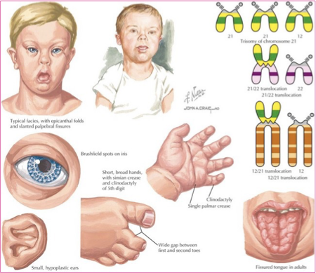
trisomy 21 - down syndrome
most common trisomy
most common chromosomal disorder
47 chromosomes
risk factors:
inc. maternal age
previous T21 pregnancy
down syndrome gene carriers
common sonographic findings:
brachycephaly
mild ventriculomegaly
flattened face
inc. nuchal thickness
cardiac defects (AVSD, VSD, ToF, echogenic intracardiac focus)
duodenal atresia
echogenic bowel
short limbs
mild hydronephrosis
absent/hypoplastic nasal bone

trisomy 18 - edwards syndrome
2nd most common autosomal trisomy
47 chromosomes
risk factors:
inc. maternal age
common sono findings:
“strawberry” shaped head
choroid plexus cysts
agenesis of corpus callosum
dandy-walker malformation
micrognathia
microphthalmos
hypertelorism
nuchal edema
clenched hands, rocker-bottom foot
cryptochidism
cardiac defects (VSD, CoAo)
omphalocele


trisomy 13 - patau syndrome
3rd most common autosomal trisomy
47 chromosomes
only compatible w/ life for a few days
risk factors:
inc. maternal age
common sono findings:
holoprosencephaly
facial abnormalities


45X/Turner syndrome
most common of the sex chromosome abnormalities in females
no assoc. w/ maternal age
45 chromosomes
common sono findings:
short stature
hydrops
cystic hygroma
generalized edema
pleural effusions
ascites
cardiac abnormalities (CoAo)
renal abnormalities
short femurs

triploidy
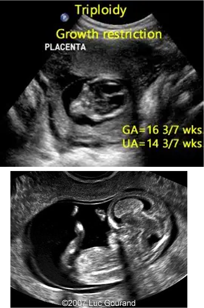
rare lethal chromosomal abnormality caused by the presence of an entire extra chromosomal set
69 chromosomes instead of the normal 46
assoc. with molar pregnancy
fusion of sperm and egg create mass-like structure instead of fetus
while it’s thought to affect as much as 1-2% of conceptions, the vast majority are thought to undergo spontaneous abortions in the early intrauterine period
common sono findings:
large or small placenta
IUGR
holoprosencephaly
agenesis of corpus callosum
dandy walker malformation
meningomyocele
hypertelorism/microphthalmia
micrognathia
inc. nuchal thickness
cystic hygroma
syndactyly (fused fingers)


noonan syndrome
single gene mutation (not a chromosomal abnormality)
mutation found in PTPN11 gene w/ a normal # of chromosomes
both sexes can be affected
affected individuals can bear some clinical features similar to that of Turner syndrome
many have normal life expectancies, depending on severity
common sono findings:
short stature
cystic hygroma
hydrops
hypertelorism
cardiac defects (usually rt-heart abnormalities)
hemivertebrae


apert syndrome
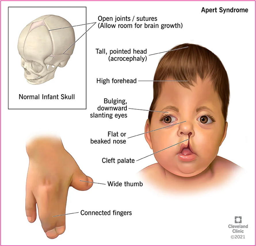
aka “acrocephalosyndactyly”
single gene mutation found in FGFR2 gene w/ normal chromosome #
assoc. w/ advanced paternal age
many have normal life expectancies, depending on severity
common sono findings:
premature fusion of skull, hands, and feet
coronal craniosynostosis (coronal sutures of skull fuse prematurely)
acrocephaly (craniosynostosis causing a cone shaped head w/ a high peak)
syndactyly
inc. nuchal thickness
“beaked” nose
hypertelorism
agenesis of corpus callosum
ventriculomegaly
frontal bossing

meckel-gruber syndrome
single gene mutation
rare, lethal multi-system disorder characterized by multicystic dysplastic kidneys and CNS/hepatic abnormalities
common sono findings:
occipital encephalocele
enlarged, cystic kidneys
polydactyly (extra fingers)


holt-oram syndrome
aka “heart-hand syndrome”
single gene mutation
condition that results in congenital heart defects and upper limb anomalies
common sono findings:
ASD/VSD
CoAo
radius abnormalities (hypoplasia, aplasia)
hand abnormalities (absent thumb, polydactyly)
phocomelia

syndrome