Approach to Anemia in Small Animals
 Call Kai
 Learn
 Practice Test
 Spaced Repetition
 Match
 Flashcards
 Knowt Play
1/29

There's no tags or description
Looks like no tags are added yet.
30 Terms
What is the definition of anemia?
“The reduction of hemoglobin concentration in the blood”
Or:
Reduced oxygen carrying capacity
What is the BIGGEST differential between the types of anemia you may be presented with?
The TIMING
Acute vs. Chronic anemia
There is a spectrum of presentation that anemia can come with depending on TIMING
Acute vs. chronic
Chronic cases of anemia allow for ADAPTATION / COMPENSATION of the hemoglobin to deliver O2 more readily to tissues as their concentration slowly decreases
Animal can be perfectly fine at rest, but with exercise can experience extreme lethargy and even faintness
What are 3 major components that are integral in the production of RBCs?
1) Erythropoietin (EPO)
Produced in the kidneys
Is released in higher concentrations when the kidney experiences a reduction in O2
Stimulates progenitor cells and encourages them to become RBCs
2) Cytokines
Produced by stromal (muscle) cells; assist in production of RBCs
3) Hemosiderin
An iron-based component from macrophages (in bone marrow) that is made to help produce hemoglobin
Allows RBCs to mature
What is the FIRST thing you need to determine when investigating an anemia case? (Essentially, what are the two different KINDS of anemia?)
Is it REGENERATIVE, or NON-REGENERATIVE ANEMIA?
Regenerative
Losing RBCs, but their production is uninhibited
EPO will be increased; evidence of premature / deformed RBCs due to sudden increase in production
Non-regenerative
Losing RBCs BECAUSE they aren’t regenerating
Issue of bone marrow, or ANYWHERE along the line of production for RBCs
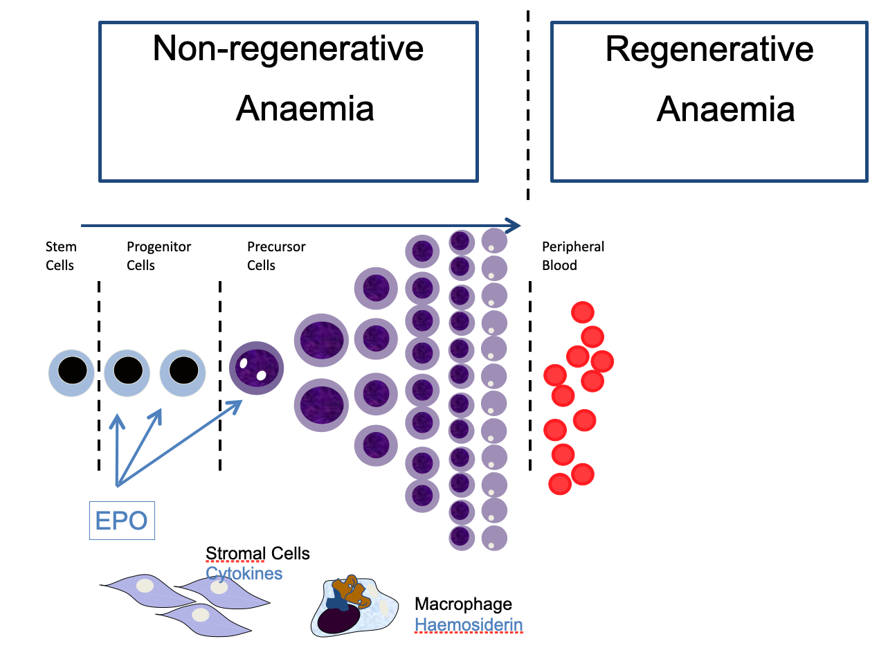
What are the two differentials that NON-REGENERATIVE anemia be split into?
Primary Bone Marrow Disease
When the bone marrow itself is the reason for the lack of RBCs
Secondary Failure of Erythropoesis
When something is going wrong with the actual prodcution of RBCs
Is ALWAYS caused by some other issue in the body, will ALWAYS be presenting with a primary condition
What are the 5 major diseases that cause Primary Bone Marrow Disease → Non-Regenerative Anemia?
PIMA = precursor immune-mediated anemia **
Immune system targets the immature RBC precursors in the bone marrow
Seen in dogs and cats
Pure red cell aplasia
ALSO immune mediated; the COMPLETE ELIMINATION of the earliest of RBC precursors (hence aplasia)
Aplastic anemia
Associated with ESTROGEN toxicity; seen in entire male dogs with sertoli cell tumors producing estrogen; causes aplastic anemia
Shuts off production of ALL cell lines -> PANCYTOPENIA (anemia + leukopenia + thrombocytopenia)
Myelofibrosis
Stromal cells / fibrosis OVERTAKES bone marrow
Bone marrow infiltration
Neoplastic infiltration of bone marrow (stage 5 lymphoma, leukemia)
What are the 5 major conditions that cause SECONDARY Failure of Erythropoiesis → Non-Regenerative Anemia?
Anaemia of inflammatory disease
A moderate anemia (PCV: high 20s in dogs, high teens in cats)
Inflammatory cytokine production prevents bone marrow / RBCs from getting ACCESS to iron needed for RBC production
So not an iron deficiency, an inability to USE said iron
Will ALWAYS have an inflammatory issue that they are presenting with as well
Chronic renal failure
Lack in production of EPO → LACK OF RBCs
Endocrine disease
Haemoglobin synthesis defects - Fe deficiency
Nuclear maturation defects - Cobalamin deficiency
What are the DISTINGUISHING clinical features that could help you determine an anemia is a Primary Bone Marrow Disease?
Clinical signs relate to gradual reduction in oxygen carrying capacity:
lethargy/weakness/exercise intolerance
Relatively BAR for degree of anaemia due to progressive reduction of RBCs over time
Other cell lines may be affected: thrombocytopenia/neutropenia
What are the DISTINGUISHING clinical features that could help you determine if an anemia is a Secondary Failure of Erythropoiesis?
**** CLINICAL SIGNS RELATE TO PRIMARY DISEASE PROCESS ****
Chronic renal failure: PUPD, reduced appetite, weight loss, vomiting
Anaemia of chronic disease: often mild to moderate anaemia and the anaemia doesn’t explain all the clinical signs
Hypothyroidism: dermatological disease
Fe deficiency: is typically associated with chronic low grade external blood loss due to parasitism (internal or external), ulcerative GI disease, chronic urinary losses
Cobalamin deficiency is typically associated with chronic GI disease, or rarely with genetic defects resulting in inability to absorb vitamin B12
What are the two different kinds of general causes that REGENERATIVE anemia can be split into?
HEMOLYTIC
vs
HEMORRHAGE
What are the 3 different differentials that HEMOLYTIC anemia is split into?
Immune-mediated
Primary (non-associative) IMHA
True autoimmune disorder
Secondary (associative) IMHA
TRIGGER causes development of anti-erythrocyte antibodies
Infections
Neoplasms
Non-immune mediated
Oxidative injury
Through TOXICITIES
Heinz Body Anemia
Infectious
Hemotrophic mycoplasms (CATS)
Babesiosis (DOGS)
What are 4 different conditions that could cause HEMORRHAGIC anemia?
1) Trauma
2) GI ulceration
3) Hemostatic disorder (coagulopothies)
4) Ruptured neoplasm (ex. splenic hemangiosarcoma)
What exactly is Immune-Mediated Hemolytic Anemia?
An anemia characterized by the presence of erythrocyte-bound antibodies
Produced due to the presence of an antigenic RBC
Antigenic either because the immune system simply recognizes it as an antigen (primary), or because an infection of the RBC caused the accidental production of an antibody against the pathogen AND RBC (secondary)
What are the two different ways RBCs get hemolyzed during IMHA? Why is this important?
Extra-vascular hemolysis
Antibody on RBC is recognized by a macrophage in the liver or spleen
Intra-vascular hemolysis
A complement-mediated lysis of RBCs inside the blood vessels
Membrane attack complexes formed
Important because they PRESENT DIFFERENTLY
Intravascular → ACUTE onset
Produce DARK DARK URINE (hemoglobinuria); owners will notice and bring dog in for that
Extravascular → JAUNDICE
+ hepatosplenomegally

What are the 3 main “triggers” that lead to the development of Secondary (associative) IMHA?
Infectious
Bacterial (leptospirosis, haemoplasmas*)
Parasitic (babesia*, leishmania, ehrlichia)
Drug-induced
Sulphonamides, penicillins, vaccines
Neoplasia
Lymphoma, leukaemias, multiple myeloma
SECONDARY IMHA INFECTIOUS CAUSES
** Keep in mind, these cause infectious hemolytic anemia on their OWN; BUT they CAN result in secondary IMHA due to the immune system. Will need to treat for both the virus and the IMHA if that happens
Describe Feline Infectious Anemia… how is it transmitted? What are the main pathogens that cause this? Clinical signs? Diagnosis? Treatment?
Pathogens:
Mycoplasma hemofelis ***
M. hemominutum (FeLV) **
M. turicensis
Is transmitted by blood-to-blood contact (FLEAS)
Mainly a disease of outdoor cats
Cause haemolysis involving immune-mediated and direct erythrocyte damage (plus sequestration)
Typically causes a regenerative anaemia (although concurrent disease may result in non-regenerative anaemia) +/- jaundice
So extra-vascular hemolysis
Often (but not always) ISA and Coombs positive
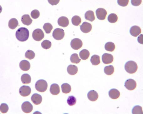
Diagnosis: Identification of haemoplasmas on blood smears (Differential dx Howell-Jolly bodies; can be seen on blue reticulocyte on image) or PCR
Treatment: doxycycline/fluoroquinolones +/- steroids/immunosuppression
Describe Canine Babesiosis… how is it transmitted? What are the main pathogens that cause this? Clinical signs? Diagnosis? Treatment?
Pathogens:
Babesia (canis) canis
Babesia (canis) vogeli
Babesia (canis) rossi
Babesia gibsoni
Is a tick-borne disease
Transmitted by Rhipicephalus & Dermacentor ticks
Clinical Presentation
Acute intravascular hemolysis
Pyrexia
Haemolytic anaemia, including IMHA
Auto-agglutination in 21%
85% Coombs positive
Thrombocytopenia
+/- splenomegaly, hyperbilirubinaemia and haemoglobinuria
+/- AKI, hepatopathy, SIRS, CNS dysfunction
Diagnosis: Identification on blood smears or PCR
Treatment: Imidocarb dipropionate (Imizol) [Unlicensed for dogs in UK]
Need to treat concurrent IMHA as well and suppress the immune system

HEMOLYTIC ANEMIA - OXIDATIVE INJURY
What are the 6 most common toxicities that cause hemolytic anemia?
Paracetamol toxicity
ESPECIALLY cats
Onion toxicity
Benzocaine
Zinc toxicity
Propofol infusion
Diabetes mellitus, hepatic lipidosis in cats
What are the classic signs of OXIDATIVE INJURY hemolytic anemia?
Heinz-bodies
Refractory blebs on RBCs
Chocolate-colored oral mucous membranes and urine
Sign of metHb, which is produced when the iron in hemoglobin is OXIDATED (i.e. why brown mm is a sign of oxidative hemolytic anemia)
Liver failure
Ascites
CARDNAL SIGNS:
Intravascular hemolysis (acute anemia)
Characteristics of oxygenated Hb in RBCs
Heinz bodies
Eccentrocytes
Denatured hemoglobin accumulating at one side of the RBC (see image)

ONTO LAB EVALUATION:
What should be your approach to assessing an emergent case that might be experiencing anemia? What are your first steps, and what do you determine first?
TAKE BLOOD SAMPLE AS YOU STABILIZE THE PATIENT
Once patient is stabilized somewhat, evaluate blood:
PCV
How severe is the anemia?
Does it explain the clinical presentation, or could there be a different cause?
Blood Smear
Is the anemia regenerative or non-regenerative?
What do the RBCs look like?
Evidence of IMHA (SPHEROCYTES)
Evidence of oxidative injury
Evidence of iron deficiency
Blood Cytology
Are other cell lines affected?
Could anemia be due to bone marrow failure?
Does the patient have enough platelets?
Could anemia be secondary to a bleeding disorder?
Does patient have other evidence of coagulopathy? (petechiation / ecchymoses)
What are the PCV values for a mild, moderate, and severe anemia?
Mild: 30-39%
Moderate: 20-30%
Severe: anything < 20%

On a blood smear, how can you tell if an anemia is regenerative vs. non-regenerative? Is this regenerative or non-regenerative anemia?
Blood smear is REGENERATIVE ANEMIA
Regenerative anemia characteristics:
Increase in RETICULOCYTES (immature RBCs)
Polychromasia - gray / blue staining of reticulocytes
Anisocytosis - variation in cell size
Nucleated RBCs
Non-regenerative anemia characteristics:
Depends on the type of non-regenerative anemia (primary BM or secondary) but generally, will always have normal-sized and normal-colored RBCs, just fewer
On blood cytology, what is regenerative anemia classified as?
Macrocytic hypochromic
MCV ^
MCHC down
MCV = mean corpuscular volume = size of the RBCs
MCHC = mean corpuscular hemoglobin concentration = color of RBCs (lower → pale)
On blood cytology, what is non-regenerative anemia classified as?
DEPENDS ON TYPE OF NON-REGENERATIVE
Primary BM Disease
Typically normocytic normochromic (looks completely normal, just less RBCs)
Will ALSO see other cell lines suffering
Pancytopenia
Bicytopenia
Anemia w/ thrombocytopenia or leukocytopenia, not both (thats pan)
Secondary Failure of Erythropoesis
Varies more depending on cause:
Iron deficiency → Microcytic hypochromic
Don’t have the proper building blocks for a mature RBC
Usually caused by chronic external blood loss in older patients (GI ulcers, parasites, clotting disorders)
Chronic Renal Failure → Normocytic normochromic
BIOCHEM evidence of renal failure (azotemia)
Anemia of Inflammation → Normocytic normochromic
Typically a mild / moderate anemia in response to a chronic inflammatory disease
IDENTIFYING IMHA
What are the DISTINGUISHING laboratory features of IMHA? What are the characteristics you’ll see on blood smear? What will be seen in the blood cytology?
IMHA → regenerative anemia → regenerative anemia characteristics
Anisocytosis
Polychomasia
Increased conc. of reticulocytes
Nucleated RBCs
Macrocytic hypochromic
BUT, will also see:
SPHEROCYTOSIS
Biggest indicator on blood smear; are spherical, densly stained RBCs
Like this due to membrane damage from phagocytes
Leukocytosis
Due to immune response to RBCs
Hyperbilirubinemia
Organ dysfunction
^ALT, ^cPLI
** side note: if patient has concurrent thrombocytopenia → EVAN’S SYNDROME
What are two confirmation tests that can be done to solidify an IMHA diagnosis?
Slide-Agglutination Test
Saline + blood on slide, watch for clumping… if clumping, positive
Direct Coomb’s Test
Adds species-specific reagent that will bind to the Abs present on the RBCs, causing agglutination (positive result)

When it comes to TREATMENT of IMHA… what are your first line medications?
STEROIDS
Specifically:
Prednisolone
Dexamethasone
Prevents the macrophages from reacting to the Ab on the RBCs
May also need to add an additional immune-suppressant on top of the steroids (i.e. Azathioprine, Ciclosporin)
May need blood transfusions if IMHA severe… may even be dependent on them
What is the prognosis of IMHA? What is most often the cause of death?
Not good; mortality rate of 25-70%
Causes of death:
Refractory to treatment
Hypoxemia
Pulmonary thromboembolism
Disseminated Intravascular Coagulation (DIC)
RESULTING PULMONARY THROMBOEMBOLISM IS THE MOST COMMON CAUSE OF DEATH
How do we prevent the development of pulmonary thromboembolic disease in patients with IMHA?
By incorporating its treatment into our IMHA treatment plan:
Oral thromboprophylaxis:
(Aspirin), clopidogrel*, or rivaroxaban
Management of Pulmonary Thromboembolism
Oxygen
Heparin in addition to clopidogrel
Really quickly, what are the distinguishing laboratory features of an animal with Hemorrhagic Anemia?
Again, depends…
May be “Pre-Regenerative”, meaning that the hemorrhage was so acute that the bone marrow didn’t even have time to try to make new RBCs
But for more chronic cases / coagulopathies…
Blood smear:
Presence of SCHISTOCYTES / RBC fragmentation
Concurrent hypoproteinemia (due to direct loss of proteins through blood)
Blood cytology for hemostatic disorders:
THROMBOCYTOPENIA will be seen
Prolonged coagulation times of blood samples