A&P Lab XII
 Call Kai
 Learn
 Practice Test
 Spaced Repetition
 Match
 Flashcards
 Knowt Play
1/92

There's no tags or description
Looks like no tags are added yet.
93 Terms
anterior cavity
filled with aqueous humor

anterior chamber

posterior chamber
blue

cornea

iris

aqueous humor

pupil

posterior cavity

vitreous humor

ciliary body

ciliary processes

lens
H

suspensory ligaments
stringlike ligaments

ora serrata

retina
M

choroid

optic disk/blind spot

fovea

macula lutea
N

optic nerve

upper eyelid

lower eyelid

sclera

ocular conjunctiva

lacrimal caruncle

lacrimal gland

lacrimal punctum
D

lacrimal sac
A

nasolacrimal duct
F

superior lacrimal canaliculus

inferior lacrimal canaliculus

choroid (retina microscope slide)
a

pigmented part of retina

rods and cones

Nuclei of rods and cones (retina)
a

bipolar cells

ganglion cells

aqueous humor
what fluid is found in anterior cavity?
vitreous humor
what fluid is found in the posterior cavity?
snellen eye chart

astigmatism chart

weber test

Rinne test

ampulla
dilated portion of a canal or duct

auditory (eustachian) tube
connects middle ear to throat, equalizes pressure in the middle ear

auricle (pinna)
collects and directs sound waves into the external acoustic meatus
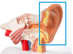
basilar membrane of cochlea
separates the cochlear duct from the scala tympani

bony labyrinth
filled with perilymph
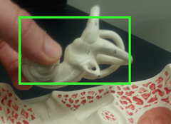
crista ampullaris
responds to changes in the velocity of rotational head movements

cochlea
tube though which sound waves trigger nerve impulses; hearing

cochlear duct
filled with endolymph; supports spiral organ

cochlear branch
hearing

cupula

endolymph in the cochlea

endolymphatic sac
stores excess endolymph

external auditory canal
transmits sound waves from the pinna to the tympanic membrane of the middle ear
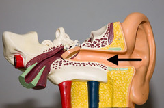
inner hair cells (cochlea)
neurons in organ of corti; responsible for auditory transduction

outer hair cells (cochlea)
neurons in the organ of corti; serve to amplify and sharpen responses of inner hair cells

incus
transmitting vibrations between malleus and stapes

macula
responsible for central vision
malleus
transmits vibrations of eardrum to incus

organ of corti (spiral organ)
organ of hearing

otolith
A
moves in response to gravity

oval window
transmits vibrations from the stapes to the perilymph of the scala vestibuli

perilymph of cochlea

round window
bulge as a pressure relief

saccule
equilibrium: static and linear (S)

semicircular canals
three canals within the inner ear that contain specialized receptor cells that generate nerve impulses with body movement

spiral ganglion
a collection of neurons in the modiolus of the cochlea that receives input from hair cells and sends output to the cochlear nuclei in the medulla via the auditory nerve

stapedius muscle
reduces movement of stapes at oval window
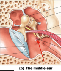
stapes
transmits sound vibrations from the incus to the oval window

Tectorial Membrane of the cochlea
A membrane located above the basilar membrane; serves as a shelf against which the cilia of the auditory hair cells move

tensor tympani muscle
stiffens tympanic membrane

tympanic duct (scala tympani)
contains perilymph
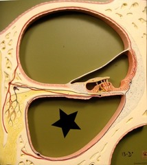
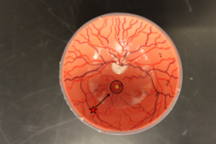
tympanic membrane
eardrum

utricle
U

Vestibular Duct (scala vestibuli)

vestibule
portion of the inner ear that senses the position of the head

Vestibular Membrane of the cochlea
separates the cochlear duct from the scala vestibuli

vestibulocochlear nerve
hearing and balance

palpebrae (sheep eye)
protect the eyes and spread lacrimal fluid with blinking

cornea (sheep eye)
forms a clear window that is the major light bending medium of the eye

iris (sheep eye)
controls the amount of light that enters the eye

ciliary body (sheep eye)
contains ciliary muscle and the ciliary process

retina (sheep eye)
begin processing of visual information

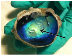
tapetum lucidum (sheep eye)
iridescent layer found in nocturnal animals for maximizing vision under low intensity light
aqueous humor (sheep eye)
fluid in anterior chamber

vitreous humor (sheep eye)
fluid in posterior cavity

optic nerve (sheep eye)
carries neural impulses from the eye to the brain

pupil (sheep eye)
opening in center of iris

sclera (sheep eye)
tough, white, outer layer of the eyeball

lens (sheep eye)
the transparent structure behind the pupil that changes shape to help focus images on the retina

choroid (sheep eye)
nourishes other layers and helps absorb excess light
